. 2020 Feb 18;25(4):901.
doi: 10.3390/molecules25040901.

Moscatilin Induces Apoptosis in Human Head and Neck Squamous Cell Carcinoma Cells via JNK Signaling Pathway

Affiliations

Moscatilin Induces Apoptosis in Human Head and Neck Squamous Cell Carcinoma Cells via JNK Signaling Pathway

Eunji Lee et al. Molecules. .

Abstract

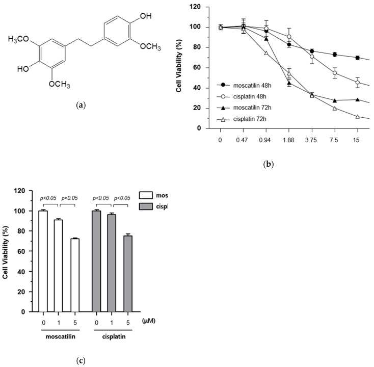
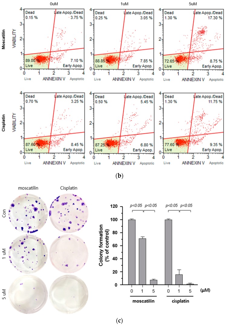
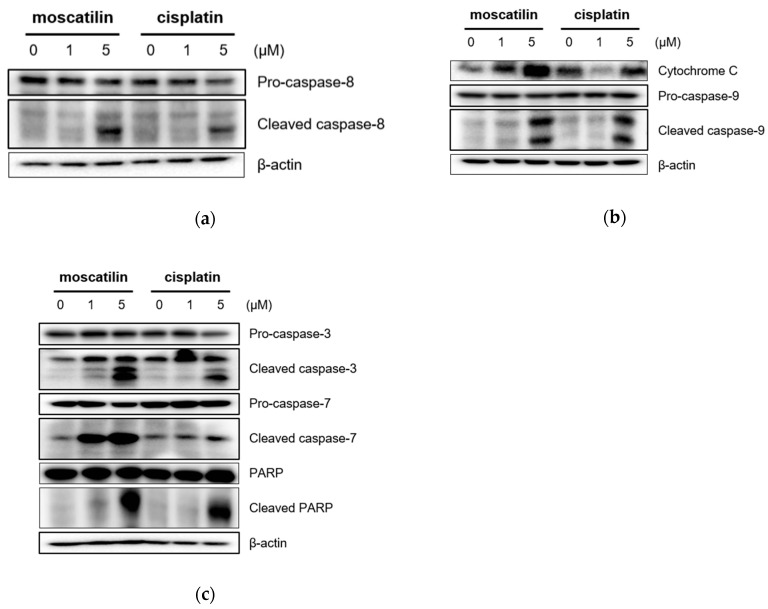
Dendrobii Herba is an herbal medicine that uses the stems of Dendrobium species (Orchidacea). It has been traditionally used to treat fever, hydrodipsomania, stomach disorders, and amyotrophia. In our previous study, a bibenzyl compound, moscatilin, which is isolated from Dendrobii Herba, showed potent cytotoxicity against a FaDu human pharyngeal squamous carcinoma cell line. Prompted by this finding, we performed additional studies in FaDu cells to investigate the mechanism of action. Moscatilin induced FaDu cell death by using 5 μM of concentration and by mediating apoptosis, whereas cell proliferation following treatment with 1 μM of moscatilin was not suppressed to the same levels as by the anti-cancer agent, cisplatin. Apoptosis-related protein expression (cleaved caspase-8, cleaved caspase-7, cytochrome c, cleaved caspase-9, cleaved caspase-3, and poly (ADP-ribose) polymerase (PARP) was increased by treating with 5 μM of moscatilin. This suggests that moscatilin-mediated apoptosis is associated with the extrinsic and intrinsic apoptotic signaling pathways. In addition, moscatilin-induced apoptosis was mediated by the c-Jun N-terminal kinase (JNK) signaling pathway. Overall, this study identified additional biological activity of moscatilin derived from natural products and suggested its potential application as a chemotherapeutic agent for the management of head and neck squamous cell carcinoma.

Keywords: Dendrobium; FaDu; apoptosis; head and neck squamous cell carcinoma; moscatilin.


Conflict of interest statement

The authors declare no conflicts of interest.

Figures

Figure 1
Figure 1
Cytotoxicity of moscatilin on FaDu cells. (a) Chemical structure of moscatilin. (b) FaDu cells (2 × 10⁴ cells/well) were seeded in 96-well plates and treated with 0.47–30 μM moscatilin or cisplatin for the indicated times. Cell viability was measured using a CCK-8 assay kit. (c) Cell viability was measured by the CCK-8 assay kit 48 h after treatment with 1 μM moscatilin, 5 μM moscatilin, or cisplatin for 48 h. The values are expressed as the mean ± SD of five independent experiments.
Figure 2
Figure 2
Moscatilin induced cell death is mediated by apoptosis. (a) FaDu cells (2 × 10⁴ cells/well) were treated with 1 μM moscatilin, 5 μM moscatilin, or cisplatin for 48 h. The live and dead cells were stained with green calcein-AM and ethidium homodimer-1 (red) and then analyzed using confocal microscopy. Scale bar is 50 μm. The values are expressed as the mean ± SD of five independent experiments. (b) FaDu cells (2 × 10⁵ cells) were seeded in six-well plates and treated with 1 μM moscatilin, 5 μM moscatilin, or cisplatin. After a 48 h incubation, the cells were collected, washed twice with ice-cold phosphate-buffered saline (PBS), resuspended in PBS containing 10% FBS, and then stained with annexin V and PI for 20 min. The apoptotic population was analyzed by the Muse Cell Analyzer. (c) Equal amounts of FaDu cells were seeded in six-well plates and treated with the indicated concentrations of moscatilin and cisplatin. After a 24-h incubation, the FaDu cells were incubated in culture media without moscatilin or cisplatin for 10 days, stained with crystal violet, and then the colonies were counted. The values are expressed as the mean ± SD of three independent experiments.
Figure 2
Figure 2
Moscatilin induced cell death is mediated by apoptosis. (a) FaDu cells (2 × 10⁴ cells/well) were treated with 1 μM moscatilin, 5 μM moscatilin, or cisplatin for 48 h. The live and dead cells were stained with green calcein-AM and ethidium homodimer-1 (red) and then analyzed using confocal microscopy. Scale bar is 50 μm. The values are expressed as the mean ± SD of five independent experiments. (b) FaDu cells (2 × 10⁵ cells) were seeded in six-well plates and treated with 1 μM moscatilin, 5 μM moscatilin, or cisplatin. After a 48 h incubation, the cells were collected, washed twice with ice-cold phosphate-buffered saline (PBS), resuspended in PBS containing 10% FBS, and then stained with annexin V and PI for 20 min. The apoptotic population was analyzed by the Muse Cell Analyzer. (c) Equal amounts of FaDu cells were seeded in six-well plates and treated with the indicated concentrations of moscatilin and cisplatin. After a 24-h incubation, the FaDu cells were incubated in culture media without moscatilin or cisplatin for 10 days, stained with crystal violet, and then the colonies were counted. The values are expressed as the mean ± SD of three independent experiments.
Figure 3
Figure 3
Moscatilin induces the apoptosis of FaDu cells through the extrinsic and intrinsic apoptotic pathways. FaDu cells were treated with 1 μM moscatilin, 5 μM moscatilin, or cisplatin. After a 24-h incubation, the cells were harvested and cell lysates were prepared. The levels of (a) pro-caspase-8 and cleaved caspase-8, (b) cytochrome C, pro-caspase-9, and cleaved caspase-9, (c) pro-caspase-3, cleaved caspase-3, pro-caspase-7, cleaved caspase-7, PARP, and cleaved PARP were compared by Western blotting. β-actin was used as a loading control.
Figure 4
Figure 4
Moscatilin-induced apoptosis in FaDu cells was regulated by the c-Jun N-terminal kinase (JNK) signaling pathway. (a) FaDu cells were treated with 1 or 5 μM of moscatilin or cisplatin. After a 24-h incubation, the cells were harvested and cell lysates were prepared. The levels of p35, phosphor-p38, ERK 1/2, phosphor-ERK 1/2, JNK, and phosphor-JNK were compared by Western blotting. β-actin was used as a loading control. FaDu cells were pre-treated with 10 μM and 50 μM of SP600125 (JNK inhibitor) (b) or Z-VAD-FMK (a pan-caspase inhibitor) (c) for 30 min, prior to treatment with 5 μM moscatilin or cisplatin. The levels of JNK, phosphor-JNK, caspase-3, cleaved caspase-3, PARP, and cleaved PARP, were compared by Western blotting. β-actin was used as a loading control.
Figure 4
Figure 4
Moscatilin-induced apoptosis in FaDu cells was regulated by the c-Jun N-terminal kinase (JNK) signaling pathway. (a) FaDu cells were treated with 1 or 5 μM of moscatilin or cisplatin. After a 24-h incubation, the cells were harvested and cell lysates were prepared. The levels of p35, phosphor-p38, ERK 1/2, phosphor-ERK 1/2, JNK, and phosphor-JNK were compared by Western blotting. β-actin was used as a loading control. FaDu cells were pre-treated with 10 μM and 50 μM of SP600125 (JNK inhibitor) (b) or Z-VAD-FMK (a pan-caspase inhibitor) (c) for 30 min, prior to treatment with 5 μM moscatilin or cisplatin. The levels of JNK, phosphor-JNK, caspase-3, cleaved caspase-3, PARP, and cleaved PARP, were compared by Western blotting. β-actin was used as a loading control.

References

    1. Leemans C.R., Braakhuis B.J., Brakenhoff R.H. The molecular biology of head and neck cancer. Nat. Rev. Cancer. 2011;11:9–22. doi: 10.1038/nrc2982. - DOI - PubMed
    1. Pezzuto F., Buonaguro L., Caponigro F., Ionna F., Starita N., Annunziata C., Buonaguro F.M., Tornesello M.L. Update on head and neck cancer: Current knowledge on epidemiology, risk factors, molecular features and novel therapies. Oncology. 2015;89:125–136. doi: 10.1159/000381717. - DOI - PubMed
    1. Park M.R., Kim S.G., Cho I.A., Oh D., Kang K.R., Lee S.Y., Moon S.M., Cho S.S., Yoon G., Kim C.S., et al. Licochalcone-A induces intrinsic and extrinsic apoptosis via ERK1/2 and p38 phosphorylation-mediated TRAIL expression in head and neck squamous carcinoma FaDu cells. Food Chem. Toxicol. 2015;77:34–43. doi: 10.1016/j.fct.2014.12.013. - DOI - PMC - PubMed
    1. Seo Y.S., Yim M.J., Kim B.H., Kang K.R., Lee S.Y., Oh J.S., You J.S., Kim S.G., Yu S.J., Lee G.J., et al. Berberine-induced anticancer activities in FaDu head and neck squamous cell carcinoma cells. Oncol. Rep. 2015;34:3025–3034. doi: 10.3892/or.2015.4312. - DOI - PubMed
    1. Crooker K., Aliani R., Ananth M., Arnold L., Anant S., Thomas S.M. A review of promising natural chemopreventive agents for head and neck cancer. Cancer Prev. Res. (Phila) 2018;11:441–450. doi: 10.1158/1940-6207.CAPR-17-0419. - DOI - PMC - PubMed

MeSH terms