. 2020 Feb 18;8(2):35.
doi: 10.3390/biomedicines8020035.

5-(Carbamoylmethylene)-oxazolidin-2-ones as a Promising Class of Heterocycles Inducing Apoptosis Triggered by Increased ROS Levels and Mitochondrial Dysfunction in Breast and Cervical Cancer

Affiliations

5-(Carbamoylmethylene)-oxazolidin-2-ones as a Promising Class of Heterocycles Inducing Apoptosis Triggered by Increased ROS Levels and Mitochondrial Dysfunction in Breast and Cervical Cancer

Biagio Armentano et al. Biomedicines. .

Abstract

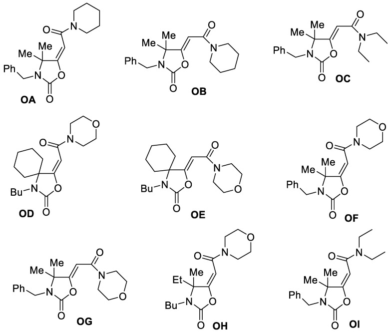
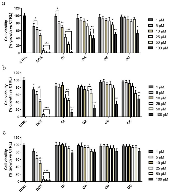
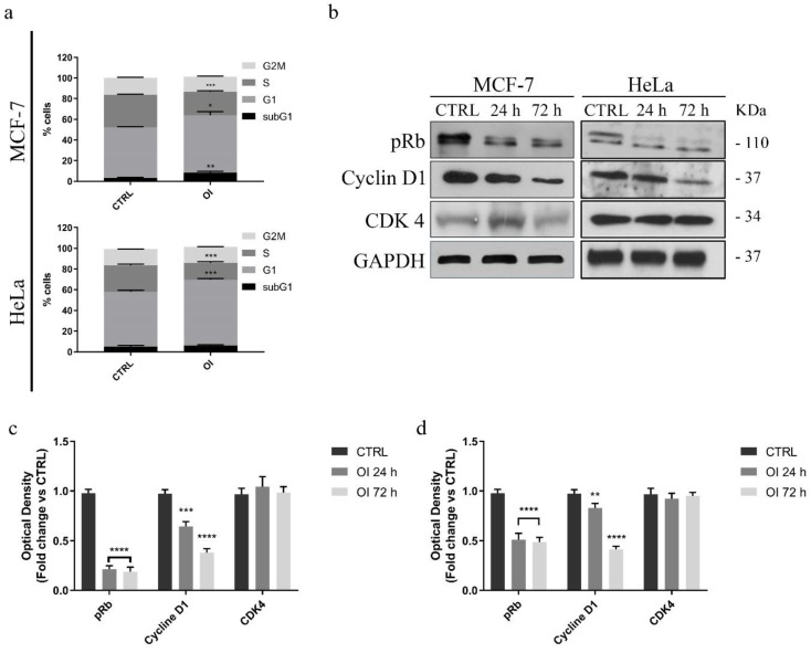
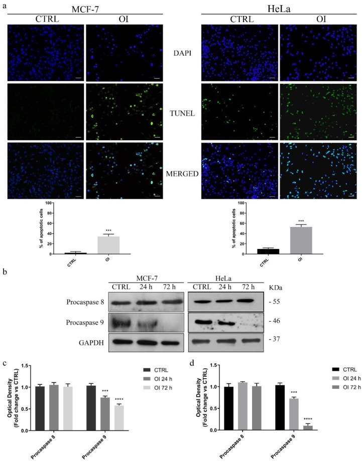
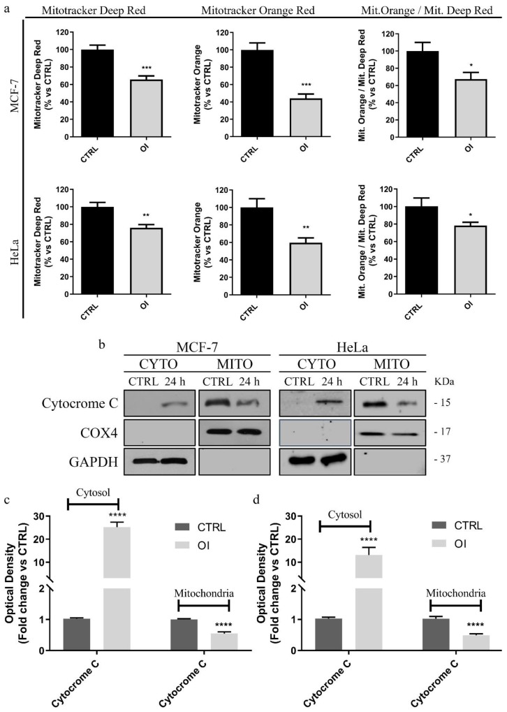
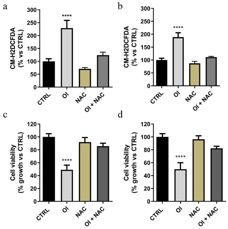
Oxazolidinones are antibiotics that inhibit protein synthesis by binding the 50S ribosomal subunit. Recently, numerous worldwide researches focused on their properties and possible involvement in cancer therapy have been conducted. Here, we evaluated in vitro the antiproliferative activity of some 5-(carbamoylmethylene)-oxazolidin-2-ones on MCF-7 and HeLa cells. The tested compounds displayed a wide range of cytotoxicity on these cancer cell lines, measured by MTT assay, exhibiting no cytotoxicity on non-tumorigenic MCF-10A cells. Among the nine tested derivatives, four displayed a good anticancer potential. Remarkably, OI compound showed IC50 values of 17.66 and 31.10 µM for MCF-7 and HeLa cancer cells, respectively. Furthermore, we assessed OI effect on the cell cycle by FACS analysis, highlighting a G1 phase arrest after 72 h, supported by a low expression level of Cyclin D1 protein. Moreover, mitochondrial membrane potential was reduced after OI treatment driven by high levels of ROS. These findings demonstrate that OI treatment can inhibit MCF-7 and HeLa cell proliferation and induce apoptosis by caspase-9 activation and cytochrome c release in the cytosol. Hence, 5-(carbamoylmethylene)-oxazolidin-2-ones have a promising anticancer activity, in particular, OI derivative could represent a good candidate for in vivo further studies and potential clinical use.

Keywords: ROS; anticancer compounds; apoptosis; mitochondria; oxazolidinones.


Conflict of interest statement

The authors declare no conflict of interest.

Figures

Scheme 1
Scheme 1
The in vitro anticancer properties of 5-(carbamoylmethylene)-oxazolidin-2-ones (OA-OI).
Figure 1
Figure 1
Effects of the compounds OA-OI on MCF-7, HeLa, and MCF-10A cell growth. (a) Breast cancer cells (MCF-7), (b) human uterine cervix adenocarcinoma cells (HeLa), and (c) non-tumorigenic breast epithelial cells (MCF-10A) were treated for 72 h with vehicle alone (control cells, CTRL) or increasing doses (1 to 100 μM) of each compounds or doxorubicin (DOX), as indicated. Cell viability was assessed by MTT assay and was expressed as percentage of growth vs. CTRL. Values represent means ± SD of three different experiments, each performed with triplicate samples. * p value < 0.05; ** p value < 0.01; *** p value < 0.001.
Figure 2
Figure 2
Effects of the compound OI on the cell cycle progression in cancer cells. (a) Quantitative analysis of the percentage of cells arrested in different phases of the cell cycle was indicated. Immunoblot and densitometric analyses of several controlling proteins of the G1/S phase transition in MCF-7 (b) and HeLa (c,d) treated cells. GAPDH was used as a loading control. Histograms represent means ± SD of three different experiments in which band intensities were assessed as optical density arbitrary units (OD), and expressed as fold change vs. control samples (CTRL). It is noteworthy that OI treatment impairs the G1/S transition of both cancer cell lines. * p value < 0.05; ** p value < 0.01; *** p value < 0.001; **** p value < 0.0001.
Figure 3
Figure 3
OI treatment triggers cell death by apoptosis. (a) TdT-mediated dUTP nick-end-labeling (TUNEL) assay in MCF-7 and HeLa cells treated for 72 h with DMSO (CTRL) or 15 and 30 µM OI, respectively. DAPI was used to visualize the cell nucleus. Scale Bars 50 µm. Histograms represent means ± SD of apoptotic cells from three independent experiments. (b) Immunoblot analysis of pro-caspases 8 and 9 after 24 and 72 h of OI treatment (see Materials and Methods, par. 2.6). GAPDH was used as a loading control. Densitometric quantification of pro-caspase expression levels in MCF-7 (c) and HeLa (d) treated cells vs. control cells. Remarkably, OI treatment induces apoptosis by activating the intrinsic caspase-dependent apoptotic pathways. Values are means ± SD of three different experiments. *** p value < 0.001; **** p value < 0.0001.
Figure 4
Figure 4
OI treatment induces mitochondrial damage in cancer cells. (a) Both MCF-7 and HeLa cells were treated for 24 h either with DMSO (CTLR) or 15 and 30 µM OI, respectively, then they were stained with different metabolic probes and analyzed by FACS. Note that the treatment decreases mitochondrial mass (MitoTracker Deep-Red) and mitochondrial membrane potential (MitoTracker Orange). Assessment of mitochondrial membrane potential vs. mitochondrial mass displayed that OI decreases mitochondrial membrane potential per mitochondria. Immunoblot and densitometric analysis of cytochrome c in treated MCF-7 (b,c) and HeLa (b,d) cells. GAPDH was used as a loading control of cytosolic fractions, whereas COX4 of the mitochondrial one. A significant release of cytochrome c into the cytosol is revealed in both tested cancer cell lines. Histograms represent means ± SD of three independent experiments in which band intensities were assessed as optical density arbitrary units (OD) and expressed as fold change vs. control samples (CTRL). **** p value < 0.0001.
Figure 5
Figure 5
The compound OI causes a significant increase in ROS levels. Both MCF-7 (a) and HeLa (b) cells were treated for 24 h with 15 and 30 µM OI, respectively, alone and/or in the presence of N-acetyl-l-cysteine (NAC); ROS levels were evaluated by FACS analysis using the CM-H2DCFDA probe. Cell proliferation assay (MTT assay) was performed in both treated MCF-7 (c) and HeLa (d) cells, in the presence or absence of 3 mM NAC. Note that after 72 h, in the presence of NAC, cells displayed recovery in growth. Values represent means ± SD of three different experiments, each performed with triplicate samples. * p value < 0.05; **** p value < 0.0001.

Similar articles

Cited by

References

    1. Cancer Fact Sheet N 297. [(accessed on 25 November 2019)]; Available online: www.who.int/mediacentre/factsheets/fs297/en/
    1. Friberg S., Nystrom A. Cancer Metastases: Early Dissemination and Late Recurrences. Cancer Growth Metastasis. 2015;8:43–49. doi: 10.4137/CGM.S31244. - DOI - PMC - PubMed
    1. Roger A.J., Munoz-Gomez S.A., Kamikawa R. The Origin and Diversification of Mitochondria. Curr. Biol. 2017;27:R1177–R1192. doi: 10.1016/j.cub.2017.09.015. - DOI - PubMed
    1. Pandit N., Singla R.K., Shrivastava B. Current updates on oxazolidinone and its significance. Int. J. Med. Chem. 2012;2012:159285. doi: 10.1155/2012/159285. - DOI - PMC - PubMed
    1. Stevens D.L., Dotter B., Madaras-Kelly K. A review of linezolid: The first oxazolidinone antibiotic. Expert Rev. Anti-Infect. Ther. 2004;2:51–59. doi: 10.1586/14787210.2.1.51. - DOI - PubMed