. 2020 Feb 18;12(2):472.
doi: 10.3390/cancers12020472.

Effect of Diphtheria Toxin-Based Gene Therapy for Hepatocellular Carcinoma

Affiliations

Effect of Diphtheria Toxin-Based Gene Therapy for Hepatocellular Carcinoma

Kenya Kamimura et al. Cancers (Basel). .

Abstract

Hepatocellular carcinoma (HCC) is a major global malignancy, responsible for >90% of primary liver cancers. Currently available therapeutic options have poor performances due to the highly heterogeneous nature of the tumor cells; recurrence is highly probable, and some patients develop resistances to the therapies. Accordingly, the development of a novel therapy is essential. We assessed gene therapy for HCC using a diphtheria toxin fragment A (DTA) gene-expressing plasmid, utilizing a non-viral hydrodynamics-based procedure. The antitumor effect of DTA expression in HCC cell lines (and alpha-fetoprotein (AFP) promoter selectivity) is assessed in vitro by examining HCC cell growth. Moreover, the effect and safety of the AFP promoter-selective DTA expression was examined in vivo using an HCC mice model established by the hydrodynamic gene delivery of the yes-associated protein (YAP)-expressing plasmid. The protein synthesis in DTA transfected cells is inhibited by the disappearance of tdTomato and GFP expression co-transfected upon the delivery of the DTA plasmid; the HCC cell growth is inhibited by the expression of DTA in HCC cells in an AFP promoter-selective manner. A significant inhibition of HCC occurrence and the suppression of the tumor marker of AFP and des-gamma-carboxy prothrombin can be seen in mice groups treated with hydrodynamic gene delivery of DTA, both 0 and 2 months after the YAP gene delivery. These results suggest that DTA gene therapy is effective for HCC.

Keywords: alpha-fetoprotein; diphtheria toxin fragment A; gene therapy; hepatocellular carcinoma; hydrodynamic gene delivery.


Conflict of interest statement

The authors declare that they have no current financial arrangement or affiliation with any organization that may have a direct influence on their work.

Figures

Figure 1
Figure 1
Development of diphtheria toxin fragment A (DTA)-expressing plasmid. The DTA expression vector, containing (a) a chicken β-actin promoter, cytomegalovirus enhancer, and an internal ribosome entry site (IRES) (pCAG–DTA), and (b) a human alpha-fetoprotein (AFP) promoter, cytomegalovirus enhancer, and an IRES (pAFP–DTA).
Figure 2
Figure 2
Effect of DTA on protein synthesis inhibition. (a) Effect of DTA on tdTomato expression in mice hepatocytes 12 h after the hydrodynamic gene delivery of pCAG–tdTomato with pCAG–DTA. (b) A quantitative analysis of tdTomato signal level determined with the ratio of the signal (T/NT ratio) in the transfected (T) liver and non-transfected (NT) liver collected from the mock transfected liver. (c) Effect of DTA on green fluorescent protein (GFP) expression in mice hepatocytes 12 h after the hydrodynamic gene delivery of pCAG–tdTomato with pCAG–DTA. (d) A quantitative analysis of the GFP signal level determined with the T/NT ratio. White arrows indicate expression of tdTomato and GFP. Scale bar represents 100 µm. The values from the two sections of three mice are shown. ** p < 0.01. Student’s t-test. Mock, empty vector; DTA, diphtheria toxin A.
Figure 3
Figure 3
Effect of DTA on hepatocellular carcinoma (HCC) cell growth. The cell growth of HCC cell lines and permanent clones overexpressing DTA determined by MTT assay. (ac) Cell growth of HLE and (df) Huh7, and (gi) HLF cell lines transfected with mock or CAG–DTA or pAFP–DTA. The values represent mean ± standard deviation (five samples from each group of three, where n = 15 for each group at different time points). ** p < 0.01 and no statistical significance (N.S.). Two-way ANOVA followed by Bonferroni’s multiple comparison test. (j) A concentration of AFP in the cell culture medium at 72 h after transfection was quantified by ELISA. The values represent mean ± standard deviation (n = 3 for each group). ** p < 0.01, *** p < 0.001, and N.S. One-way ANOVA followed by Bonferroni’s multiple comparison test.
Figure 4
Figure 4
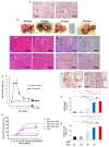
Effect of DTA on HCC mice model. (a) Expression of yes-associated protein (YAP) in the liver 3 days after the hydrodynamic gene delivery of YAP-expressing plasmid (5SA). (b) Representative images of time-dependent liver–tumor occurrence and sizes. (cf) Hematoxylin and eosin staining and (gj) immunohistochemical staining of YAP of the liver tumor. (k) Time dependent changes in YAP expression in the liver after 5SA injection. Quantification was performed by measuring the integrated density in pixels using the ImageJ software (version 1.6.0_20, National Institutes of Health, Bethesda, MD, USA). The values represent mean ± standard deviation (n = 5 for each group). (l,m) Immunohistochemical staining of the AFP of the liver–tumor developed 40 (l) and 100 days (m) after the delivery of 5SA. (n) Cumulative HCC occurrence curve in the liver of 5SA-injected mice generated by the Kaplan–Meier method. The occurrence rate of HCC was compared with 5SA injected with no treatment (5SA, red solid line), treated with pAFP–DTA immediately after treatment (5SA + DTA (0 M), black dot line), 2 months after treatment (5SA + DTA (2 M), blue dot line), 4 months after treatment (5SA + DTA (4 M), blue solid line), and control (pAFP–DTA with no ¬YAP induction, DTA, black solid line). n = 15 for each group. ** p < 0.01 and N.S. compared with the 5SA-delivered mice with no treatment group (red solid line). Log-rank test. Quantitative analysis of area with positive staining for AFP (o) and proliferating cell nuclear antigen (PCNA) (p) in the tumors of each group developed 180 days after YAP-expressing plasmid (5SA) delivery with/without gene therapy of pAFP–DTA. Quantification was performed measuring the integrated density in pixels using the ImageJ software (version 1.6.0_20, National Institutes of Health). The values represent mean ± standard deviation (n = 5 for each group; same number of liver tissue specimens were assessed for no 5SA-delivered group). * p < 0.05; ** p < 0.01; *** p < 0.001; and N.S. One-way ANOVA followed by Bonferroni’s multiple comparison test. 0 M, 2 M, and 4 M, at 0, 2, and 4 months after the delivery of pAFP–DTA.
Figure 5
Figure 5
Tumor markers in YAP-induced HCC mice model treated with pAFP–DTA. Serum concentration of AFP and des-gamma carboxyprothrombin (DCP) quantified at 60 days (a,b), 120 days (c,d), and 180 days (e,f) after YAP-expressing plasmid (5SA) delivery with/without the gene therapy of pAFP–DTA by ELISA. The values represent mean ± standard deviation (n = 5 for each group). * p < 0.05; ** p < 0.01; *** p < 0.001; and N.S. One-way ANOVA followed by Bonferroni’s multiple comparison test. 0 M, 2 M, and 4 M, at 0, 2, and 4 months after the delivery of pAFP–DTA.
Figure 5
Figure 5
Tumor markers in YAP-induced HCC mice model treated with pAFP–DTA. Serum concentration of AFP and des-gamma carboxyprothrombin (DCP) quantified at 60 days (a,b), 120 days (c,d), and 180 days (e,f) after YAP-expressing plasmid (5SA) delivery with/without the gene therapy of pAFP–DTA by ELISA. The values represent mean ± standard deviation (n = 5 for each group). * p < 0.05; ** p < 0.01; *** p < 0.001; and N.S. One-way ANOVA followed by Bonferroni’s multiple comparison test. 0 M, 2 M, and 4 M, at 0, 2, and 4 months after the delivery of pAFP–DTA.
Figure 6
Figure 6
Time-dependent course of serum biochemical factors. Serum concentrations of aspartate transaminase (AST), alanine aminotransferase (ALT), lactate dehydrogenase (LDH), and total bilirubin (T-Bil) in (a) mice delivered with YAP-expressing plasmid (5SA) and (b) mice treated with pAFP–DTA just after the delivery of 5SA. The values represent mean ± standard deviation (n = 3 for each group).

Similar articles

Cited by

References

    1. Bray F., Ferlay J., Soerjomataram I., Siegel R.L., Torre L.A., Jemal A. Global cancer statistics 2018: GLOBOCAN estimates of incidence and mortality worldwide for 36 cancers in 185 countries. CA Cancer J. Clin. 2018;68:394–424. doi: 10.3322/caac.21492. - DOI - PubMed
    1. Yang J.D., Hainaut P., Gores G.J., Amadou A., Plymoth A., Roberts L.R. A global view of hepatocellular carcinoma: trends, risk, prevention and management. Nat. Rev. Gastroenterol. Hepatol. 2019;16:589–604. doi: 10.1038/s41575-019-0186-y. - DOI - PMC - PubMed
    1. Bruix J., Reig M., Sherman M. Evidence-based diagnosis, staging, and treatment of patients with hepatocellular carcinoma. Gastroenterology. 2016;150:835–853. doi: 10.1053/j.gastro.2015.12.041. - DOI - PubMed
    1. Llovet J.M., Zucman-Rossi J., Pikarsky E., Sangro B., Schwartz M., Sherman M., Gores G. Hepatocellular Carcinoma. Nat. Rev. Dis. Primers. 2016;2:16018. doi: 10.1038/nrdp.2016.18. - DOI - PubMed
    1. European Association for the Study of the Liver European association for the study of the liver EASL clinical practice guidelines: management of hepatocellular carcinoma. J. Hepatol. 2018;69:182–236. - PubMed

LinkOut - more resources