Review
. 2020 Feb 18;9(2):470.
doi: 10.3390/cells9020470.

Flexible Players within the Sheaths: The Intrinsically Disordered Proteins of Myelin in Health and Disease

Affiliations
Review

Flexible Players within the Sheaths: The Intrinsically Disordered Proteins of Myelin in Health and Disease

Arne Raasakka et al. Cells. .

Erratum in

Abstract

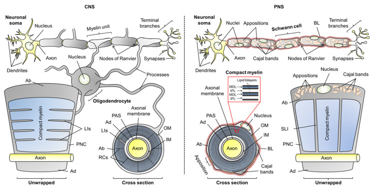
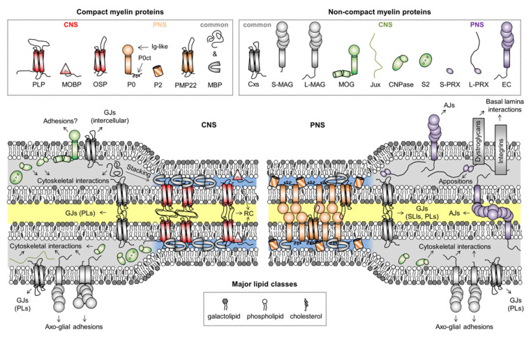
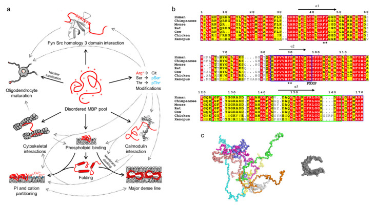
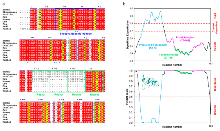
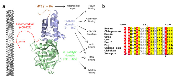
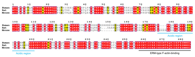
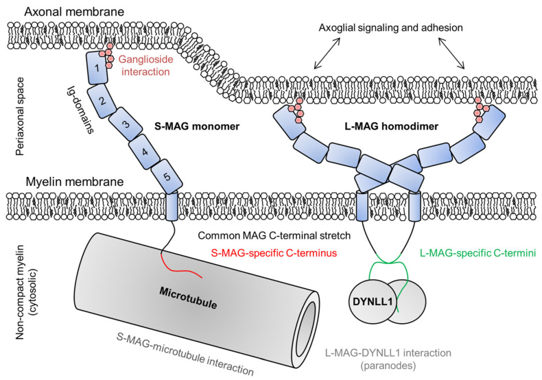
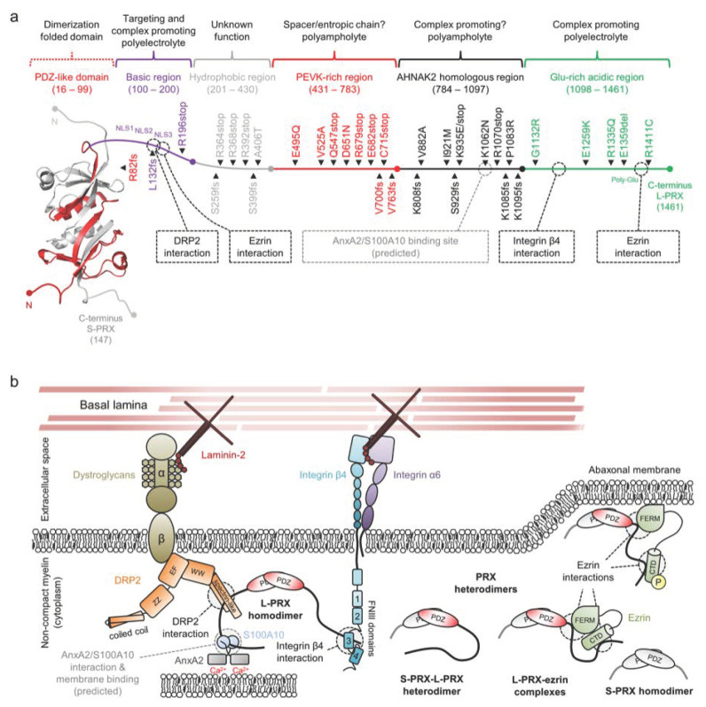
Myelin ensheathes selected axonal segments within the nervous system, resulting primarily in nerve impulse acceleration, as well as mechanical and trophic support for neurons. In the central and peripheral nervous systems, various proteins that contribute to the formation and stability of myelin are present, which also harbor pathophysiological roles in myelin disease. Many myelin proteins have common attributes, including small size, hydrophobic segments, multifunctionality, longevity, and regions of intrinsic disorder. With recent advances in protein biophysical characterization and bioinformatics, it has become evident that intrinsically disordered proteins (IDPs) are abundant in myelin, and their flexible nature enables multifunctionality. Here, we review known myelin IDPs, their conservation, molecular characteristics and functions, and their disease relevance, along with open questions and speculations. We place emphasis on classifying the molecular details of IDPs in myelin, and we correlate these with their various functions, including susceptibility to post-translational modifications, function in protein-protein and protein-membrane interactions, as well as their role as extended entropic chains. We discuss how myelin pathology can relate to IDPs and which molecular factors are potentially involved.

Keywords: intrinsically disordered protein; multiple sclerosis; myelin; myelination; peripheral neuropathies; protein folding; protein–membrane interaction; protein–protein interaction.


Conflict of interest statement

The authors declare no conflict of interest.

Figures

Figure 1
Figure 1
The anatomy of oligodendrocytic and Schwann cell myelin sheaths in the central nervous system (CNS) and peripheral nervous system (PNS), respectively. The arrangement of multiple myelin units along an axon is illustrated, as well as unwrapped myelin units and cross sections. Compact myelin and non-compact myelin are colored blue and gray, respectively. Abbreviations: Ab, abaxonal layer; Ad, adaxonal layer; BL, basal lamina; IM, inner mesaxon; IPL, intraperiod line; LIs, longitudinal incisures; MDL, major dense line; OM, outer mesaxon; PAS, periaxonal space; PNC, paranodal collar; RCs, radial components; SLI, Schmidt–Lanterman incisures.
Figure 2
Figure 2
Myelin proteins and their compartmentalization. Some proteins are shared between CNS and PNS myelin, but their expression levels can vary drastically between the two, like in the case of CNPase, which is predominantly a CNS enzyme. The intracellular compartment is colored blue and gray for compact and non-compact myelin, respectively. The extracellular and intramyelinic compartments are colored white and yellow, respectively. Note that cytoskeletal elements, other common proteins, and cell organelles are not included for clarity. Abbreviations: AJs, adherens junctions; CNPase, 2′,3′-cyclic nucleotide 3′-phosphodiesterase; Cxs, connexins; EC, epithelial cadherin; GJs, gap junctions; Ig-like, immunoglobulin-like; Jux, juxtanodin; LIs, longitudal incisures; MAG, myelin-associated glycoprotein; MBP, myelin basic protein; MOBP, myelin-associated oligodendrocytic basic protein; MOG, myelin/oligodendrocyte glycoprotein; OSP, oligodendrocyte-specific protein/claudin 11; P0, myelin protein zero; P0ct, the cytoplasmic domain of P0; P2, peripheral myelin protein 2; PLP, proteolipid protein; PLs, paranodal loops; PMP22, peripheral myelin protein 22; PRX, periaxin; RC, radial component; S2, sirtuin 2; SLIs, Schmidt–Lanterman incisures. In MAG and PRX, the L- and S- prefixes indicate long and short isoforms, respectively.
Figure 3
Figure 3
The multifunctionality, conformations, and conservation of MBP. (a) Schematic of the multifunctionality (solid arrows) of MBP, which arises through its disordered nature. Various PTMs, especially citrullination and phosphorylation, regulate the known functions of MBP (dashed arrows). The panel was inspired by Vassall et al. [65]. (b) Sequence alignment of 18.5-kDa MBP from vertebrates generated using ESPript [66]. MBP is highly conserved, especially all helically folding, lipid-interacting segments (black arrows; α1-α3), one of which overlaps with the immunogenic region (blue outlines). A noteworthy feature is the conservation of Arg residues, most of which are targets for citrullination. Black asterisks denote double-Phe motifs that are required for the phase transition of MBP upon lipid binding. Residue numbering corresponds to human MBP. (c) Conformational ensemble of 18.5-kDa MBP as determined using SAXS [24] (left) in comparison to a model of lipid-bound MBP [67] (right). Each colored chain in the ensemble represents a single conformational subpopulation in the pool of disordered MBP.
Figure 4
Figure 4
The conservation and predicted flexibility of MOBP isoform 1. (a) Sequence alignment of mammalian MOBPs generated using ESPript [66]. MOBP is highly conserved throughout mammals and especially within primates. Residue numbering corresponds to human MOBP. The residues predicted [48] to interact with Zn2+ in the putative FYVE domain have been indicated with blue asterisks. The tandem repeats within the Pro-rich region are indicated with green outlines. (b) DynaMine [132] (top) and PONDR [133] (bottom) predict human MOBP to be mostly disordered, with a folded N-terminal FYVE domain. The various compositional regions have been indicated. The structure in the PONDR inset represents the Phyre2 [134] prediction of the FYVE domain.
Figure 5
Figure 5
The domain structure of CNPase. (a) CNPase consists of two folded domains and a C-terminal 20-residue disordered tail, which mediates membrane interactions via the lipidated residue Cys418. Isoform 2 contains an additional N-terminal mitochondrial targeting sequence (MTS). The C-terminal tail tethers CNPase to the membrane, while it carries out its various functions [44,141]. (b) The C-terminal tail is conserved within several vertebrates, but is lost e.g. in fishes [141]. The lipidated Cys residue is indicated with an asterisk. Residue numbering corresponds to human CNPase. Orca, killer whale; Devil, Tasmanian devil; Xenopus, African clawed frog.
Figure 6
Figure 6
Comparison of human, rat, and mouse Jux. Jux is less conserved than MBP and MOBP, human Jux sharing only ~60% homology to mouse and rat Jux. An acidic region (blue) separates the protein into N- and C-terminal halves, the latter of which contains the fully conserved ERM-type actin binding domain (black). Residue numbering corresponds to human Jux.
Figure 7
Figure 7
The roles of S- and L-MAG on both sides of the adaxonal layer. MAG is able to dimerize through Ig-domains 4 and 5, which determines the dimensions of MAG and thus the intermembrane distance within the periaxonal space [179]. MAG interacts with gangliosides on the axonal membrane and mediates bidirectional axoglial signaling [180], maintaining the width of the periaxonal space. In the cytosol beneath the adaxonal membrane, S-MAG interacts with microtubules and L-MAG with DYNLL1 [178,181].
Figure 8
Figure 8
The structure and interactions of periaxin. (a) A schematic of a PRX heterodimer, with S-PRX in gray and L-PRX colored based on region, with the PDZ domain in red. L-PRX, apart from the PDZ-like domain, is predicted to be disordered [45], and can be divided into separate regions based on sequence composition. Peripheral neuropathy mutations are indicated alongside L-PRX. Dashed boxes and lines denote protein–protein interactions. L-PRX contains a predicted AnxA2 and S100A10 binding region, as reported earlier for AHNAK [195,196]. See Table 1 for mutation details. (b) L-PRX is an assembler within abaxonal non-compact myelin, linking dystroglycans and integrins together in membrane appositions, forming the periaxinosome. These interaction partners connect the Schwann cell basal lamina to the Schwann cell cytoplasm. S-PRX forms heterodimers with L-PRX, which might allow regulation of the cytoplasmic assembly as well as the nuclear export of L-PRX. Ezrin in complex with hetero- or homodimeric L-PRX might have relevance in such regulations, especially considering its phosphoregulated membrane-binding activity [197]. The function of the S-PRX homodimer is unknown. The significance of the putative L-PRX/AnxA2/Sl00A10 ternary complex could involve linking the entire assembly via AnxA2 and Ca2+ to the underlying membrane, possibly forming a structural basis for membrane appositions that line Cajal bands in myelinating Schwann cells.
Figure 9
Figure 9
Ternary complex between AHNAK peptide (sticks), S100A10 (blue ribbon), and the acetylated N terminus of AnxA2 (black ribbon) in a 1:2:2 stoichiometry (PDB ID 4ftg [196]; top). Sequence alignment of the binding motif of AHNAK with L-PRX predicts a similar binding site in L-PRX (bottom). Coloring of the bound AHNAK peptide corresponds to residue conservation between AHNAK and L-PRX as evident from the sequence alignment.
Figure 10
Figure 10
The fate of the MDL formation is governed by the concentration of active MBP (red), which normally would associate with membranes, form a pre-stack state, and continue to accumulate until stable membrane stacks form. MS is linked to an autoimmune response and loss of myelin, which could stem from molecular mimicry. The immune system recognizes antigenic MBP peptides formed via proteolysis of free or membrane-bound MBP. Changes in membrane lipid composition or concentration of intracellular ions could pre-expose to this process [53,90]. Changes in the PTMs of MBP have additionally been shown to play a role [60,226,239], as might lowered MBP expression levels [24].

Similar articles

Cited by

References

    1. Wang S.S.H., Shultz J.R., Burish M.J., Harrison K.H., Hof P.R., Towns L.C., Wagers M.W., Wyatt K.D. Shaping of white matter composition by biophysical scaling constraints. J. Neurosci. 2008;28:4047–4056. doi: 10.1523/JNEUROSCI.5559-05.2008. - DOI - PMC - PubMed
    1. Lundgaard I., Luzhynskaya A., Stockley J.H., Wang Z., Evans K.A., Swire M., Volbracht K., Gautier H.O.B., Franklin R.J.M., ffrench-Constant C., et al. Neuregulin and BDNF Induce a Switch to NMDA Receptor-Dependent Myelination by Oligodendrocytes. PLoS. Biol. 2013;11:e1001743. doi: 10.1371/journal.pbio.1001743. - DOI - PMC - PubMed
    1. Salzer J.L. Schwann Cell Myelination. Cold Spring Harbor Perspect. Biol. 2015;7:a020529. doi: 10.1101/cshperspect.a020529. - DOI - PMC - PubMed
    1. Nave K. Myelination and the trophic support of long axons. Nat. Rev. Neurosci. 2010;11:275–283. doi: 10.1038/nrn2797. - DOI - PubMed
    1. Young J.Z. The functioning of the giant nerve fibres of the squid. J. Exp. Biol. 1938;15:170–185. - PubMed

MeSH terms

Substances