. 2020 Feb 18;12(2):476.
doi: 10.3390/cancers12020476.

Plasma-activated Ringer's Lactate Solution Displays a Selective Cytotoxic Effect on Ovarian Cancer Cells

Affiliations

Plasma-activated Ringer's Lactate Solution Displays a Selective Cytotoxic Effect on Ovarian Cancer Cells

Alina Bisag et al. Cancers (Basel). .

Abstract

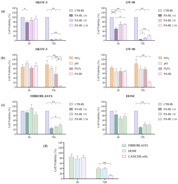
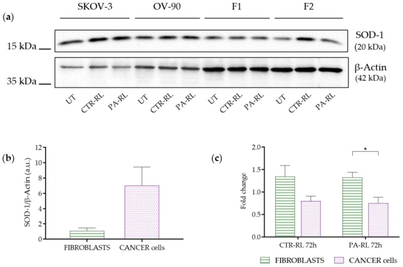
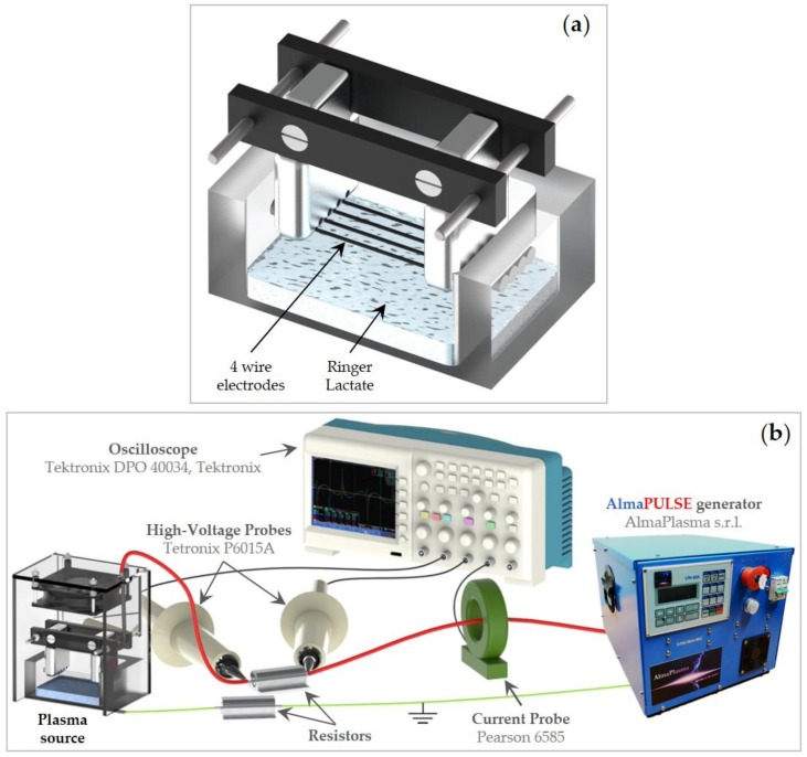
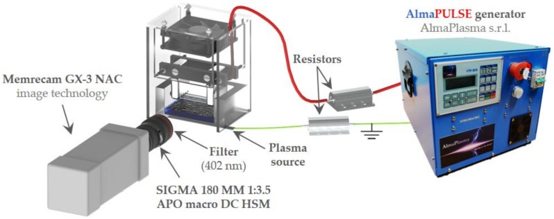
Epithelial Ovarian Cancer (EOC) is one of the leading causes of cancer-related deaths among women and is characterized by the diffusion of nodules or plaques from the ovary to the peritoneal surfaces. Conventional therapeutic options cannot eradicate the disease and show low efficacy against resistant tumor subclones. The treatment of liquids via cold atmospheric pressure plasma enables the production of plasma-activated liquids (PALs) containing reactive oxygen and nitrogen species (RONS) with selective anticancer activity. Thus, the delivery of RONS to cancer tissues by intraperitoneal washing with PALs might be an innovative strategy for the treatment of EOC. In this work, plasma-activated Ringer's Lactate solution (PA-RL) was produced by exposing a liquid substrate to a multiwire plasma source. Subsequently, PA-RL dilutions are used for the treatment of EOC, non-cancer and fibroblast cell lines, revealing a selectivity of PA-RL, which induces a significantly higher cytotoxic effect in EOC with respect to non-cancer cells.

Keywords: cold atmospheric pressure plasma; cytotoxicity; ovarian cancer; plasma medicine; plasma-activated Ringer’s lactate solution; selectivity.


Conflict of interest statement

The authors declare no conflict of interest.

Figures

Figure 1
Figure 1
Electrical characterization of plasma source during treatment of Ringer’s Lactate (RL) solution: (a) representative voltage (red) and current (blue) waveforms at 18 kV and 1 kHz and (b) power values as a function of the applied voltage. Data are presented as mean ± SEM (n = 3).
Figure 2
Figure 2
Plasma treatment leads to the formation of H2O2 and NO2. (a) Reactive oxygen and nitrogen species (RONS) concentration as a function of the average power after 10 min of plasma treatment. Data are presented as mean ± SEM (n = 3). (b) H2O2 and NO2 concentrations as a function of treatment time. Data are presented as mean ± SEM (n = 3) and statistical significance is specified with asterisks (** p ≤ 0.001 as determined by a paired Student’s t-test, versus the 5 min treatment).
Figure 3
Figure 3
Chemical characterization of plasma-activated RL (PA-RL) and its dilutions after 10 min of plasma treatment at 18 kV. (a) pH and (b) conductivity as a function of serial dilutions. Data are presented as mean ± SEM (n = 3).
Figure 4
Figure 4
Low-speed images and high-speed (HS) filter images of the multiwire plasma discharge during RL treatment. (a) Picture of plasma generated during the treatment of PA-RL with an applied voltage of 18 kV and 30 fps. (b) HS filter images of plasma filaments for different values of applied voltage (between 15 to 18 kV) and 100 fps.
Figure 5
Figure 5
PA-RL displays a selective cytotoxic effect on Epithelial Ovarian Cancer (EOC) cell lines. (a) Viability of SKOV-3 (n = 7) and OV-90 (n = 9) cell lines treated with PA-RL dilutions (1:4, 1:8 and 1:16). Data are mean ± SEM normalized on the corresponding control in RL (CTR-RL). (b) Viability of SKOV-3 and OV-90 cell lines treated with PA-RL 1:16 and synthetic solutions at dilution 1:16. H2O2-supplemented RL, NO2-supplemented RL and pH-adjusted RL solutions were diluted in RL to obtain the final treatment solutions. Data are mean ± SEM (n = 3) normalized on the corresponding CTR-RL. (c) Viability of non-cancer cells, namely human fibroblasts (n = 9) and HOSE (n = 4) treated with different PA-RL dilutions (1:4, 1:8 and 1:16). Data are mean ± SEM normalized on the corresponding CTR-RL. (d) PA-RL 1:16 efficacy on cell viability in non-cancer and EOC cell lines. Cell viability was normalized to the CTR-RL at 2 h and plotted as percentage relative to the corresponding CTR-RL, for both time points. In each panel, data are mean ± SEM and statistical significance is specified with asterisks (* p ≤ 0.05, ** p ≤ 0.001 as determined by a paired Student’s t-test).
Figure 6
Figure 6
PA-RL solution induces an increase in Superoxide Dismutase-1 (SOD-1) expression in fibroblasts but not in EOC cell lines. (a) Western blot analysis of EOC cell lines and fibroblasts (F1 and F2) at 72 h after treatment with PA-RL 1:16 (UT, untreated cells). A representative experiment of three is shown. (b) SOD-1 levels in untreated fibroblasts and cancer cell lines. Histograms show densitometric values of the SOD-1 protein normalized to the β-actin used as a loading control. All data are presented as mean ± SEM of three independent experiments. (c) Relative densities of SOD-1 and β-actin were measured using densitometric analysis. SOD-1 levels of CTR-RT and PA-RL 1:16 after 72 h of treatment were normalized to β-actin and plotted as fold change relative to the untreated (UT) sample. All data are presented as mean ± SEM of three independent experiments. Statistical significance is specified with asterisks (* p ≤ 0.05 as determined by a paired Student’s t-test).
Figure 7
Figure 7
(a) Illustration of the high voltage electrodes and RL and (b) layout of the setup used for electrical characterization.
Figure 8
Figure 8
High-speed filter imaging setup.

References

    1. Reid B.R., Permuth J.B., Sellers T.A. Epidemiology of ovarian cancer: A review. Cancer Biol. Med. 2017;14:9–32. doi: 10.20892/j.issn.2095-3941.2016.0084. - DOI - PMC - PubMed
    1. Van Baal J.O.A.M., Van Noorden C.J.F., Nieuwland R., Van De Vijver K.K., Sturk A., Van Driel W.J., Kenter G.G., Lok C.A.R. Development of Peritoneal Carcinomatosis in Epithelial Ovarian Cancer: A Review. J. Histochem. Cytochem. 2018;66:67–83. doi: 10.1369/0022155417742897. - DOI - PMC - PubMed
    1. Prat J. FIGO’s staging classification for cancer of the ovary, fallopian tube, and peritoneum: Abridged republication. J. Gynecol. Oncol. 2015;26:87. doi: 10.3802/jgo.2015.26.2.87. - DOI - PMC - PubMed
    1. Jewell A., McMahon M., Khabele D. Heated Intraperitoneal Chemotherapy in the Management of Advanced Ovarian Cancer. Cancers. 2018;10:296. doi: 10.3390/cancers10090296. - DOI - PMC - PubMed
    1. Lheureux S., Gourley C., Vergote I., Oza A.M. Epithelial ovarian cancer. Lancet. 2019;393:1240–1253. doi: 10.1016/S0140-6736(18)32552-2. - DOI - PubMed

LinkOut - more resources