Comparative Study
. 2020 Feb 18;11(2):211.
doi: 10.3390/genes11020211.

Inhibition of Angiotensin-Converting Enzyme Ameliorates Renal Fibrosis by Mitigating DPP-4 Level and Restoring Antifibrotic MicroRNAs

Affiliations
Comparative Study

Inhibition of Angiotensin-Converting Enzyme Ameliorates Renal Fibrosis by Mitigating DPP-4 Level and Restoring Antifibrotic MicroRNAs

Swayam Prakash Srivastava et al. Genes (Basel). .

Abstract

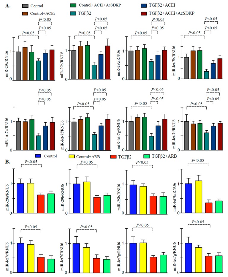
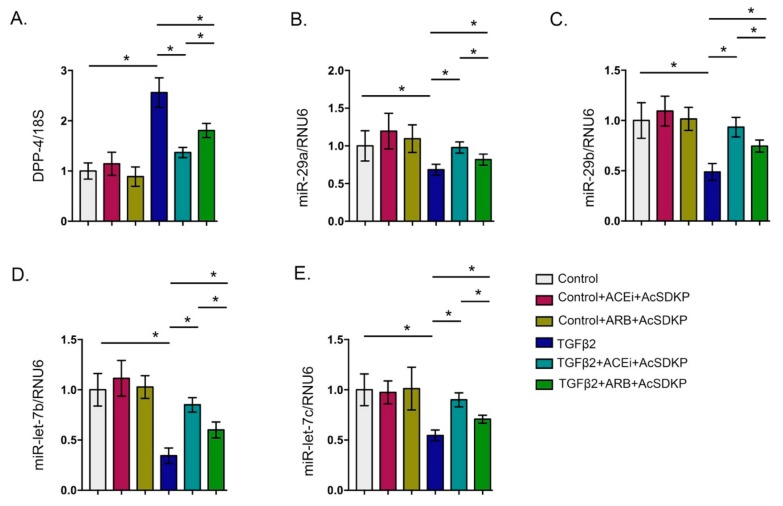
Two class of drugs 1) angiotensin-converting enzyme inhibitors (ACEis) and 2) angiotensin II receptor blockers (ARBs) are well-known conventional drugs that can retard the progression of chronic nephropathies to end-stage renal disease. However, there is a lack of comparative studies on the effects of ACEi versus ARB on renal fibrosis. Here, we observed that ACEi ameliorated renal fibrosis by mitigating DPP-4 and TGFβ signaling, whereas, ARB did not show. Moreover, the combination of N-acetyl-seryl-aspartyl-lysyl-proline (AcSDKP), one of the substrates of ACE, with ACEi slightly enhanced the inhibitory effects of ACEi on DPP-4 and associated-TGFβ signaling. Further, the comprehensive miRome analysis in kidneys of ACEi+AcSDKP (combination) treatment revealed the emergence of miR-29s and miR-let-7s as key antifibrotic players. Treatment of cultured cells with ACEi alone or in combination with AcSDKP prevented the downregulated expression of miR-29s and miR-let-7s induced by TGFβ stimulation. Interestingly, ACEi also restored miR-29 and miR-let-7 family cross-talk in endothelial cells, an effect that is shared by AcSDKP suggesting that AcSDKP may be partially involved in the anti-mesenchymal action of ACEi. The results of the present study promise to advance our understanding of how ACEi regulates antifibrotic microRNAs crosstalk and DPP-4 associated-fibrogenic processes which is a critical event in the development of diabetic kidney disease.

Keywords: ACE; ARBs; AcSDKP; DPP-4; EMT; EndMT; diabetic nephropathy; kidney fibrosis.


Conflict of interest statement

The authors have no conflicts of interest to declare.

Figures

Figure 1
Figure 1
Inhibition of ACE suppresses DPP-4 and associated TGFβ signaling in diabetic kidneys (A) Quantitative analysis of DPP-4 mRNA expression by real time PCR using specific primers in the kidney of control, DM, DM+ACEi, DM+combination and DM+ARB treated mice. N = 6 were analyzed in each group. 18S was used as internal control to normalize the expression data. (B) Western blot analysis of DPP-4, TGFβR1, p-smad3, smad3, FSP-1, αSMA, Colla1a and fibronectin (FN) in the kidney of control, DM, DM + ACEi, DM + combination treatment and DM + ARB treated diabetic mice. Representative blots are shown. Quantification of DPP-4, TGFβR1, smad3 phosphorylation, FSP-1, αSMA, Colla1a and FN by densitometry. The data were normalized by β-actin. N = 5 were analyzed in each group. (C) Co-immunofloroscence analysis of DPP-4/CD31 and DPP-4/ αSMA in the kidney of control, DM, DM + ACEi, DM + ACEi + AcSDKP and DM + ARB, the representative pictures are shown. Scale bar 50 µm. DPP-4 FITC (green) labeled whereas, CD31 and αSMA are rhodamine labeled and DAPI blue. N = 5 were analyzed in each group. (D) DPP activity analysis by fluorimeter in kidney homogenate of control, DM, DM + ACEi, DM + combination and DM + ARB treated mice. N = 6 were analyzed in each group. (E) DPP activity analysis in the plasma of control, DM, DM + ACEi, DM + combination and DM + ARB treated mice. N = 6 were analyzed in each group. Data in the graph are presented as mean ± SEM. One-way Anova Tukey test was performed for calculation of statistical significance. C = control (non-diabetic), DM = diabetic group, combination = (ACEi + AcSDKP), Colla1 = collagen I, FN = fibronectin.
Figure 2
Figure 2
miRome analysis reveal up-regulated expression of miR-29 and miR-let-7 family members in the kidneys of ACE inhibitor or combination treated diabetic mice. (A) microRNA-array analysis in the diabetic group vs ACEi + AcSDKP treatment in diabetic mice revealed alteration in the expression level of pro and antifibrotic microRNAs. N = 3 were analyzed in each group. (B) miR-29 and miR-let-7 family members emerged as important regulatory antifibrotic molecules and validation by the real time PCR using specific primers in the kidney of control, DM, ACEi, combination treatment and ARB group. N = 6 were analyzed in each group. Hs_RNU6 was used as internal control to normalize the expression data. Data in the graph are presented as mean±SEM. One-way Anova Tukey test was performed for calculation of statistical significance.
Figure 3
Figure 3
Inhibition of ACE inhibit DPP-4 level and TGFβ signaling in endothelial cells. (A) Western blot analysis in the ACEi and ACEi+AcSDKP treated HMVECs in presence and absence of TGFβ2. Quantification of DPP-4, TGFβR1, FSP-1, p-smad3 and smad3 respectively by densitometry. Representative blots are shown. The data was normalized by β-actin. N = 3 were analyzed in each group. (B) Western blot analysis in the ARB treated HMVECs in presence and absence of TGFβ2. Quantification of DPP-4, TGFβR1, FSP-1, p-smad3 and smad3 respectively by densitometry. Representative blots are shown. The data was normalized by β-actin. N = 3 were analyzed in each group. Data in the graph are presented as mean ± SEM. One-way Anova Tukey test was performed for calculation of statistical significance.
Figure 4
Figure 4
ACEi and combination treatment restore the downregulated level of miR-29 and miR-let-7 family members in the TGFβ2-stimulated HMVECs. (A) qPCR analysis of miR-29 and miR-let-7 family members in the control, ACEi, and ACEi+AcSDKP in the presence and absence of TGFβ2 in the HMVECs. N = 4 were analyzed in each group. Hs_RNU6 was used as internal control to normalize the expression data. (B) qPCR analysis of miR-29 and miR-let-7 family members in the control and ARB stimulation in the presence and absence of TGFβ2. N = 4 were analyzed in each group. Hs_RNU6 was used as internal control to normalize the expression data. Data in the graph are presented as mean±SEM. One-way Anova Tukey test was performed for calculation of statistical significance.
Figure 5
Figure 5
Gene expression analysis of DPP-4 and antifibrotic microRNAs in the combination treatments (ACEi + AcSDKP and ARB + AcSDKP). (A) Gene expression analysis of DPP-4 mRNA. (BC) Gene expression analysis of miR-29 family members and miR-let-7 family member in the TGFβ2-stimulated HMVECs. N = 5 were analyzed in each group Data in the graph are presented as mean±SEM. One-way Anova Tukey test was performed for calculation of statistical significance.
Figure 6
Figure 6
Inhibition of ACE restored the TGFβ2-associated disruption of cross-talk regulation between miR-29 and miR-let-7 family members in the endothelial cells. (A) Gene expression analysis of miR-29 family members in the anti-miR-let-7b transfected HMVECs, ACEi+anti-miR-let-7b transfected, and ARB+anti-miR-let-7b transfected HMVECs. N = 4 were analyzed in each group. (B) Gene expression studies of miR-let-7b and miR-let-7c in the anti-miR-29b transfected HMVECs, ACEi+anti-miR-29b transfected, and ARB+anti-miR-29b transfected HMVECs. N = 4 were analyzed in each group. Data in the graph are presented as mean ± SEM. One-way Anova Tukey test was performed for calculation of statistical significance.
Figure 7
Figure 7
Working hypothesis for ACEi action on the suppression of DPP-4 associated fibrogenic program and restoration of antifibrotic microRNAs.

References

    1. Held P.J., Port F.K., Webb R.L., Wolfe R.A., Garcia J.R., Blagg C.R., Agodoa L.Y. The united states renal data system’s 1991 annual data report: An introduction. Am. J. Kidney Dis. 1991;18:1–16. - PubMed
    1. Liu Y. Cellular and molecular mechanisms of renal fibrosis. Nat. Rev. Nephrol. 2011;7:684–696. doi: 10.1038/nrneph.2011.149. - DOI - PMC - PubMed
    1. Srivastava S.P., Hedayat F.A., Kanasaki K., Goodwin J.E. Microrna crosstalk influences epithelial-to-mesenchymal, endothelial-to-mesenchymal, and macrophage-to-mesenchymal transitions in the kidney. Front. Pharmacol. 2019;10:904. doi: 10.3389/fphar.2019.00904. - DOI - PMC - PubMed
    1. Ganesh J., Viswanathan V. Management of diabetic hypertensives. Indian J. Endocrinol. Metab. 2011;15(Suppl 4):S374–379. - PMC - PubMed
    1. Hsu F.Y., Lin F.J., Ou H.T., Huang S.H., Wang C.C. Renoprotective effect of angiotensin-converting enzyme inhibitors and angiotensin ii receptor blockers in diabetic patients with proteinuria. Kidney Blood Press Res. 2017;42:358–368. doi: 10.1159/000477946. - DOI - PubMed

Publication types

MeSH terms

LinkOut - more resources