. 2020 Sep;39(9):2750-2759.
doi: 10.1109/TMI.2020.2974844. Epub 2020 Feb 18.

Deformable Slice-to-Volume Registration for Motion Correction of Fetal Body and Placenta MRI

Deformable Slice-to-Volume Registration for Motion Correction of Fetal Body and Placenta MRI

Alena Uus et al. IEEE Trans Med Imaging. 2020 Sep.

Abstract

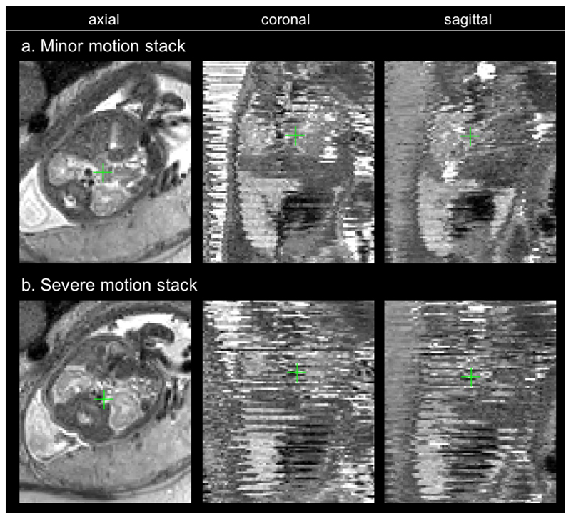
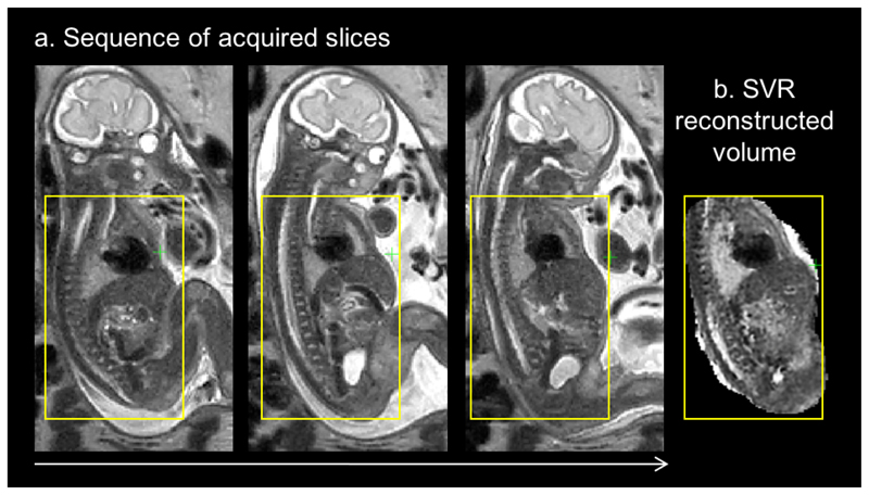
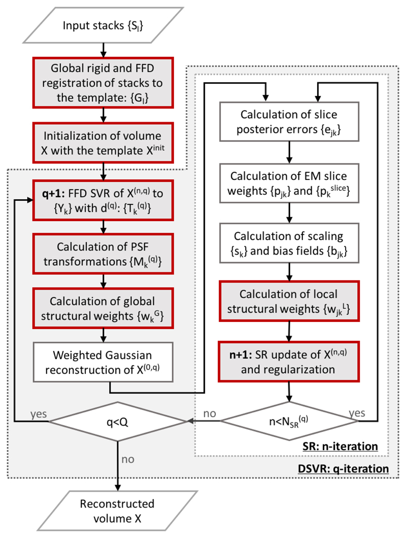
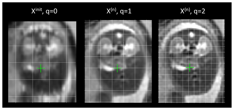
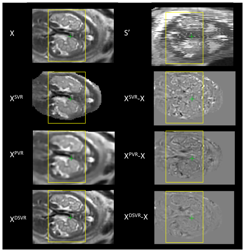
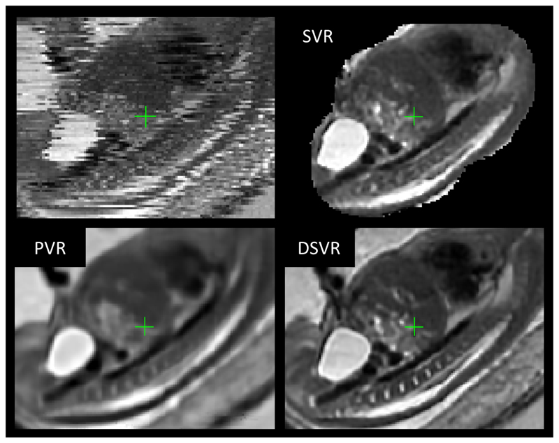
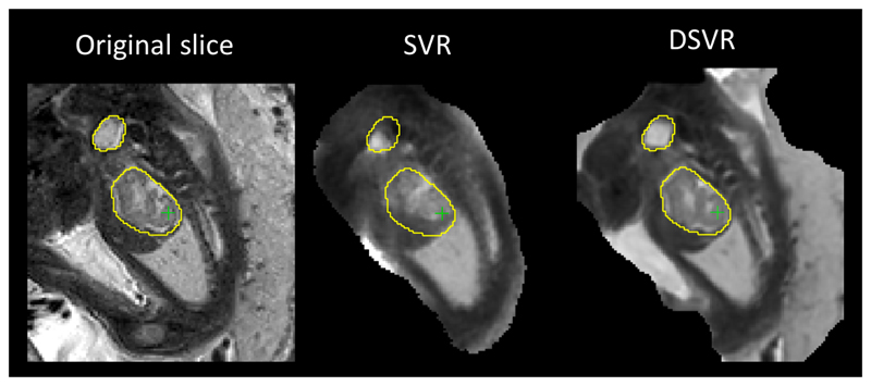
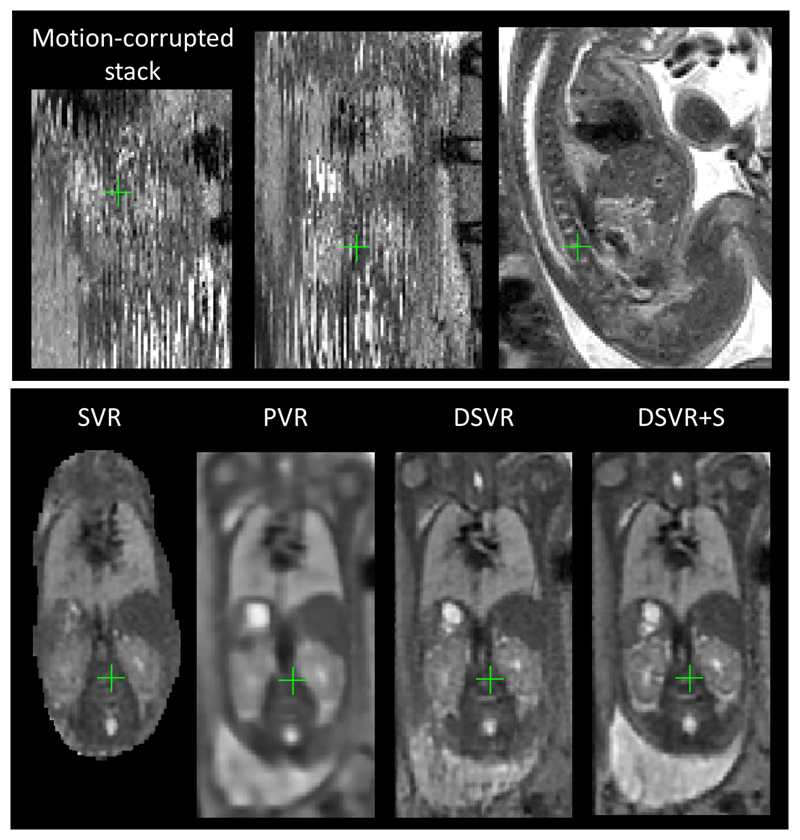
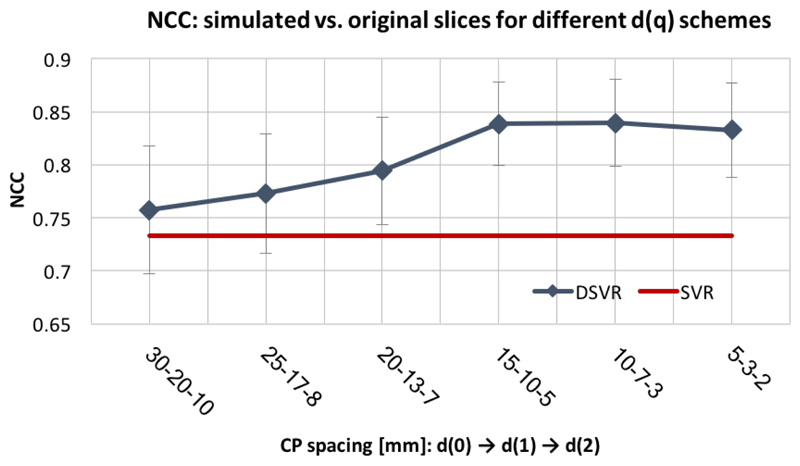
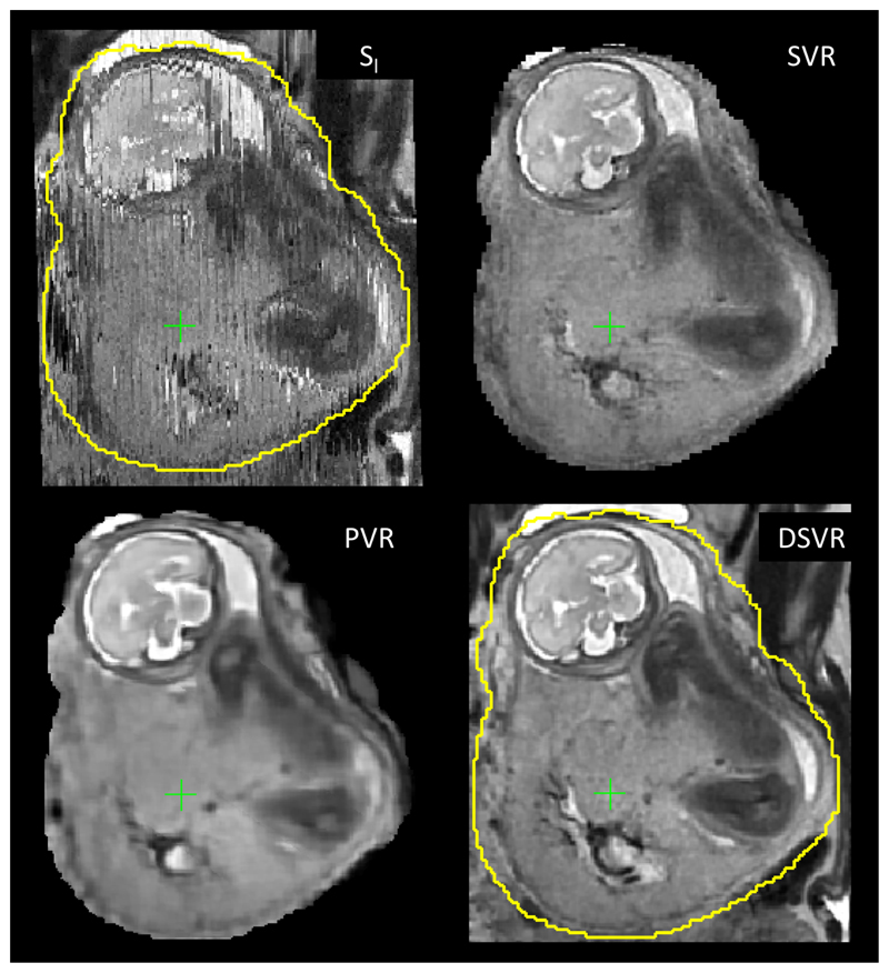
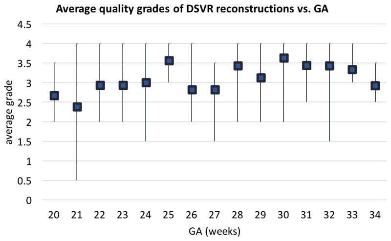
In in-utero MRI, motion correction for fetal body and placenta poses a particular challenge due to the presence of local non-rigid transformations of organs caused by bending and stretching. The existing slice-to-volume registration (SVR) reconstruction methods are widely employed for motion correction of fetal brain that undergoes only rigid transformation. However, for reconstruction of fetal body and placenta, rigid registration cannot resolve the issue of misregistrations due to deformable motion, resulting in degradation of features in the reconstructed volume. We propose a Deformable SVR (DSVR), a novel approach for non-rigid motion correction of fetal MRI based on a hierarchical deformable SVR scheme to allow high resolution reconstruction of the fetal body and placenta. Additionally, a robust scheme for structure-based rejection of outliers minimises the impact of registration errors. The improved performance of DSVR in comparison to SVR and patch-to-volume registration (PVR) methods is quantitatively demonstrated in simulated experiments and 20 fetal MRI datasets from 28-31 weeks gestational age (GA) range with varying degree of motion corruption. In addition, we present qualitative evaluation of 100 fetal body cases from 20-34 weeks GA range.


Figures

Fig. 1
Fig. 1
Fetal MRI: examples of stacks with minor (a) and severe (b) motion corruption acquired during the same fetal exam and under the same orientation at different time points.
Fig. 2
Fig. 2
Example of a sequence of slices (a) acquired during the change of fetal trunk position and the corresponding SVR reconstructed volume (b). Note that bending deforms the shape of the spine and internal organs in both in-plane and through-plane directions.
Fig. 3
Fig. 3
Proposed DSVR reconstruction algorithm. The novel elements are highlighted by red outline.
Fig. 4
Fig. 4
An example of refinement of B-spline control point grid (d (q)) at each DSVR iteration (sagittal plane with respect to uterus).
Fig. 5
Fig. 5
Simulated experiment: original reference volume (X), one of the generated motion-corrupted stacks (S′), SVR (XSV R), PVR (XPV R) and DSVR (XDSV R) reconstruction results and their difference with the reference (coronal plane).
Fig. 6
Fig. 6
Example of motion correction for a minor motion dataset: motion corrupted stack, SVR, PVR and DSVR reconstructions (sagittal plane).
Fig. 7
Fig. 7
Comparison of SVR and DSVR in presence of non-rigid motion: original acquired slice (Yk) vs. slices simulated from SVR and DSVR (Ȳk). The yellow isolines delineate the structure in the original slice, and show misalignment with SVR reconstruction due to limitation of rigid motion correction. The problem is resolved by non-rigid motion correction in DSVR.
Fig. 8
Fig. 8
Example of motion correction for a severe motion dataset: motion corrupted stack, SVR, PVR and DSVR reconstructions (coronal plane).
Fig. 9
Fig. 9
CP spacing analysis: NCC between the original (Yk*) vs. simulated (Ȳk) slices.
Fig. 10
Fig. 10
Example of motion correction for placenta: motion-corrupted stack, SVR, PVR and DSVR+S reconstructed volumes (coronal plane).
Fig. 11
Fig. 11
Quality of DSVR reconstructions of fetal body region for 100 reconstructed iFIND cases vs. GA. Squares represent the average grade per week of GA, and bars represent the range of values.

References

    1. Malamateniou C, et al. Motion-compensation techniques in neonatal and fetal MR imaging. American Journal of Neuroradiology. 2013;34(6):1124–1136. - PMC - PubMed
    1. Story L, Rutherford M. Advances and applications in fetal magnetic resonance imaging. The Obstetrician & Gynaecologist. 2015;17(3):189–199.
    1. Gholipour A, Estroff JA, Warfield SK. Robust super-resolution volume reconstruction from slice acquisitions: Application to fetal brain MRI. IEEE Transactions on Medical Imaging. 2010;29(10):1739–1758. - PMC - PubMed
    1. Rousseau F, Kim K, Studholme C, Koob M, Dietemann J-L. On Super-Resolution for Fetal Brain MRI. Med Image Comput Comput Assist Interv. 2010;13(Pt 2):355–362. - PMC - PubMed
    1. Kuklisova-Murgasova M, Quaghebeur G, Rutherford MA, Hajnal JV, Schnabel JA. Reconstruction of fetal brain MRI with intensity matching and complete outlier removal. Medical Image Analysis. 2012;16(8):1550–1564. - PMC - PubMed

Publication types