. 2020 Apr;32(4):1081-1101.
doi: 10.1105/tpc.19.00631. Epub 2020 Feb 21.

Pathogen-Associated Molecular Pattern-Triggered Immunity Involves Proteolytic Degradation of Core Nonsense-Mediated mRNA Decay Factors During the Early Defense Response

Affiliations

Pathogen-Associated Molecular Pattern-Triggered Immunity Involves Proteolytic Degradation of Core Nonsense-Mediated mRNA Decay Factors During the Early Defense Response

Ho Won Jung et al. Plant Cell. 2020 Apr.

Abstract

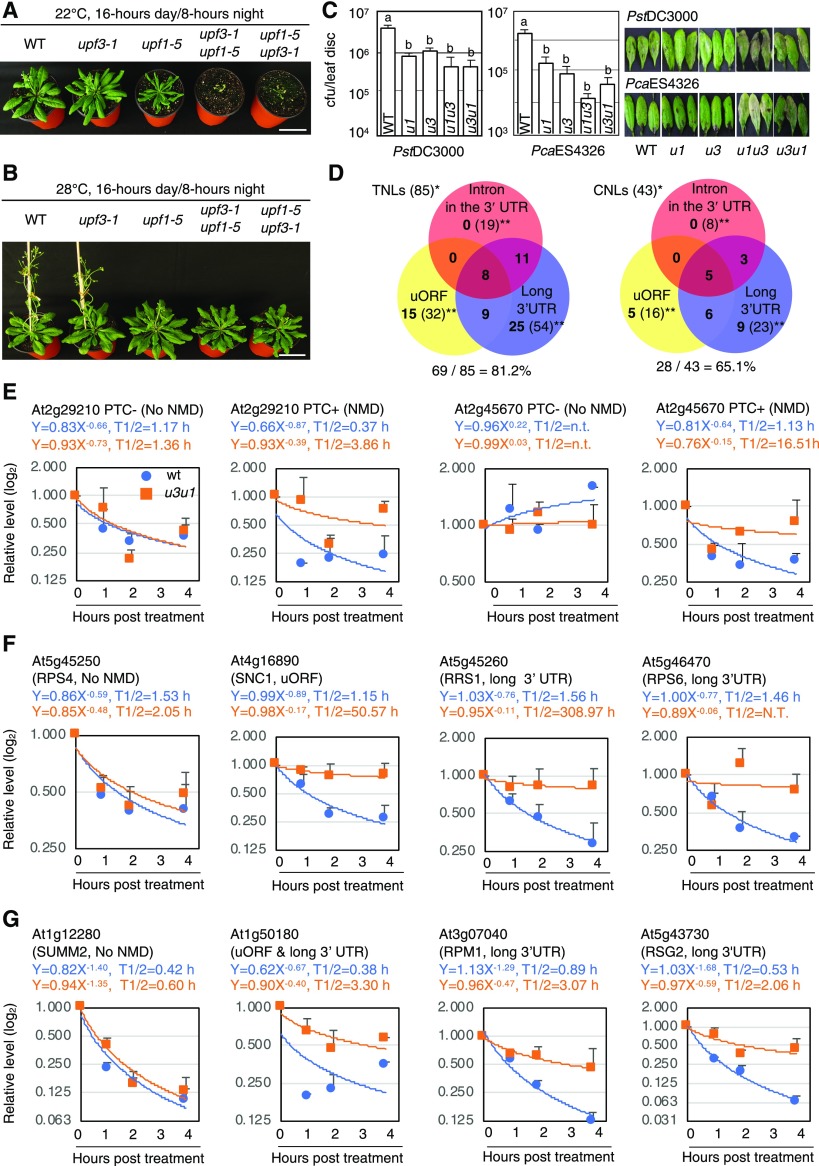
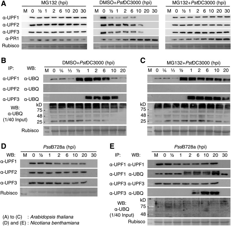
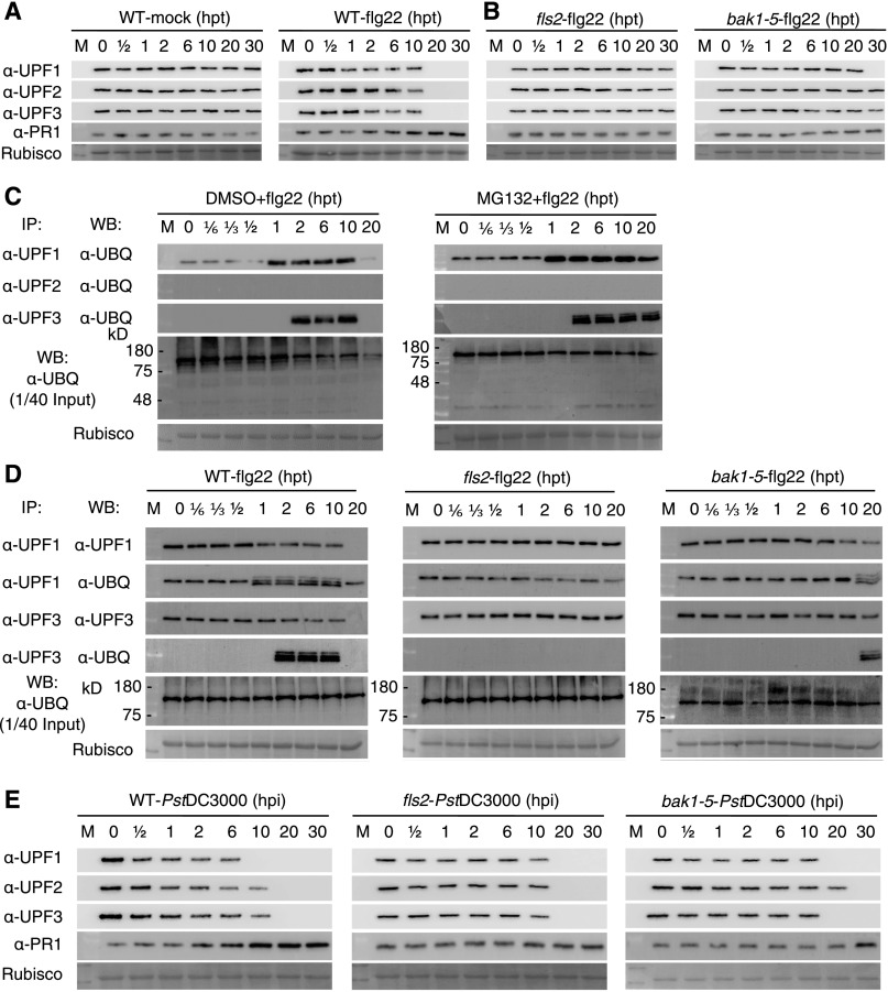
Nonsense-mediated mRNA decay (NMD), an mRNA quality control process, is thought to function in plant immunity. A subset of fully spliced (FS) transcripts of Arabidopsis (Arabidopsis thaliana) resistance (R) genes are upregulated during bacterial infection. Here, we report that 81.2% and 65.1% of FS natural TIR-NBS-LRR (TNL) and CC-NBS-LRR transcripts, respectively, retain characteristics of NMD regulation, as their transcript levels could be controlled posttranscriptionally. Both bacterial infection and the perception of bacteria by pattern recognition receptors initiated the destruction of core NMD factors UP-FRAMESHIFT1 (UPF1), UPF2, and UPF3 in Arabidopsis within 30 min of inoculation via the independent ubiquitination of UPF1 and UPF3 and their degradation via the 26S proteasome pathway. The induction of UPF1 and UPF3 ubiquitination was delayed in mitogen-activated protein kinase3 (mpk3) and mpk6, but not in salicylic acid-signaling mutants, during the early immune response. Finally, previously uncharacterized TNL-type R transcripts accumulated in upf mutants and conferred disease resistance to infection with a virulent Pseudomonas strain in plants. Our findings demonstrate that NMD is one of the main regulatory processes through which PRRs fine-tune R transcript levels to reduce fitness costs and achieve effective immunity.


Figures

Figure 1.
Figure 1.
The upf1 upf3 Double Mutants Exhibit Intensified Autoimmunity. (A) and (B) Five-week–old Arabidopsis ecotype Columbia-0 (wild type [WT]), upf1-5, upf3-1, upf1-5 upf3-1, and upf3-1 upf1-5 plants were grown at 22 ± 1°C (A) or 28 ± 1°C (B) with a 16-h d/8-h night photoperiod. Simultaneous mutation of UPF1 and UPF3 arrested Arabidopsis growth at 22°C, while the growth defect was moderately recovered by a temperature shift to 28°C. Scale bars = 5 cm. (C) Bacterial counts and disease symptoms in the wild type (WT) and NMD-compromised mutants 3 d after PstDC3000 and PcaES4326 infection. Different letters (a and b) indicate statistically significant differences. P < 0.01; one-way ANOVA. Error bars indicate se (n = 8). (D) Venn diagrams of TNL- and CNL-type R genes illustrating the number of genes carrying different NMD features. Numbers in parentheses are the total number of expressed genes (*) and the total number of genes bearing each NMD event (**). (E) to (G) Stability of NMD reporters (E), representative TNL transcripts (F), and representative CNL transcripts (G). RT-qPCR analysis was performed using total RNA extracted from ActD-treated leaves of the wild type (WT) and upf3-1 upf1-5 (u3u1) mutants that had been collected at 0, 1, 2, and 4 hpt. The half-lives were calculated by nonlinear least-squares regression analysis (average ± sd, n = 3; three biological replicates with three technical repeats). Stability analyses of other R transcripts are provided in Supplemental Figure 3. Sequences of the individual primers used in this study are presented in Supplemental Data Set 3.
Figure 2.
Figure 2.
UPF1, UPF2, and UPF3 Decay during an Early Phase of PstDC3000 Infection. (A) Dynamics of UPF1, UPF2, and UPF3 proteins up to 30 hpi (top) and 50 mpi (bottom). Immunoblot analyses (left) were performed for leaf samples taken from wild-type Col-0 plants that had been infected with Pseudomonas and collected at the indicated time points using an anti-UPF1 monoclonal antibody (α-UPF1), α-UPF2, or α-UPF3. The right shows UPF protein levels in infected leaves at the indicated time points (average ± sd, n = 4). Different letters above the bars (i.e., a, b, c, d, ab, and bc) indicate statistically significant differences (P < 0.05, one-way ANOVA). Successful infection was verified by examining the levels of PR1. Both experiments were performed with four biological replicates. (B) The NMD factor SMG7 and the EJC core protein components, but not UPF1, UPF2, or UPF3, remained stable during PstDC3000 infection. The leaves of transgenic Arabidopsis plants (T3) stably expressing protein A-fused UPF1, UPF2, UPF3, SMG7, Y14, MAGO, elF4A-III, BTZ1, and BTZ2 were infected with PstDC3000 and collected at the indicated time points after infection for immunoblot analysis. GFP-protein A was used as a representative stable protein. The recombinant proteins were detected using an α-PAP antibody. M, prestained protein ladder.
Figure 3.
Figure 3.
P. syringae Infection Induces Ubiquitination of UPF1 and UPF3 in Arabidopsis and N. benthamiana Plants. (A) Effects of the 26S proteasome inhibitor MG132 on the decay of UPF1, UPF2, and UPF3 during infection. DMSO alone or MG132 dissolved in DMSO was coinfiltrated into wild-type leaves with PstDC3000. Immunoblot analyses were performed using α-UPF1, α-UPF2, or α-UPF3. Successful infection was verified by examining the levels of PR1. (B) Ubiquitination of UPF1 and UPF3, but not UPF2, in wild-type leaves after PstDC3000 infection. Immunocomplexes with the α-UPF1, α-UPF2, or α-UPF3 antibody were subjected to immunoblotting with α-UBQ. (C) IP assay with proteins extracted from MG132+PstDC3000-treated wild-type plants. The levels of UPF proteins in IP complexes in (B) and (C) are shown in Supplemental Figure 8A and 8B, respectively. (D) Degradation of UPF proteins in N. benthamiana leaves infected with P. syringae pv syringae B728a (PssB728a). (E) Induction of UPF1 and UPF3 ubiquitination in N. benthamiana leaves during PssB728a infection. Levels of ubiquitinated UPF1 and UPF3 were determined by IP with α-UPF1 or α-UPF3 and subsequent immunoblot analysis with α-UPF1, α-UPF3, and α-UBQ.
Figure 4.
Figure 4.
Steady-State Levels and the Stability of R Transcripts in PstDC3000-Infected Wild-Type Plants. (A) Stability of NMD markers At2g29210 PTC+ and LPEAT2 PTC+ in infected wild-type leaves. (B) and (C) Transcript levels and half-lives of representative TNL transcripts, the non-NMD target RPS4, and SIKIC3 and RPS6 bearing NMD-sensitive features (B), and CNL transcripts, the non-NMD target SUMM2, and the NMD-targets RPM1 and RPP7 (C). Left, steady-state levels of selected R transcripts were measured in infected wild-type leaves during PstDC3000 infection without ActD treatment. Data points are the average ± sd of three biological replicates with two technical repeats (n = 3). Different letters (i.e., a, b, c, ab, and bc) above the bars indicate statistically significant differences (P < 0.01, one-way ANOVA). Right, either ActD alone (cyan) or ActD together with PstDC3000 (magenta) was infiltrated into wild-type leaves, and the treated leaves were collected at the indicated time points. Data points are the average ± sd of three biological replicates with three technical repeats (n = 3). Sequences of the gene-specific primers are presented in Supplemental Data Set 3.
Figure 5.
Figure 5.
Induction of UPF1 Ubiquitination Occurs Independently of UPF2 or UPF3, While UPF1 Positively Affects the Induction of UPF3 Ubiquitination. (A) Levels of UPF1, UPF2, and UPF3 in the leaves of the wild type (UPF1 UPF3 and UPF2) and the mutant (upf1-1 UPF3, UPF1 upf3-1, and upf2-12) plants before and after PstDC3000 infection (1 hpi). M, prestained protein ladder. (B) Induction of UPF1 and UPF3 ubiquitination in wild-type and mutant plants. Protein complexes with the α-UPF1 or α-UPF3 antibody were subjected to immunoblot analyses with α-UBQ. Notably, depletion of UPF3 did not affect the induction of UPF1 ubiquitination (second row), while the induced ubiquitination signal of UPF3 was reduced in the upf1-1 allele (fourth row). The experiment was performed with three biological replicates, which showed similar results. M, prestained protein ladder (C) Depletion of UPF1 reduces the ubiquitination level of UPF3 during PstDC3000 infection. Total proteins were extracted from the wild type (WT) and upf1-1 that had been infected with PstD3000 and immunoprecipitated with α-UPF3 to examine whether UPF3 is differentially ubiquitinated between the wild type and upf1-1 mutant at the indicated time points. The immunocomplexes were subjected to immunoblotting with α-UPF3 or α-UBQ. M, prestained protein ladder.
Figure 6.
Figure 6.
The Recognition of the Representative MAMP flg22 Is Sufficient To Trigger UPF Protein Decay in Arabidopsis. (A) Dynamics of UPF1, UPF2, and UPF3 proteins in wild-type (WT) leaves treated with 10 mM of MgSO4 (WT-mock) or 1 μM of flg22 (WT-flg22) up to 30 hpt. M, prestained protein ladder. (B) The extracellular immune receptor (FLS2) and the coreceptor (BAK1) are required to initiate degradation of the UPF proteins upon flg22 treatment. M, prestained protein ladder. (C) Perception of flg22 induces ubiquitination of UPF1 and UPF3 in wild-type (WT) leaves. Levels of the UPF proteins in the IP complexes are shown in Supplemental Figures 8C and 8D. M, prestained protein ladder. (D) No or delayed induction of UPF1/UPF3 ubiquitination is observed after flg22 treatment in the fls2 and bak1-5 mutants, respectively. M, prestained protein ladder. (E) Levels of UPF1, UPF2, and UPF3 proteins in wild-type (WT), fls2, and bak1-5 leaves during PstDC3000 infection. M, prestained protein ladder.
Figure 7.
Figure 7.
Initiation of UPF Protein Decay Involves a MAPK Pathway. (A) Levels of UPF1, UPF2, and UPF3 in the leaves of mpk3, mpk6 (defective in the MAPK cascade), and rbohD rbohF (defective in the ROS burst) mutants after recognition of flg22 and PstDC3000 infection. M, prestained protein ladder; WT, wild type. (B) MPK3 and MPK6, but not RBOHD or RBOHF, are genetically required for the induction of UPF1 and UPF3 ubiquitination. M, prestained protein ladder. (C) EDS1 (a key player in TNL-type R-triggered immunity and SA regulation), ICS1/SID2 (SA biosynthesis), and NPR1 (SA response) are dispensable for the decay of UPF1, UPF2, and UPF3. M, prestained protein ladder; WT, wild type. Wild-type and mutant plants were either inoculated with PstDC3000 or treated with 1 μM of flg22 and then used for immunoblotting. Col-0 is the parental line of mpk3, mpk6, rbohD rbohF, sid2-1, and npr1-1, while Wassilewskija (Ws) is the parental line of eds1-1.
Figure 8.
Figure 8.
Stable Expression of Previously Uncharacterized Fusion Transcripts Enhances the Basal Defense Response to P. syringae Infection in Arabidopsis. (A) Dynamics of fusion TNL-type R transcript accumulation in wild type upon PstDC3000 infection. Data points are the average ± sd with three biological replicates with two technical repeats (n = 3). Statistically significant differences are shown using different letters (i.e., a, b, c, and ab) above the bars (P < 0.01, one-way ANOVA). (B) RNAPII enrichment at the genomic loci containing the split genes in the wild type (WT; white bars) and upf3-1 upf1-5 (gray bars). Four independent biological experiments were performed in technical triplicates (average ± se, **P < 0.01, two-tailed Student’s t test, n = 4). (C) Bacterial growth in the wild type (WT; white bars) and in two independent transgenic Arabidopsis Col-0 plants expressing either At1g57630-At1g57650 or At5g38344-At5g38350 (light and dark gray bars, respectively) 3 d after PstDC3000 and PstDC3000 hrcC infection. Average ± se values are plotted. Different letters (a and b) above the bars indicate statistically significant differences (P < 0.01, one-way ANOVA, n = 8). The numbers of PstDC3000 in wild-type plants were compared with those in independent transgenic lines expressing the same fusion transcript, as different transgenic plants had been grown separately. (D) PR1 mRNA expression in noninfected wild-type (WT) and transgenic Arabidopsis plants used in (C). Different letters (a and b) above the bars indicate statistically significant differences among the genotypes (average ± sd, P < 0.05, one-way ANOVA, n = 3). Three biological experiments were performed with three technical repeats.

Comment in

References

    1. Alcázar R., Parker J.E.(2011). The impact of temperature on balancing immune responsiveness and growth in Arabidopsis. Trends Plant Sci. 16: 666–675. - PubMed
    1. Arciga-Reyes L., Wootton L., Kieffer M., Davies B.(2006). UPF1 is required for nonsense-mediated mRNA decay (NMD) and RNAi in Arabidopsis. Plant J. 47: 480–489. - PubMed
    1. Banihashemi L., Wilson G.M., Das N., Brewer G.(2006). Upf1/Upf2 regulation of 3′ untranslated region splice variants of AUF1 links nonsense-mediated and A+U–rich element-mediated mRNA decay. Mol. Cell. Biol. 26: 8743–8754. - PMC - PubMed
    1. Berrocal-Lobo M., Stone S., Yang X., Antico J., Callis J., Ramonell K.M., Somerville S.(2010). ATL9, a RING zinc finger protein with E3 ubiquitin ligase activity implicated in chitin- and NADPH oxidase-mediated defense responses. PLoS One 5: e14426. - PMC - PubMed
    1. Boccara M., Sarazin A., Thiébeauld O., Jay F., Voinnet O., Navarro L., Colot V.(2015). Correction: The Arabidopsis miR472-RDR6 silencing pathway modulates PAMP- and effector-triggered immunity through the post-transcriptional control of disease resistance genes. PLoS Pathog. 11: e1004814. - PMC - PubMed

Publication types

MeSH terms

Substances

LinkOut - more resources