. 2020 Mar 10;117(10):5472-5477.
doi: 10.1073/pnas.1907297117. Epub 2020 Feb 21.

HNRNPA1-induced spliceopathy in a transgenic mouse model of myotonic dystrophy

Affiliations

HNRNPA1-induced spliceopathy in a transgenic mouse model of myotonic dystrophy

Moyi Li et al. Proc Natl Acad Sci U S A. .

Abstract

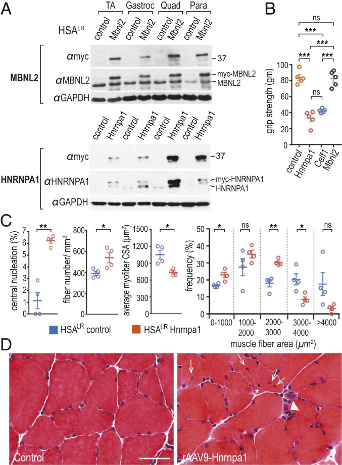
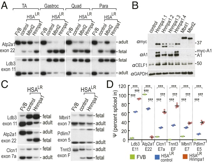
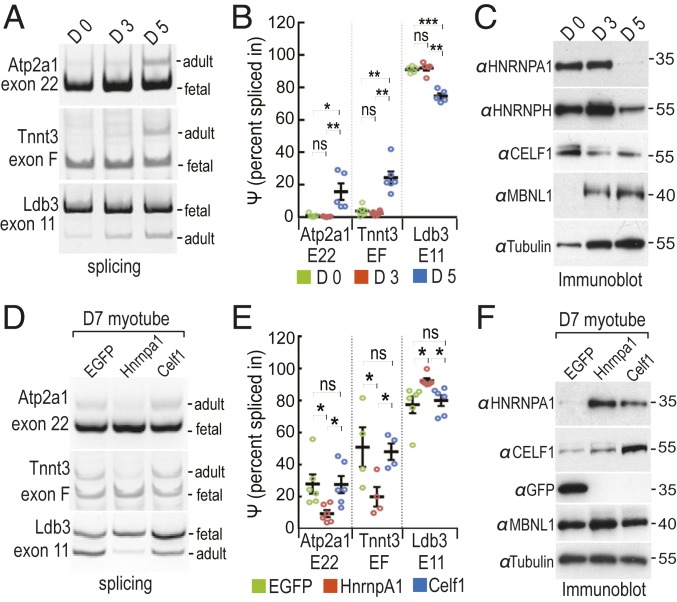
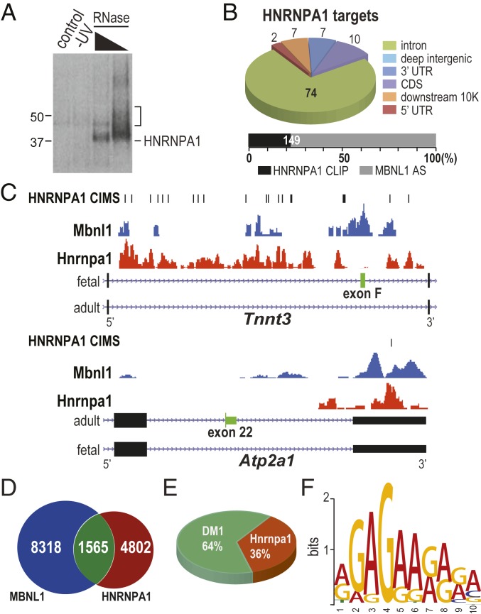
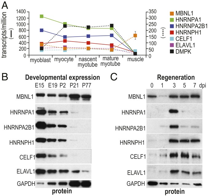
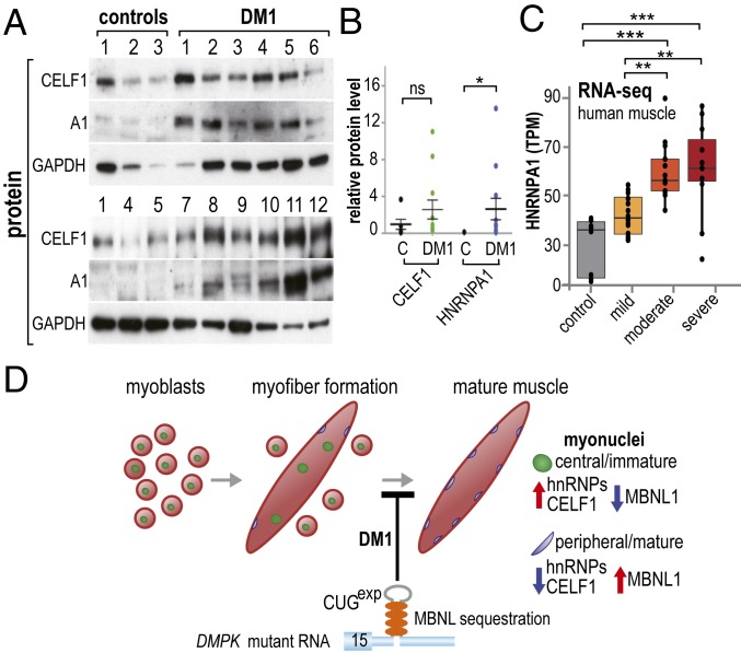
Studies on myotonic dystrophy type 1 (DM1) have led to the RNA-mediated disease model for hereditary disorders caused by noncoding microsatellite expansions. This model proposes that DM1 disease manifestations are caused by a reversion to fetal RNA processing patterns in adult tissues due to the expression of toxic CUG RNA expansions (CUGexp) leading to decreased muscleblind-like, but increased CUGBP1/ETR3-like factor 1 (CELF1), alternative splicing activities. Here, we test this model in vivo, using the mouse HSALR poly(CUG) model for DM1 and recombinant adeno-associated virus (rAAV)-mediated transduction of specific splicing factors. Surprisingly, systemic overexpression of HNRNPA1, not previously linked to DM1, also shifted DM1-relevant splicing targets to fetal isoforms, resulting in more severe muscle weakness/myopathy as early as 4 to 6 wk posttransduction, whereas rAAV controls were unaffected. Overexpression of HNRNPA1 promotes fetal exon inclusion of representative DM1-relevant splicing targets in differentiated myoblasts, and HITS-CLIP of rAAV-mycHnrnpa1-injected muscle revealed direct interactions of HNRNPA1 with these targets in vivo. Similar to CELF1, HNRNPA1 protein levels decrease during postnatal development, but are elevated in both regenerating mouse muscle and DM1 skeletal muscle. Our studies suggest that CUGexp RNA triggers abnormal expression of multiple nuclear RNA binding proteins, including CELF1 and HNRNPA1, that antagonize MBNL activity to promote fetal splicing patterns.

Keywords: CELF1; HNRNPA1; MBNL1; microsatellite; splicing.


Conflict of interest statement

The authors declare no competing interest.

Figures

Fig. 1.
Fig. 1.
AAV9-mediated systemic overexpression of HNRNPA1 in a mouse model of DM1 leads to disease-associated pathology. (A) HSALR P0-P2 mice (n = 4 each) were injected (i.v.) with PBS (control), AAV9/Mbnl2 (Mbnl2), or AAV9/HnrnpA1 (Hnrnpa1) and protein levels were assessed by immunoblotting using antibodies against both endogenous (MBNL2, HNRNPA1) and exogenous (myc-MBNL2, myc-HNRNPA1) proteins in tibialis anterior (TA), gastrocnemius (Gastroc), quadriceps (Quad) and paraspinal (Para) muscles. GAPDH served as the loading control. (B) AAV9-mediated overexpression of either CELF1 (AAV9-Celf1, n = 5) or HNRNPA1 (AAV9-Hnrnpa1 n = 5) led to reductions in grip strength compared with PBS (n = 5), GFP (AAV9-GFP, n = 5), or Mbnl2-injected (AAV9-Mbnl2, n = 5) mice. (C) Statistical analysis and (D) representative muscle cross-sections indicated an earlier onset of DM1-relevant myopathic changes including centralized myonuclei (white arrows), atrophic (white arrowhead) myofibers, and split (asterisk) fibers in HNRNPA1 (rAAV9-Hnrnpa1) overexpression quadriceps. P values were calculated using a one-way ANOVA with Tukey’s HSD post hoc test or an unpaired two-tailed Student’s t test. Data are SEM and significant. *P < 0.05; **P < 0.01; ***P < 0.001; ns, not significant. (Scale bar, 50 µm.)
Fig. 2.
Fig. 2.
Increased expression of HNRNPA1 in skeletal muscles promotes fetal splicing patterns for DM1 targets. (A) Systemic HNRNPA1 overexpression exacerbated, while MBNL2 partially reversed, DM1 fetal splicing patterns in HSALR muscles. RT-PCR analysis of Atp2a1 exon 22 and Ldb3 exon 11 splicing patterns were used as representative DM1 targets and positions of the fetal and adult exon splicing RT-PCR products are indicated (Right). Alternative cassette exon splicing was determined in wild-type (FVB) and HSALR mice without injection (control) or following AAV9-Mbnl2 (Mbnl2) or AAV9-HnrnpA1 HSALR injections (n = 4 each). (B) Immunoblotting for myc-tagged HNRNPA1 (αmyc), HNRNPA1 (αA1), CELF1 (αCELF1), and the loading control GAPDH (αGAPDH) following direct i.m. injections of AAV9-mycHnrnpa1 (n = 4 mice Hnrnp1.1-1.4), AAV9-mycMbnl1, or AAV9-mycMbnl2. The positions of myc-HNRNPA1 (myc-A1) and endogenous HNRNPA1 (A1) are indicated. (C) RT-PCR splicing analysis of six DM1 targeted gene transcripts demonstrated that HNRNPA1 overexpression in HSALR TA promoted fetal splicing events. The wild-type splicing pattern for each gene is shown in the FVB lanes. (D) Fetal exon inclusion was determined using percentage spliced in (Ψ; n = 4). P values were calculated using a one-way ANOVA with Tukey’s HSD post hoc test. Data are SEM and significant. ***P < 0.001.
Fig. 3.
Fig. 3.
Alternative splicing assay in myoblast/myotube cultures confirms DM1 fetal exon splicing is promoted by HNRNPA1. (A) Splicing patterns of three endogenous DM1 targets (Atp2a1 exon 22, Tnnt3 exon F, and Ldb3 exon 11) in myoblasts at day 0 (D0), day 3 (D3), and day 5 (D5) after differentiation. (B) Endogenous DM1 exon splicing in A was determined using percentage spliced in (Ψ) (n = 5). (C) Western blot analysis of four RNA binding proteins (HNRNPA1, HNRNPH, CELF1, and MBNL1) in myoblasts at D0, D3, and D5. (D) Effects of lenti-induced overexpression HnrnpA1 and Celf1 on endogenous DM1 targets in differentiated myofibers harvested at day 7 (D7). AAV9/EGFP is used as a negative control. (E) DM1 exon splicing in D was determined using Ψ (n = 4). (F) Immunoblot of four RNA binding proteins (HNRNPA1, HNRNPH, CELF1, and MBNL1) in AAV9/EGFP (EGFP), AAV9/HnRNPA1 (HnrnpA1), and AAV9/CELF1 (Celf1) transduced myoblasts harvested at D7. P values were calculated using a one-way ANOVA (or RM one-way ANOVA) with Tukey’s HSD post hoc test. Data are SEM and significant. *P < 0.05; **P < 0.01; ***P < 0.001; ns, not significant.
Fig. 4.
Fig. 4.
HITS-CLIP confirms that MBNL1 and HNRNPA1 proteins bind a set of overlapping RNA targets. HNRNPA1 binds directly to MBNL-regulated exons. (A) Protein gel autoradiograph of HNRNPA1-RNA 32P-labeled complexes after cross-linking, RNase A digestion and immunopurification with anti-HNRNPA1 mAb 4B10 from myc-HNRNPA1-injected HSALR quadriceps. PBS-injected (control) and uncrosslinked (-UV) controls are also shown. (B) Pie chart of HNRNPA1-CLIP tag distribution. (Lower) A stacked bar chart of the fraction of total MBNL1-regulated alternatively spliced exons bound by HNRNPA1 (±500 bp): MBNL1-regulated AS events (gray, n = 666) (45), AS events covered by HNRNPA1 CLIP peaks (black, n = 149). (C) Wiggle tracks of HNRNPA1 CLIP tag density for Tnnt3, and Atp2a1 for MBNL-regulated alternative exons. The MBNL1 CLIP-seq FVB WT quadriceps muscle dataset has been reported previously (46). (D) Venn diagram of overlap between HNRNPA1 binding sites and MBNL1 binding sites in FVB quadriceps. (E) Pie chart of overlap between HNRNPA1-CLIP tags and DM1 relative AS events in HSALR transgenic mice. (F) De novo motif search using sequences from top 500 CLIP clusters and MEME.
Fig. 5.
Fig. 5.
HNRNPA1 expression declines during mouse postnatal muscle development. (A) Human HNRNPA1 RNA levels decline as muscle precursor cells differentiate into mature skeletal muscle. Previously reported RNA-seq datasets were analyzed, including myoblasts, myocytes, nascent myotubes, and mature myotubes (22) and mature skeletal muscle (http://www.dmseq.org/; n = 3 for each sample). TPM (transcripts per million) values are shown for interested proteins. (B) Developmental expression of MBNL1 and hnRNP proteins (HNRNPA1, HNRNPA2/B1, HNRNPH), as well as CELF1 and ELAVL1, show opposite expression trends during muscle development from embryonic day (E)15 to postnatal day (P)77. (C) Immunoblot showing that HNRNPA1 is only transiently expressed at day 3 postinjection (dpi) during muscle regeneration after Notexin-induced muscle ablation and regeneration compared with other RNA binding proteins.
Fig. 6.
Fig. 6.
Up-regulation of both HNRNPA1 and CELF1 in DM1 muscle. (A) Immunoblots of control (n = 5) and DM1 (n = 12) muscle biopsy samples (GAPDH, loading control). (B) Quantitative analysis of relative protein levels (CELF/GAPDH, HNRNPA1/GAPDH) between control (C) and DM1 muscle biopsies. Note the very low level of HNRNPA1 in control adult muscles. P values were calculated using unpaired one-tailed Student’s t test. Data are SEM and significant. *P = 0.0268. (C) Box plots of HNRNPA1 mRNA levels (transcripts per million, TPM) in tibialis anterior muscle biopsies from control or DM1 patients. DM1 patients were classified as mild, moderate, or severe, as previously described (47). Patient RNA-seq data were acquired from the Myotonic Dystrophy Deep Sequencing Data Repository (http://www.dmseq.org/). P values were calculated using a one-way ANOVA with Tukey’s HSD post hoc test. Data are SEM and significant. **P < 0.01; ***P < 0.001. (D) Model for RBP misregulation in DM1 pathogenesis. Normally, myoblasts (Left, red circles with green central/immature nuclei) fuse to form immature myofibers (Middle, red ellipsoid) followed by nuclear migration to the subsarcolemmal region resulting in mature myofibers (Right, blue peripheral/mature nuclei). In DM1, the CUG expansion (gray hairpin in DMPK 3′ UTR) inhibits this normal differentiation process so DM1 myofibers are characterized by centralized myonuclei. At the molecular level, MBNL (orange ovals) splicing activity is down-regulated (blue arrow) due to sequestration by CUGexp RNAs, while hnRNPs and CELF1 are up-regulated (red arrow) either at the transcriptional (HNRNPA1) or posttranslational (CELF1) levels, leading to coordinate misregulation of these RNA splicing factors and DM1 spliceopathy.

References

    1. Iyer R. R., Pluciennik A., Napierala M., Wells R. D., DNA triplet repeat expansion and mismatch repair. Annu. Rev. Biochem. 84, 199–226 (2015). - PMC - PubMed
    1. Goodwin M., et al. , MBNL sequestration by toxic RNAs and RNA misprocessing in the myotonic dystrophy brain. Cell Rep. 12, 1159–1168 (2015). - PMC - PubMed
    1. Echeverria G. V., Cooper T. A., RNA-binding proteins in microsatellite expansion disorders: Mediators of RNA toxicity. Brain Res. 1462, 100–111 (2012). - PMC - PubMed
    1. Kuyumcu-Martinez N. M., Wang G. S., Cooper T. A., Increased steady-state levels of CUGBP1 in myotonic dystrophy 1 are due to PKC-mediated hyperphosphorylation. Mol. Cell 28, 68–78 (2007). - PMC - PubMed
    1. Scotti M. M., Swanson M. S., RNA mis-splicing in disease. Nat. Rev. Genet. 17, 19–32 (2016). - PMC - PubMed

Publication types

LinkOut - more resources