. 2020 Mar;30(3):244-255.
doi: 10.1038/s41422-020-0283-z. Epub 2020 Feb 21.

In vitro testicular organogenesis from human fetal gonads produces fertilization-competent spermatids

Affiliations

In vitro testicular organogenesis from human fetal gonads produces fertilization-competent spermatids

Yan Yuan et al. Cell Res. 2020 Mar.

Abstract

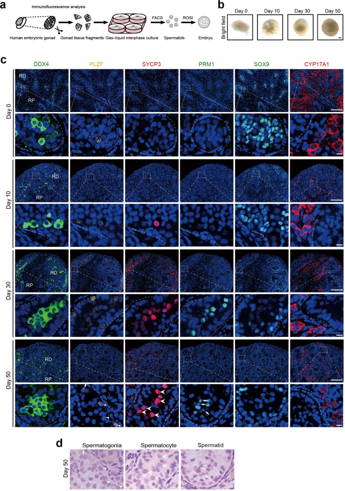
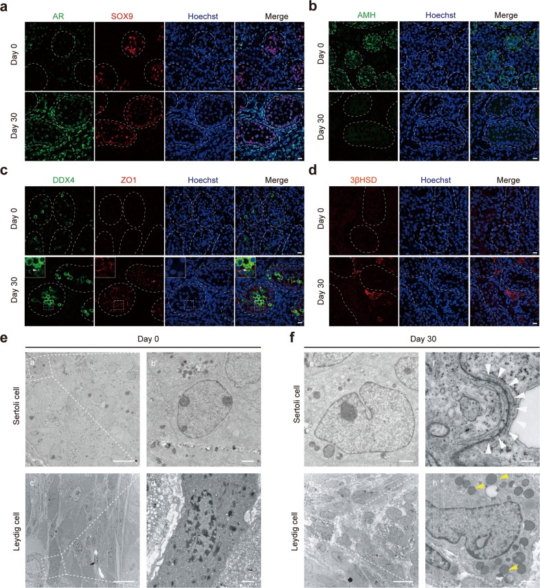
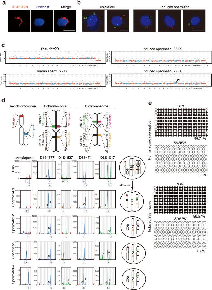
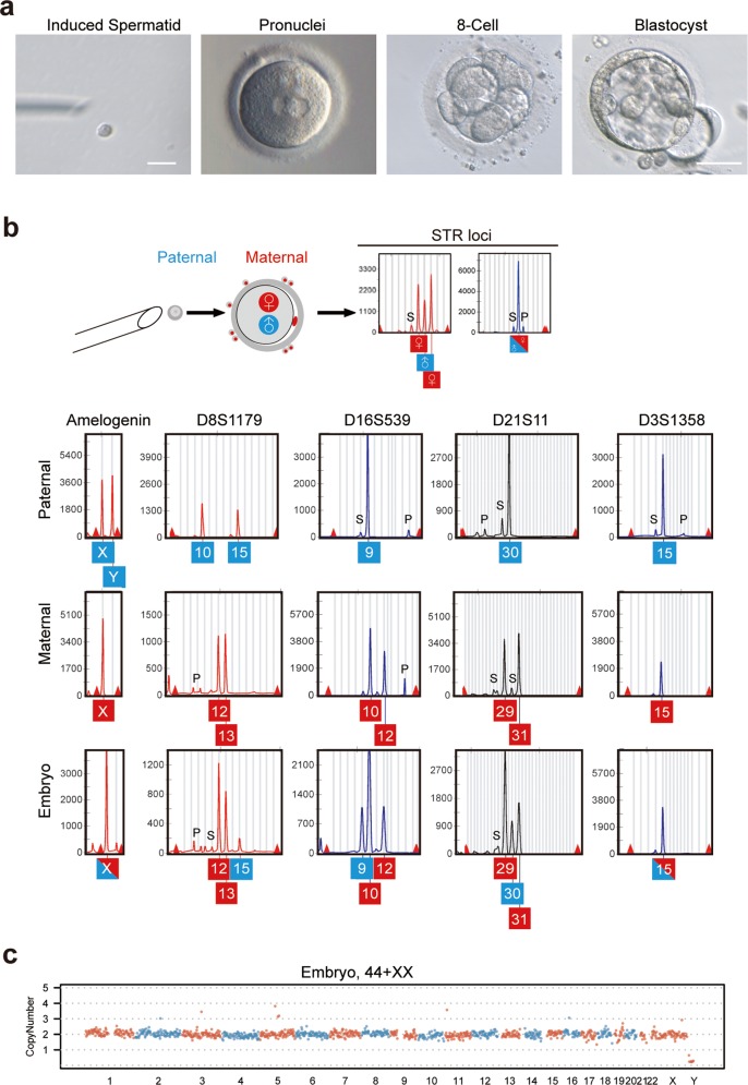
Unlike most organs that mature during the fetal period, the male reproductive system reaches maturity only at puberty with the commencement of spermatogenesis. Robust modelling of human testicular organogenesis in vitro would facilitate research into mechanisms of and factors affecting human spermatogenic failure and male fertility preservation in prepubertal tumor patients. Here, we report successful recapitulation of human testicular organogenesis in vitro from fetal gonadal ridge. Our model displayed the formation of mature seminiferous epithelium and self-renewing spermatogonia. Remarkably, in vitro-derived haploid spermatids have undergone meiotic recombination, and showed increased genetic diversity as indicated by genetic analysis. Moreover, these spermatids were able to fertilize oocytes and support subsequent blastocyst formation. The in vitro testicular organogenesis system described here will play an important role in elucidating the regulation of human testis development and maintaining male fertility in prepubertal cancer patients.


Conflict of interest statement

The authors declare no competing interests.

Figures

Fig. 1
Fig. 1. In vitro organogenesis of male human fetus gonads.
a Schematic representation of the technique used for human testicular organogenesis from embryonic gonads. b Bright-field images of 0-, 10-, 30-, and 50-day cultures. Scale bar = 1 mm. c Immunostaining of 0-, 10-, 30-, 50-day cultured gonadal tissues for DDX4 (green), PLZF (yellow) (triangle), SYCP3 (red) (arrowhead), PRM1 (green) (arrow), SOX9 (green), and CYP17A1 (red). Scale bars of low magnification = 100 µm. Scale bars of partial magnification = 10 µm. DNA was counterstained with Hoechst 33342 (blue). RP, Region proximal to mesonephros; RD, Region distal to mesonephros. d Consecutive H&E staining of the same section of 50-day cultured tissue used in IF analysis (c). Spermatogonia (triangle), spermatocytes (arrowhead), and spermatid-like cell (arrow) are indicated; Scale bars, 10 μm.
Fig. 2
Fig. 2. Sertoli and Leydig cell maturation during human testis organogenesis in vitro.
a Immunostaining of 0- and 30-day cultured gonadal tissues for AR (green) and SOX9 (red). Scale bars = 10 µm. DNA was counterstained with Hoechst 33342 (blue). b Detection of Sertoli cells in 0- and 30-day cultured tissues by immunostaining for AMH (green). Scale bars = 10 µm. DNA was counterstained with Hoechst 33342 (blue). c Immunostaining of 0- and 30-day cultured tissues for DDX4 (green) and ZO1 (red). Scale bars = 10 µm. DNA was counterstained with Hoechst 33342 (blue). Chromatoid body (white triangle). d Immunostaining of Leydig cells for 3β-HSD (red) after 0 and 30 days in culture. Scale bars = 10 µm. DNA was counterstained with Hoechst 33342 (blue). e, f Transmission electron microscopy shows ultrastructural features of Sertoli and Leydig cells from day 0 and 30 cultured gonadal tissues. Tight junction between Sertoli cells (white triangles) and lipid droplets in the Leydig cells (yellow triangles) are indicated. Scale bars = 10 µm (a′, c′, g′); 1 µm (b′, d′, e′, h′); 200 nm (f′).
Fig. 3
Fig. 3. Successful spermatogenesis of germ cells during human testis organogenesis.
a Immunofluorescent labeling of 0-, 10-, 30- and 50-day cultured gonadal tissues co-labeled for PLZF (red) and Ki67 (green). PLZF+Ki67+ cells (white triangles). Scale bar = 10 µm. b Percentage of PLZF-positive KI67-positive cells from three biological replicates. (Each dot represents the percentage in the cross section of a seminiferous tubule, different colors of the dots represent different specimens). c Immunostaining of cultured tissues for SYCP3 (green), γH2AX (red), MLH1 (red) and SYCP1 (green). DNA was counterstained with Hoechst 33342 (blue). Scale bar = 10 µm. d Transmission electron microscopy shows ultrastructural findings of seminiferous epithelium. Spermatids: white stars; spermatogonia: yellow stars; primary spermatocytes with nuclear synaptonemal complexes: blue stars; spermatids with acrosome vesicles: yellow triangle; and early tail structure: white triangles. Scale bars, 10 µm (a); 2 µm (b, d); 200 nm (c); 1 µm (e). e DNA content distribution of cells from 0- to 50-day cultured tissues by FACS. Haploid (1C), diploid (2C), and tetraploid (4C) cells are indicated. f Percentages of haploid cells in FACS analysis of 11 cultured samples.
Fig. 4
Fig. 4. Key genetic and epigenetic properties of induced spermatids.
a Immunostaining for ACROSIN in haploid cells isolated from cultured tissues by FACS. Scale bars = 10 µm. b FISH analysis using somatic probes specific to chromosomes 13 and 21, or sex probes specific to chromosomes X and Y for diploid and induced spermatids. Scale bars = 10 µm. c Genomic DNA sequencing of diploid cells from aborted fetal skin cell, in vivo spermatid and in vitro-derived single spermatid sorted by FACS. Abnormal copy number changes in chromosome 12: black arrow. d Alleles of STR loci in aborted fetal somatic skin cells and induced spermatids depict chromosome separation and homologous recombination (left). Schematic map of chromosomal STR loci and separation of chromosomes after meiosis (right). S: Stutter peak; P: Pull down peak. e Bisulfite sequencing of differentially methylated regions (DMRs) of H19 and SNRPN in human round spermatids, and induced spermatids. White and black circles indicate unmethylated and methylated CpGs, respectively.
Fig. 5
Fig. 5. Functional analysis of spermatids derived from in vitro testicular organogenesis.
a A MII oocyte derived through IVM was injected with an induced spermatid, followed by development to pronuclear stage with two pronuclei, 8-cell and blastocyst stages. Scale bars = 10 µm (left); 100 µm (right). b STR and sex loci of embryo, and fetal skin (paternal) that contributed the round spermatid and granulosa cells (maternal) of oocyte. S: Stutter peak; P: Pull down peak. c CNV analysis of the embryo (b) by genomic DNA sequencing.

References

    1. Chemes HE. Infancy is not a quiescent period of testicular development. Int. J. Androl. 2001;24:2–7. doi: 10.1046/j.1365-2605.2001.00260.x. - DOI - PubMed
    1. Rey RA, Musse M, Venara M, Chemes HE. Ontogeny of the androgen receptor expression in the fetal and postnatal testis: its relevance on Sertoli cell maturation and the onset of adult spermatogenesis. Microsc. Res. Tech. 2009;72:787–795. doi: 10.1002/jemt.20754. - DOI - PubMed
    1. Stukenborg JB, Colon E, Soder O. Ontogenesis of testis development and function in humans. Sex. Dev. 2010;4:199–212. doi: 10.1159/000317090. - DOI - PubMed
    1. Clermont Y. Kinetics of spermatogenesis in mammals: seminiferous epithelium cycle and spermatogonial renewal. Physiol. Rev. 1972;52:198–236. doi: 10.1152/physrev.1972.52.1.198. - DOI - PubMed
    1. Inhorn MC, Patrizio P. Infertility around the globe: new thinking on gender, reproductive technologies and global movements in the 21st century. Hum. Reprod. Update. 2015;21:411–426. doi: 10.1093/humupd/dmv016. - DOI - PubMed

Publication types

LinkOut - more resources