. 2020 Apr 17;48(7):3525-3541.
doi: 10.1093/nar/gkaa101.

FIGLA, LHX8 and SOHLH1 transcription factor networks regulate mouse oocyte growth and differentiation

Affiliations

FIGLA, LHX8 and SOHLH1 transcription factor networks regulate mouse oocyte growth and differentiation

Zhengpin Wang et al. Nucleic Acids Res. .

Abstract

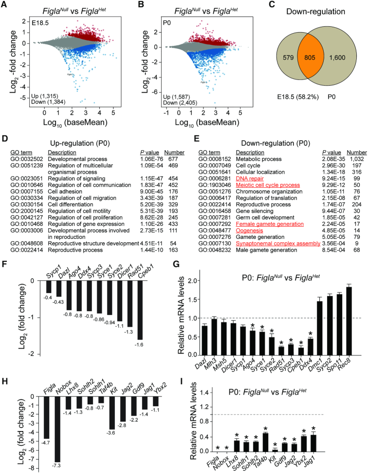
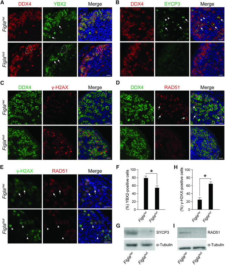
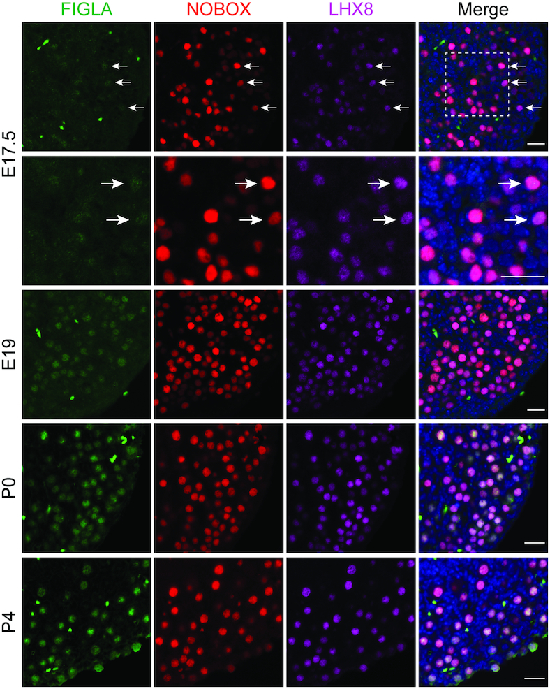
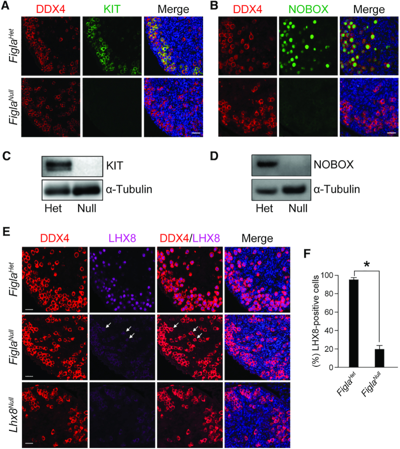
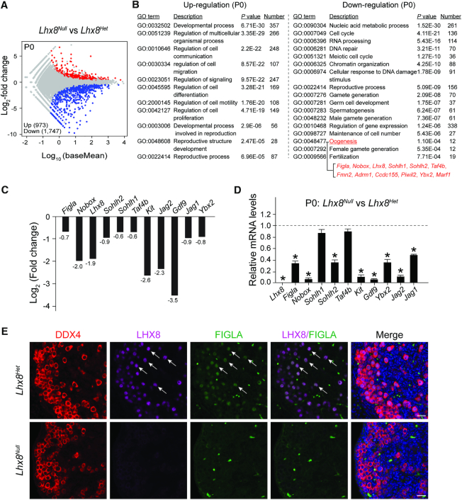
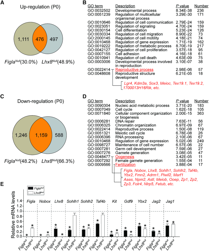
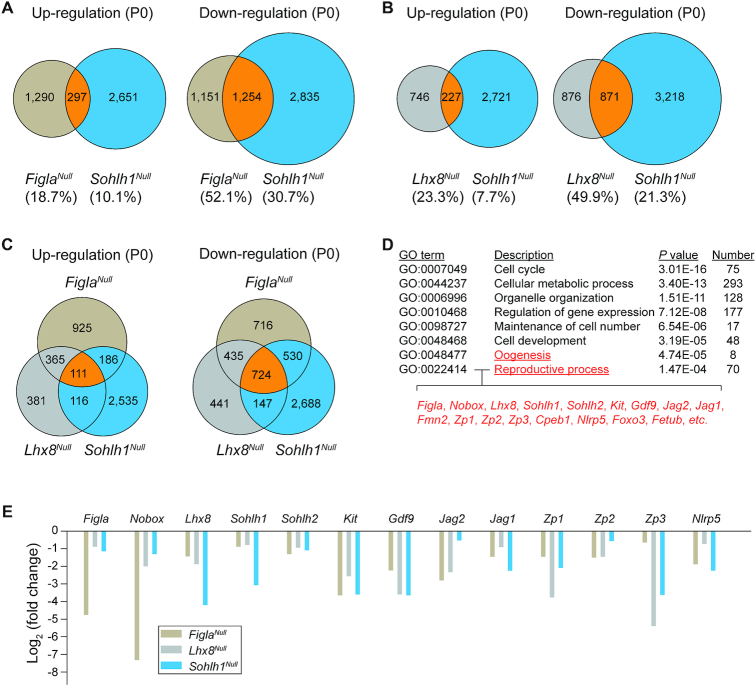
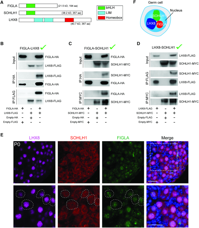
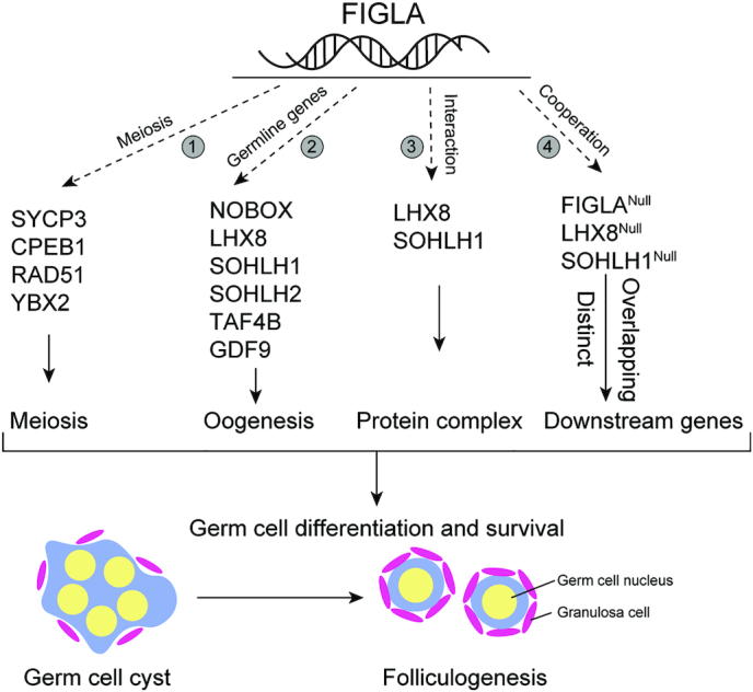
Germ-cell transcription factors control gene networks that regulate oocyte differentiation and primordial follicle formation during early, postnatal mouse oogenesis. Taking advantage of gene-edited mice lacking transcription factors expressed in female germ cells, we analyzed global gene expression profiles in perinatal ovaries from wildtype, FiglaNull, Lhx8Null and Sohlh1Null mice. Figla deficiency dysregulates expression of meiosis-related genes (e.g. Sycp3, Rad51, Ybx2) and a variety of genes (e.g. Nobox, Lhx8, Taf4b, Sohlh1, Sohlh2, Gdf9) associated with oocyte growth and differentiation. The absence of FIGLA significantly impedes meiotic progression, causes DNA damage and results in oocyte apoptosis. Moreover, we find that FIGLA and other transcriptional regulator proteins (e.g. NOBOX, LHX8, SOHLH1, SOHLH2) are co-expressed in the same subset of germ cells in perinatal ovaries and Figla ablation dramatically disrupts KIT, NOBOX, LHX8, SOHLH1 and SOHLH2 abundance. In addition, not only do FIGLA, LHX8 and SOHLH1 cross-regulate each other, they also cooperate by direct interaction with each during early oocyte development and share downstream gene targets. Thus, our findings substantiate a major role for FIGLA, LHX8 and SOHLH1 as multifunctional regulators of networks necessary for oocyte maintenance and differentiation during early folliculogenesis.


Figures

Figure 1.
Figure 1.
Figla deficiency alters expression patterns of meiotic and oogenic genes. (A, B) MA plot (log ratio RNA abundance versus abundance) of RNA-seq data from FiglaHet and FiglaNull ovaries at E18.5 (A) and P0 (B), respectively, using adjusted P < 0.1 as the cut off. (C) Venn diagram depicting the overlap of down-regulated genes examined at two developmental time points, E18.5 and P0. (D, E) The enriched Gene Ontology terms of biological process categories of up- (D) and down-regulated (E) transcripts in P0 FiglaNull ovaries. (F) RNA-seq results of selected transcripts (log2-fold change) related to meiosis in P0 FiglaNull ovaries. (G) Quantitative RT-PCR validation of meiosis-related genes in FiglaNull ovaries at P0. The control genes relative to β-actin were set to 1. Data are presented as mean ± S.D. for n = 3 biologically independent experiments. *P <0.05 by two-tailed Student's t test. (H) Same as (F), but of oogenesis-related transcripts. (I) Same as (G), but of genes related to oogenesis.
Figure 2.
Figure 2.
Figla deficiency impairs meiotic progression and causes DNA damage. (A) Immunofluorescence staining with antibodies to DDX4 (red) and YBX2 (green) on ovarian sections from P0 FiglaHet and FiglaNull mice. DNA was stained with Hoechst 33342. Arrows indicate YBX2-positive oocytes. The dashed lines indicate DDX4-positive, but YBX2-negative oocytes. Scale bar, 20 μm. (B) Same as (A), but with antibodies to DDX4 (red) and SYCP3 (green). Some oocytes showed visible nucleoli (arrows) after staining with SYCP3 antibodies indicating that these oocytes had reached the dictyate stage. Oocytes that were negative for nucleoli (arrowheads) had not reached the dictyate. (C) Same as (A), but with antibodies to DDX4 (green) and γ-H2AX (red). (D) Same as (A), but with antibodies to DDX4 (green) and RAD51 (red). Arrows indicate oocytes expressing RAD51 foci in the nucleus. (E) Same as (A), but with antibodies to γ-H2AX (green) and RAD51 (red). Arrows indicate γ-H2AX-positive and RAD51-positive oocytes. Arrowheads indicate γ-H2AX-positive, but RAD51-negative oocytes. (F, H) Statistical analysis of the percentage of YBX2-positive cells (F) and γ-H2AX-positive cells (H) per section in FiglaHet and FiglaNull ovaries. Mean ± s.d, n = 3 biologically independent ovaries from three different animals. *P <0.05 by two-tailed Student's t test. (G, I) Immunoblot assay of SYCP3 (G) and RAD51 (I) in P0 FiglaHet and FiglaNull ovaries. α-Tubulin was used as an internal control. Representative of n = 3 (A–E, G and I) independent biological replicates with similar results per condition.
Figure 3.
Figure 3.
Co-expression of FIGLA, LHX8 and NOBOX in perinatal ovaries. Images showing immunofluorescence results from FiglaFLAG ovarian sections stained with antibodies against FLAG (FIGLA, green), NOBOX (red) and LHX8 (purple) at the indicated time points. DNA was stained with Hoechst 33342. Arrows indicate that FIGLA is expressed solely in a small subset of NOBOX- and LHX8-positive oocytes in E17.5 ovaries. FIGLA was co-expressed with NOBOX and LHX8 in the great majority of oocytes in E19, P0 and P4 ovaries. Scale bar, 20 μm. Data are representative of n = 3 independent ovaries from three different animals with similar results per condition.
Figure 4.
Figure 4.
Figla deficiency disrupts KIT, NOBOX and LHX8 expression. (A) Immunofluorescence staining with DDX4 (red) and KIT (green) in FiglaHet and FiglaNull ovaries at P0. DNA was stained with Hoechst 33342. KIT expression was completely abolished in FiglaNull ovaries. Scale bar, 20 μm. (B) Same as (A), but with antibodies to DDX4 (red) and NOBOX (green). NOBOX expression was significantly diminished in Figla-deficient ovaries. (C) Immunoblot analysis of KIT protein expression in FiglaHet and FiglaNull ovaries at P0. α-Tubulin was used as a load control. (D) Same as (C), but with antibodies to NOBOX which was absent in Figla-deficient ovaries. (E) Immunostaining of DDX4 (red) and LHX8 (purple) in P0 FiglaHet, FiglaNull and Lhx8Null ovaries. DNA was stained with Hoechst 33342. Arrows indicate weak expression of LHX8 in the nucleus of Figla-deficient oocytes. Scale bar, 20 μm. (F) Statistical analysis of LHX8-positive cells per section in FiglaHet and FiglaNull ovaries at P0. Mean ± S.D., n = 3 biologically independent ovaries from three different animals. *P <0.05 by two-tailed Student's t test. Representative of n = 3 (A–E) independent biological replicates with similar results per condition.
Figure 5.
Figure 5.
Lhx8 deficiency disrupts oogenesis-associated genes. (A) MA plot of RNA-seq data from Lhx8Het and Lhx8Null ovaries at P0, using adjusted P < 0.1 as the cut off. (B) Enriched Gene Ontology terms of the biological process categories for up- and down-regulated transcripts in Lhx8Null ovaries. (C) RNA-seq results of selected transcripts (log2-fold change) related to oogenesis in Lhx8Null ovaries. (D) Quantitative RT-PCR validation of oogenesis-related genes in Lhx8Null ovaries at P0. Control genes relative to β-actin were set to 1. Data are presented as mean ± s.d for n = 3 biologically independent experiments. *P <0.05 by two-tailed Student's t test. (E) Immunofluorescence staining with antibodies to DDX4 (red), LHX8 (purple) and FIGLA (green) on ovarian sections from P0 Lhx8Het and Lhx8Null mice after crossing with FiglaFLAG mice. The DNA was stained with Hoechst 33342. Arrows indicate both LHX8- and FIGLA-positive germ cells. Scale bar, 20 μm. Representative of n = 3 independent biological replicates with similar results per condition.
Figure 6.
Figure 6.
FIGLA and LHX8 coordinate oocyte differentiation. (A) Venn diagram showing overlap and difference in the number of up-regulated transcripts from FiglaNull and Lhx8Null ovaries when compared with control newborn ovary transcriptomes. (B) The enriched Gene Ontology terms of the biological process categories of up-regulated genes in both FiglaNull and Lhx8Null ovaries at P0. (C) Same as (A) but showing the down-regulated transcripts. (D) Same as (B), but of down-regulated transcripts. (E) Quantitative RT-PCR validation of oogenesis-related genes in both FiglaNull and Lhx8Null ovaries at P0. The control genes relative to β-actin were set to 1. Data are presented as mean ± S.D. for n = 3 biologically independent experiments. *P <0.05 by two-tailed Student's t test.
Figure 7.
Figure 7.
Transcriptome profiles and networks in P0 FiglaNull, Lhx8Null and Sohlh1Null ovaries. (A) Venn diagram of overlap and difference in the number of up-regulated and down-regulated transcripts in FiglaNull and Sohlh1Null ovaries. (B) Same as (A), but in Lhx8Null and Sohlh1Null ovaries. (C) Same as (A), but in FiglaNull, Lhx8Null and Sohlh1Null ovaries. (D) The enriched Gene Ontology terms of the biological process categories of down-regulated genes from FiglaNull, Lhx8Null and Sohlh1Null ovaries. (E) RNA-seq results of selected transcripts (log2-fold change) associated with oogenesis and reproductive processes from FiglaNull, Lhx8Null and Sohlh1Null ovaries.
Figure 8.
Figure 8.
Interactions of FIGLA, LHX8 and SOHLH1. (A) Representative protein domains (bHLH, LIM, homeobox) of FIGLA, SOHLH1 and LHX8. (B) FIGLAHA and LHX8FLAG expression vectors were co-transfected into HEK-293T cells. Cell lysates were probed with HA and FLAG antibodies to detect input protein FIGLA and LHX8, respectively. After immunoprecipitation with HA and FLAG antibody, immunoblots were performed to detect FIGLA and associated LHX8 protein or LHX8 and associated FIGLA protein, respectively. (C) Same as (B) except that FIGLAHA and SOHLH1MYC were co-transfected into HEK-293T cells. Antibodies to HA and MYC were used to detect input proteins and immunoprecipitate FIGLA and SOHLH1, respectively, and their associated proteins. (D) Same as (B) except that LHX8FLAG and SOHLH1MYC were co-transfected into HEK-293T cells. Antibodies to FLAG and MYC were used to detect input proteins and immunoprecipitate LHX8 and SOHLH1, respectively, and their associated proteins. (E) Co-expression of FIGLA, LHX8 and SOHLH1 in P0 ovaries from FiglaFLAG mice. The dashed circles indicate co-expression of FIGLA, LHX8 and SOHLH1 in the same oocytes. Scale bar, 20 μm. (F) FIGLA, LHX8 and SOHLH1 appear to form a nuclear complex in oocytes. Representative of n = 3 (B–E) independent biological replicates with similar results per condition.
Figure 9.
Figure 9.
Hypothetical model of FIGLA function in early folliculogenesis. FIGLA concomitantly functions in multiple pathways during early oogenesis: 1, FIGLA regulates meiotic genes (e.g. Sycp3, Cpeb1, Rad51, Ybx2) to ensure proper meiotic progression; 2, FIGLA up-regulates a subset of germline genes (Nobox, Lhx8, Sohlh1, Sohlh2, Taf4b, Gdf9) and their downstream genes that are essential for early oocyte development; 3, FIGLA, LHX8 and SOHLH1 interact to form a nuclear complex in the oocytes which could recruit transcriptional co-activators or co-repressors during oocyte growth and differentiation and 4, FIGLA regulates the transcription of downstream targets in conjunction with LHX8 and SOHLH1, but each factor also has distinct, non-overlapping target genes.

Similar articles

Cited by

References

    1. Pepling M.E., Spradling A.C.. Female mouse germ cells form synchronously dividing cysts. Development. 1998; 125:3323–3328. - PubMed
    1. Pepling M.E., Spradling A.C.. Mouse ovarian germ cell cysts undergo programmed breakdown to form primordial follicles. Dev. Biol. 2001; 234:339–351. - PubMed
    1. Pepling M.E. From primordial germ cell to primordial follicle: mammalian female germ cell development. Genesis. 2006; 44:622–632. - PubMed
    1. Zhang H., Liu L., Li X., Busayavalasa K., Shen Y., Hovatta O., Gustafsson J.A., Liu K.. Life-long in vivo cell-lineage tracing shows that no oogenesis originates from putative germline stem cells in adult mice. Proc. Natl. Acad. Sci. U.S.A. 2014; 111:17983–17988. - PMC - PubMed
    1. Lei L., Spradling A.C.. Female mice lack adult germ-line stem cells but sustain oogenesis using stable primordial follicles. Proc. Natl. Acad. Sci. U.S.A. 2013; 110:8585–8590. - PMC - PubMed

Publication types

MeSH terms

Substances