. 2020 Apr;34(4):5563-5577.
doi: 10.1096/fj.201903134R. Epub 2020 Feb 21.

A novel postsynaptic signal pathway of sympathetic neural regulation of murine colonic motility

Affiliations

A novel postsynaptic signal pathway of sympathetic neural regulation of murine colonic motility

Masaaki Kurahashi et al. FASEB J. 2020 Apr.

Abstract

Transcriptome data revealed α1 adrenoceptors (ARs) expression in platelet-derived growth factor receptor α+ cells (PDGFRα+ cells) in murine colonic musculature. The role of PDGFRα+ cells in sympathetic neural regulation of murine colonic motility was investigated. Norepinephrine (NE), via α1A ARs, activated a small conductance Ca2+ -activated K+ (SK) conductance, evoked outward currents and hyperpolarized PDGFRα+ cells (the α1A AR-SK channel signal pathway). α1 AR agonists increased intracellular Ca2+ transients in PDGFRα+ cells and inhibited spontaneous phasic contractions (SPCs) of colonic muscle through activation of a SK conductance. Sympathetic nerve stimulation inhibited both contractions of distal colon and propulsive contractions represented by the colonic migrating motor complexes (CMMCs) via the α1A AR-SK channel signal pathway. Postsynaptic signaling through α1A ARs in PDGFRα+ cells is a novel mechanism that conveys part of stress responses in the colon. PDGFRα+ cells appear to be a primary effector of sympathetic neural regulation of murine colonic motility.

Keywords: PDGFRα+ cells; colonic motility; sympathetic neural regulation; α1 adrenoceptor.


Figures

Figure 1:
Figure 1:
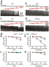
Capability of PDGFRα+ cells to responding to sympathetic neural signaling. A: Whole tissue immunohistochemical labeling of PDGFRα (green) and tyrosine hydroxylase (TH) (red) in circular muscle layer (a) and plane of the myenteric plexus (b) in the mouse distal colon. PDGFRα+ cells were closely associated with TH+ varicose nerve fibers. White scale bars represent 10 μm. B: Gene expression in PDGFRα+ cells and unsorted cells, which were the dispersed cells of colon before FACS, analyzed by qPCR. α1 Adrenoceptors (ARs) (Adra1a, Adra1b and Adra1d), especially Adra1a were expressed in PDGFRα+ cells. C-E: Patch clamp data to PDGFRα+ cells. Norepinephrine (NE; 10 μM) was applied repetitively (black bars). NE evoked transient outward currents in voltage-clamp mode at the holding potential of −50 mV (C and D). NE caused transient hyperpolarization in current-clamp mode (I = 0) in PDGFRα+ cells (Ea). Outward currents and hyperpolarization responses evoked by NE were blocked by RS100329 (RS; 100nM) (C and E) or apamin (Apa; 300nM) (D). Eb summarizes ΔAUC (mV·min) (AUC for 1 min after applying NE - AUC for 1min before applying NE) in the absence or the presence of RS in the current-clamp mode. * P = 0.0084.
Figure 2:
Figure 2:
Ca2+ transients in response to NE in PDGFRα+ cells. A Representative raw image showing PDGFRα cells in the intramuscular region of the colon expressing the Ca2+ sensor, GCaMP6f. A purple hue was added as an overlay to enhance visualization in B; color scale indicates intensity of Ca2+ transients (i.e. dark blue is low florescence; light yellow to white indicate high florescence levels). C Image showing enhanced Ca2+ transients in PDGFRα+ cells in response to NE (10 μM). Scale bar in A is 20 μm and pertains to all images. D Representative traces showing Ca2+ transient activity in PDGFRα+ cells elicited by MRS2365 (P2Y1 receptor specific agonist; 1 μM). This is considered a signature response that identifies PDGFRα+ cells. E Representative Ca2+ transient activity in PDGFRα+ cells in response to NE (10 μM). F Ca2+ responses to NE were inhibited by RS100329 (100 nM, red trace). Arrows (blue) in D, E and F indicate points of NE application. G Frequency, H Ca2+ transient area, I duration and J spatial spread. * denotes significant difference between control and after NE application. The numbers of asterisks mean: * 0.05 > P ≥ 0.01; ** 0.01 > P ≥ 0.001; *** 0.001 > P ≥ 0.0001; **** 0.0001 > P.
Figure 3:
Figure 3:
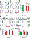
Effects of Phenylephrine (PE) on membrane potentials recorded from circular SMCs of mouse distal colon. Intracellular recordings were performed in the presence of TTX (1 μM) to inhibit neural activity. A: PE (1 and 10 μM) induced a hyperpolarization. B: PE-induced hyperpolarization was abolished by pretreatment of RS100329 (100nM). C: Apamin (Apa) 300nM inhibited PE-induced hyperpolarization. The resting membrane potentials were: A: −47 mV; B: −42 mV; C: −46 mV. A-C were recorded from different tissues. Each record in a given set of two was obtained from the same impalement. D: Summary showing the effects of RS100329 and Apa on PE-induced hyperpolarization. * P < 0.0001, significant difference from ΔV of PE (1 μM).†P < 0.0001, significant difference from ΔV of PE (10 μM).
Figure 4:
Figure 4:
Spontaneous phasic contractions (SPCs) of distal colonic circular muscle in the presence of tetrodotoxin (TTX; 1 μM). A: Phenylephrine (PE), a selective α1 AR ligand, inhibited SPCs in a concentration-dependent manner (Aa). RS100329 (RS; 100nM) (Ab), apamin (Apa; 300nM) (Ac), or the deletion of the gene encoding α1A ARs (Ad) inhibited the effects of PE. B: Summary of 4 contractile parameters, area under the curve (AUC) (Ba), amplitude (Bb), tone (Bc), and frequency (Bd) were tabulated as the ratio of SPCs before application of PE (control) to after application of PE. Amplitude of SPCs was most explicitly inhibited by PE alone compared to those in the presence of RS, apamin, or in the colonic muscles of Adra1a−/− mouse (Bb). * denote significant difference between PE alone and the other protocol. The numbers of asterisks mean the same as those in Fig. 2.
Figure 5:
Figure 5:
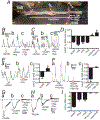
Responses of the distal colon to sympathetic nerve stimulation (SNS) with atropine (1 μM) (A), L-NNA (100 μM) (L), MRS2500 (1 μM) (M), hexamethonium (100 μM) (H) (ALMH) in the bath solution. All red bars represent SNS at 20 Hz at 150 V for 10 s (pulse duration: 0.3 ms). All black dot-lines represent the control baseline of contractions or the resting membrane potentials. A: A preparation for the tension recordings with the lumber colonic nerve (LCN) and the inferior mesenteric ganglion (IMG) intact. B and C: Contractile experiments of WT mouse. SNS caused inhibition of contractions (Ba and Ca). Propranolol (Pro; 10 μM) didn’t block inhibitions by SNS (Bb and Cb), but Pro+ RS100329 (RS; 100 nM) (Bc) or + apamin (Apa; 300nM) (Cb) blocked them (Bc and Cc). D: Summary of ΔAUC (mN•min) by SNS (AUC during SNS - the 10s average of AUC before SNS). * and†P < 0.0005, significant difference from ΔAUC with any single reagent. E: Contractile experiments of Adra1a−/− mouse. SNS relaxed distal colon (Ea) and Pro blocked inhibitions by SNS (Eb). Ec shows summary of ΔAUC (mN•min) by SNS. * P < 0.05. F: Contractile experiments of WT mouse with Pro and substance P (Sp; 1 μM). SNS relaxed distal colon (Fa). 18β-Glycyrrhetinic acid (18β-Gly; 100 μM) blocked inhibitions by SNS (Fb). Fc showed summary of ΔAUC (mN•min) by SNS. ** P < 0.005. G-H: Intracellular electrical recordings of SMCs of WT mouse. SNS induced fast and slow phases of hyperpolarization (Ga and Ha). Prazosin (Praz; 1 μM) or Apa inhibited the fast hyperpolarization (Gb and Hb), and Pro inhibited the residual slow hyperpolarization (Gc and Hc). Resting membrane potential were: G, −48 mV; H, −51mV. G and H were recorded from different tissues. Each record in a given set of three was obtained from the same impalement. I: Summary of ΔV (mV) (induced hyperpolarization by SNS). * and†P < 0.0001, significant difference from ΔV of control. ** and††P < 0.0001, significant difference from Praz and Apa respectively.
Figure 6:
Figure 6:
Tension recordings at proximal (Prox), mid and distal colon in the preparation shown in Figure 5A. In this experiment, transmural nerve stimulation (TNS) across wall of proximal colon (blue bars in A and B) elicited the colonic migrating motor complex (CMMC; indicated by *). A: Sympathetic nerve stimulation (SNS; 2 Hz; red bars in A and B) inhibited CMMC at mid and distal colon (Ab). B: The inhibitory effects of SNS (2Hz) on CMMC were blocked by RS100329 (RS; 100nM) (Ba and c). C: Summary of normalized amplitude of CMMC with SNS. RS attenuated the inhibitory effects of SNS. D: Spontaneous CMMC were inhibited by SNS (2 Hz) in WT mouse (Da), however CMMC were not affected by SNS (2 Hz) in the colon of Adra1a−/− mouse (Db). E: Summary of normalized amplitude of CMMC with SNS at 2 Hz (Ea) and 5 Hz (Eb). Deletion of the gene encoding α1A ARs blocked the inhibitory effects of SNS. In panels of C and E, Black asterisks denote statistically significant differences between the values connected by black lines and white asterisks mean statistically significant differences between the amplitude with SNS and the control. The numbers of asterisks mean the same as those in Fig. 2.
Figure 7:
Figure 7:
Schematic diagram depicting a novel signal pathway of sympathetic neural regulation of murine colon. Sympathetic nerve fibers, a blood vessel and the cells of the SIP syncytium (SMCs, ICC and PDGFRα+ cells) are displayed. Gap junctions (GJ) are shown between SMCs and ICC and PDGFRα+ cells in dark green. PDGFRα+ cells (pale green cell) express α1A ARs. Neuronal or hormonal norepinephrine (NE) (and possibly epinephrine; Epi) (orange circles) bind to α1A ARs, enhance Ca2+ release from endoplasmic reticulum (ER) via generation of inositol triphosphate (IP3) and activate small conductance Ca2+ -activated K+ channels type 3 (SK3; expressed robustly in PDGFRα+ cells; Fig. 1B). SK3 channels generate outward currents and induce hyperpolarization (Hyperpol.) in PDGFRα+ cells, which conducts via GJ to SMCs (pale blue cell) and probably ICC (pink cell). Hyperpolarization of SMCs reduces the open probability of L-type Ca2+ channels (L-VDCC) and decreases intracellular [Ca2+], leading to inhibition of contractions. ICC (pale pink cell) are not involved in this novel signal pathway.

Similar articles

Cited by

References

    1. Norberg KA and Sjoqvist F (1966) New possibilities for adrenergic modulation of ganglionic transmission. Permacol. Rev 18, 743–751 - PubMed
    1. Burnstock G and Costa M (1973) Inhibitory innervation of the gut. Gastroenterology 64, 141–144 - PubMed
    1. De Ponti F, Giaroni C, Cosentino M, Lecchini S and Frigo G (1996) Adrenergic mechanisms in the control of gastrointestinal motility: from basic science to clinical applications. Pharmacol. Ther 69, 59–78 - PubMed
    1. Lomax AE, Sharkey KA and Furness JB (2010) The participation of the sympathetic innervation of the gastrointestinal tract in disease states. Neurogastroenterol. Motil 22, 7–18 - PubMed
    1. Gagnon DJ, Devroede G. and Belisle S. (1972) Excitatory effects of adrenaline upon isolated preparations of human colon. Gut 13, 654–657 - PMC - PubMed

Publication types

MeSH terms

Substances

LinkOut - more resources