Review
. 2020 Jun;1867(6):118688.
doi: 10.1016/j.bbamcr.2020.118688. Epub 2020 Feb 19.

Targeting apoptotic caspases in cancer

Affiliations
Review

Targeting apoptotic caspases in cancer

Ashley Boice et al. Biochim Biophys Acta Mol Cell Res. 2020 Jun.

Abstract

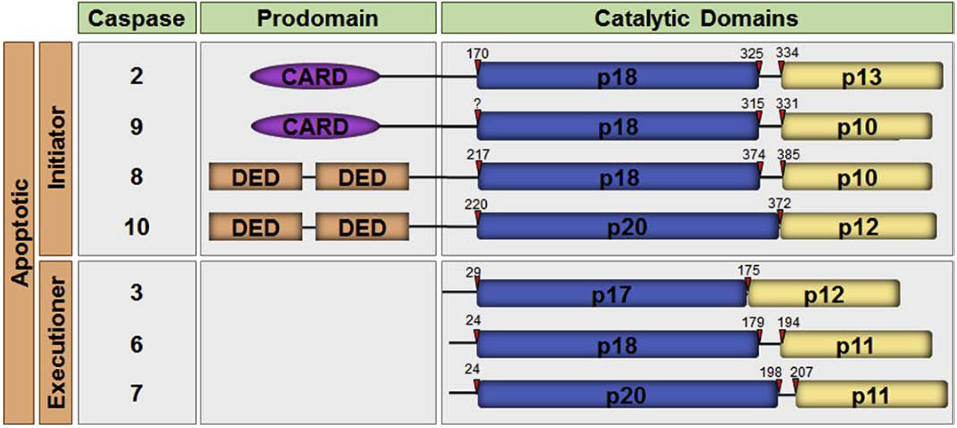
Members of the caspase family of proteases play essential roles in the initiation and execution of apoptosis. These caspases are divided into two groups: the initiator caspases (caspase-2, -8, -9 and -10), which are the first to be activated in response to a signal, and the executioner caspases (caspase-3, -6, and -7) that carry out the demolition phase of apoptosis. Many conventional cancer therapies induce apoptosis to remove the cancer cell by engaging these caspases indirectly. Newer therapeutic applications have been designed, including those that specifically activate individual caspases using gene therapy approaches and small molecules that repress natural inhibitors of caspases already present in the cell. For such approaches to have maximal clinical efficacy, emerging insights into non-apoptotic roles of these caspases need to be considered. This review will discuss the roles of caspases as safeguards against cancer in the context of the advantages and potential limitations of targeting apoptotic caspases for the treatment of cancer.

Keywords: Apoptosis; Cancer; Caspase; IAPs; Mitochondria.


Conflict of interest statement

Declaration of competing interest The authors declare that they have no known competing financial interests or personal relationships that could have appeared to influence the work reported in this paper.

Figures

Figure 1.
Figure 1.. The human apoptotic caspases.
A schematic representation of the domain organization of the human apoptotic caspases is shown. The caspases are divided into the initiator caspases and the executioner caspases. Each caspase comprises a prodomain and a large and small catalytic subunit. Initiator caspases have long prodomains containing the protein:protein interaction motifs, caspase recruitment domain (CARD) or death effector domain (DED), as indicated. The cleavage sites for each caspase are depicted with red arrows.
Figure 2.
Figure 2.. Structure of the caspase-3 heterotetramer.
The crystal structure of the active caspase-2 heterodimer (left) and a ribbon diagram of the structure (right) are shown. The large (p20) and small (p10) subunits of the heterodimer are depicted in blue and yellow respectively. Caspase-3 is shown bound to the inhibitor z-DEVD-cmk (cyan). The catalytic histidine and cysteine are shown in red. The loops, L1 (magenta), L2 (black), LH (green), L3 (orange), and L4 (pale pink) make up the active pocket. Caspase-3 is activated by cleavage of the intersubunit linker (L2). Cleavage of the intersubunit linker opens up the active pocket, exposing the active cysteine (red star). For clarity, the loops, catalytic residues, and inhibitor are only highlighted in one half of the heterotetramer. The molecular models of the protein structures were created using PyMOL v1.3.Edu with the full length caspase-3 crystal structure (PDB:2DKO) [38].
Figure 3.
Figure 3.. The mitochondrial pathway of apoptosis.
Diverse cellular stresses such as DNA damage, ER stress, and metabolic stress activate the intrinsic pathway leading to mitochondrial outer membrane permeabilization (MOMP). MOMP allows for the release of cytochrome c, second mitochondria-derived activator of caspases/ direct IAP binding protein with low pI (Smac/DIABLO), and Omi/ high temperature requirement protein A2 (HtrA2) from the mitochondrial intermembrane space. Cytochrome c binds to the WD repeats (red rectangle) of apoptotic peptidase activating factor 1 (APAF1), inducing a conformational change that opens up the molecule. This allows multiple molecules of APAF1 to oligomerize and assemble into the APAF1 apoptosome. The apoptosome recruits and activates caspase-9, which then activates caspase-3 and caspase-7 leading to apoptosis. Smac/DIABLO and Omi/HtrA2 bind to XIAP to prevent it from inhibiting active caspases.
Figure 4.
Figure 4.. The death receptor pathway of apoptosis.
At the plasma membrane, the ligands CD95L, TNF-related apoptosis-inducing ligand (TRAIL), and tumor necrosis factor α (TNFα) induce multimerization and activation of the death receptors CD95, TRAIL R1/R2, and TNFR1 respectively leading to death-inducing signaling complex (DISC) assembly. The CD95 or TRAILR1/R2 DISC leads to caspase-8 induced proximity via fas-associated protein with death domain (FADD)binding the intracellular death domain (DD) of the receptor and the death effector domain (DED) of caspase-8. Caspase-8 activates the downstream caspases directly or indirectly through BID cleavage and mitochondrial outer membrane permeabilization (MOMP). TNFR1 recruits receptor-interacting serine/threonine-protein kinase 1 (RIPK1), TNFR1-associated death domain protein (TRADD), TNF receptor associated factor 2 (TRAF2), cellular inhibitor of apoptosis protein 1/2 (cIAP1/2), and linear ubiquitin assembly complex (LUBAC), comprised of HOIL1-interacting protein (HOIP), heme-oxidized IRP2 ubiquitin ligase 1 (HOIL1), and SHANK Associated RH Domain Interactor (Sharpin). LUBAC adds linear ubiquitin chains (Ub) to RIPK1 and NF-kappa-B essential modulator (NEMO)to recruit the inhibitor of nuclear factor kappa-B kinase (IKK)complex to activate nuclear factor kappa-light-chain-enhancer of activated B cells (NFκB) (p50/p65). cIAP1/2 induce NIK degradation to inhibit non-canonical NFκB (RelB/p50). In the absence of cIAP1/2, TRADD/RIPK1/TRAF2 disassociates from the plasma membrane and recruits FADD and caspase-8 (Complex IIa) to induce apoptosis. In the absence of caspase-8, RIPK1 forms a complex with RIPK3 (Complex IIb). RIPK3 induces phosphorylation of mixed lineage kinase domain like pseudokinase (MLKL), resulting in necroptosis. Cellular-FLICE inhibitory protein long or short (c-FLIPL/s) can form heterodimers with caspase-8. At high concentrations c-FLIPL and c-FLIPs inhibit caspase-8 activation. c-FLIPL interaction with caspase-8 within complex IIa destabilizes the complex IIb, promoting survival. For simplicity, c-FLIPs is shown in the DISC and c-FLIPL is shown in complex IIa but the isoforms can interact with both complexes.

References

    1. Martin SJ, Green DR, Protease activation during apoptosis: death by a thousand cuts?, Cell, 82 (1995) 349–352. - PubMed
    1. Kerr JF, Wyllie AH, Currie AR, Apoptosis: a basic biological phenomenon with wide-ranging implications in tissue kinetics, Br J Cancer, 26 (1972) 239–257. - PMC - PubMed
    1. Arandjelovic S, Ravichandran KS, Phagocytosis of apoptotic cells in homeostasis, Nature immunology, 16 (2015) 907–917. - PMC - PubMed
    1. McIlwain DR, Berger T, Mak TW, Caspase functions in cell death and disease, Cold Spring Harb Perspect Biol, 5 (2013) a008656. - PMC - PubMed
    1. Thompson CB, Apoptosis in the pathogenesis and treatment of disease, Science, 267 (1995) 1456–1462. - PubMed

Publication types

MeSH terms