Review
. 2020 Jun:139:104820.
doi: 10.1016/j.nbd.2020.104820. Epub 2020 Feb 19.

Impact of sleep disturbances on neurodegeneration: Insight from studies in animal models

Affiliations
Review

Impact of sleep disturbances on neurodegeneration: Insight from studies in animal models

Jessica E Owen et al. Neurobiol Dis. 2020 Jun.

Abstract

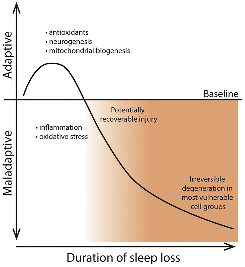
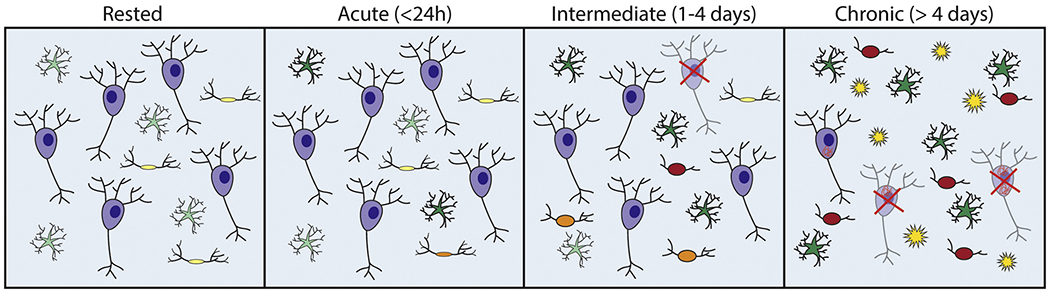
Chronic short sleep or extended wake periods are commonly observed in most industrialized countries. Previously neurobehavioral impairment following sleep loss was considered to be a readily reversible occurrence, normalized upon recovery sleep. Recent clinical studies suggest that chronic short sleep and sleep disruption may be risk factors for neurodegeneration. Animal models have been instrumental in determining whether disturbed sleep can injure the brain. We now understand that repeated periods of extended wakefulness across the typical sleep period and/or sleep fragmentation can have lasting effects on neurogenesis and select populations of neurons and glia. Here we provide a comprehensive overview of the advancements made using animal models of sleep loss to understand the extent and mechanisms of chronic short sleep induced neural injury.

Keywords: Amyloid-beta; Neural injury; Neurodegeneration; Neurogenesis; Sleep loss; Tau.


Figures

Figure 1
Figure 1. Time dependent effects of sleep disruption.
Very brief (≤ 3 h) wakefulness extension may be adaptive by increasing antioxidants, hippocampal neurogenesis and mitochondrial biogenesis, longer durations are maladaptive with increased inflammation and oxidative stress which may be reversible. Even longer durations of sleep disruption ultimately result in degeneration, irreversible injury and impairment.
Figure 2
Figure 2. Cellular response to sleep disruption over time
Diagrammatic representation of injury to different cell types with increasing durations of sleep disruption. The rested or baseline condition shows neurons (purple), astrocytes (green) and microglia (yellow). Acute sleep disruption shows minor changes to astrocytes (darker green) and microglia (yellow & orange) which include upregulation of some neuroinflammatory markers. Intermediate sleep disruption shows increased neuroinflammatory changes in astrocytes and microglia as well as some neuronal loss. Chronic sleep disruption shows many reactive astrocytes and microglia (red) and further neuronal loss. Additionally, increased amyloid beta plaques (yellow stars) and tauopathy (orange squiggles) can be seen in models that predispose animals to developing this pathology.

References

    1. Acosta-peña E, Camacho-Abrego I, Melgarejo-Gutiérrez M, Flores G, Drucker-Colín R, & García-García F (2015). Sleep deprivation induces differential morphological changes in the hippocampus and prefrontal cortex in young and old rats. Synapse, 69(1), 15–25. 10.1002/syn.21779 - DOI - PubMed
    1. Artamokhina IV, Belova VA, & Romanova IV (2011). Immunohistochemical investigation of bcl-2 and p53 levels in rat hypothalamus after sleep deprivation. Journal of Evolutionary Biochemistry and Physiology, 47(5), 458–463. 10.1134/s0022093011050082 - DOI - PubMed
    1. Asai H, Ikezu S, Tsunoda S, Medalla M, Luebke J, Haydar T, … Ikezu T (2015). Depletion of microglia and inhibition of exosome synthesis halt tau propagation. Nature Neuroscience, 18(11), 1584–1593. 10.1038/nn.4132 - DOI - PMC - PubMed
    1. Belenky G, Wesensten NJ, Thorne DR, Thomas ML, Sing HC, Redmond DP, … Balkin TJ (2003). Patterns of performance degradation and restoration during sleep restriction and subsequent recovery: a sleep dose-response study. Journal of Sleep Research, 12(1), 1–12. 10.1046/j.1365-2869.2003.00337.x - DOI - PubMed
    1. Bellesi M, de Vivo L, Chini M, Gilli F, Tononi G, & Cirelli C (2017). Sleep Loss Promotes Astrocytic Phagocytosis and Microglial Activation in Mouse Cerebral Cortex. The Journal of Neuroscience : The Official Journal of the Society for Neuroscience, 37(21), 5263–5273. 10.1523/JNEUROSCI.3981-16.2017 - DOI - PMC - PubMed

Publication types

MeSH terms

Substances