. 2020 Feb 3:2020:8595940.
doi: 10.1155/2020/8595940. eCollection 2020.

Comparative Analysis of Adipose-Derived Stromal Cells and Their Secretome for Auricular Cartilage Regeneration

Affiliations

Comparative Analysis of Adipose-Derived Stromal Cells and Their Secretome for Auricular Cartilage Regeneration

Se-Joon Oh et al. Stem Cells Int. .

Abstract

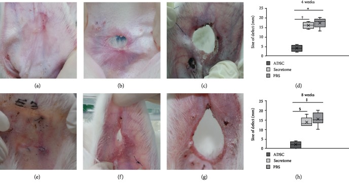
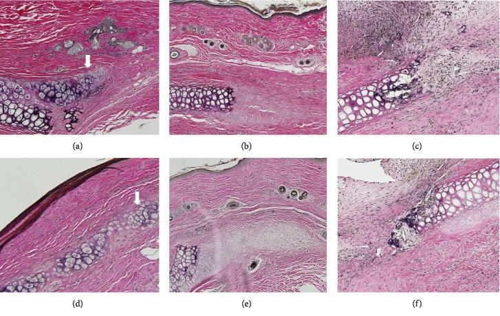
Adipose-derived stromal cells (ADSCs) can repair auricular cartilage defects. Furthermore, stem cell secretome may also be a promising biological therapeutic option, which is equal to or even superior to the stem cell. We explored the therapeutic efficacies of ADSCs and their secretome in terms of rabbit auricular cartilage regeneration. ADSCs and their secretome were placed into surgically created auricular cartilage defects. After 4 and 8 weeks, the resected auricles were histopathologically and immunohistochemically examined. We used real-time PCR to determine the levels of genes expressing collagen type II, transforming growth factor-β1 (TGF-β1), and insulin-like growth factor-1 (IGF-1). ADSCs significantly improved auricular cartilage regeneration at 4 and 8 weeks, compared to the secretome and PBS groups, as revealed by gross examination, histopathologically and immunohistochemically. ADSCs upregulated the expression of collagen type II, TGF-β1, and IGF-1 more so than did the secretome or PBS. The expression levels of collagen type II and IGF-1 were significantly higher at 8 weeks than at 4 weeks after ADSC injection. Although ADSCs thus significantly enhanced new cartilage formation, their secretome did not. Therefore, ADSCs may be more effective than their secretome in the repair of auricular cartilage defect.


Conflict of interest statement

The authors declare that they have no competing interests.

Figures

Figure 1
Figure 1
The experimental protocol and method. (a) Purified adipose-derived stromal cells (ADSCs; 2 × 10⁷ cells/mL) or 10 μg/50 μL of the secretome was injected subperichondrially (S.P.) into 2 cm diameter, surgically created, auricular cartilage defects (b) on days 0, 2, 4, 6, and 8. The materials were injected (using a 26-gauge needle) at the edge of the excision site (c), and Spongostan was sutured to each defect (d).
Figure 2
Figure 2
Gross auricular cartilage findings. (a) Four weeks after ADSC injection, the defects were filled with soft pinkish tissue. The defects of the secretome (b) and PBS (c) groups remained evident. The defect size decreased significantly in the ADSC compared to the secretome or the PBS group at 4 (d) and 8 weeks (H) after ADSC injection. (e) In ADSC-treated ears, the defects were completely healed by 8 weeks; the tissue surface was smooth and of similar color to that of the surrounding tissue. The secretome (f) and PBS (g) groups exhibited minimal defect repair. ,†,‡,§p < 0.001. ADSCs: adipose-derived stromal cells; PBS: phosphate-buffered saline.
Figure 3
Figure 3
Immunofluorescence of adipose-derived stromal cells (ADSCs) in auricular cartilage defects. ADSCs labeled with the red Cell Tracker CM-Dil dye were detected in the ADSC group at 4 (a) and 8 weeks (b) after ADSC injection.
Figure 4
Figure 4
Histopathological features of auricular cartilage defects at 4 (a–c) and 8 (d–f) weeks after adipose-derived stromal cell (ADSC) injection. Chondroblasts and cartilage-specific extracellular matrix (ECM) were evident in the ADSC group (a). ECM was deposited in the secretome group (b), and fibrous tissue formation was apparent in the PBS group (c). Typical cartilaginous features (chondrocytes, chondroblasts, and cartilage-specific ECM (arrow)) were observed in the ADSC group (d). Small numbers of chondroblasts and a little collagenous tissue were evident at the defect edges of the secretome (e) and PBS groups (f), respectively. The sections were stained with hematoxylin and eosin (magnification 200x).
Figure 5
Figure 5
Immunohistochemical detection of elastin. New cartilage (arrow) was evident in the adipose-derived stromal cell (ADSC) group at 4 (a) and 8 weeks (d) after ADSC injection. Small amounts of ECM and fibrous tissue were observed, respectively, in the secretome (b, e) and PBS (c, f) groups at 4 and 8 weeks (magnification, 200x).
Figure 6
Figure 6
The expression levels of genes encoding collagen type II and various growth factors at 4 (a) and 8 (b) weeks after ADSC injection. The collagen type II expression level was significantly higher in the ADSC than in the secretome or PBS group 8 weeks after ADSC injection. In the ADSC group, the expression levels of transforming growth factor- (TGF-) β1 and insulin-like growth factor (IGF) were significantly higher than in the secretome or PBS group at 4 and 8 weeks postinjection. ,‡‡p = 0.002; †,§§p = 0.001; p = 0.023; §p = 0.011; ǁ,¶,∗∗,††p < 0.001. ADSCs: adipose-derived stromal cells; PBS: phosphate-buffered saline.

Similar articles

Cited by

References

    1. Toh W. S., Foldager C. B., Pei M., Hui J. H. P. Advances in mesenchymal stem cell-based strategies for cartilage repair and regeneration. Stem Cell Reviews. 2014;10(5):686–696. doi: 10.1007/s12015-014-9526-z. - DOI - PubMed
    1. Pleumeekers M. M., Nimeskern L., Koevoet W. L. M., Karperien M., Stok K. S., van Osch G. J. V. M. Cartilage regeneration in the head and neck area: combination of ear or nasal chondrocytes and mesenchymal stem cells improves cartilage production. Plastic and Reconstructive Surgery. 2015;136(6):762e–774e. doi: 10.1097/PRS.0000000000001812. - DOI - PubMed
    1. Delorme B., Charbord P. Culture and characterization of human bone marrow mesenchymal stem cells. Methods in Molecular Medicine. 2007;140:67–81. doi: 10.1007/978-1-59745-443-8_4. - DOI - PubMed
    1. Liu T. M., Martina M., Hutmacher D. W., Hui J. H., Lee E. H., Lim B. Identification of common pathways mediating differentiation of bone marrow- and adipose tissue-derived human mesenchymal stem cells into three mesenchymal lineages. Stem Cells. 2007;25(3):750–760. doi: 10.1634/stemcells.2006-0394. - DOI - PubMed
    1. Titorencu I., Jinga V., Constantinescu E., et al. Proliferation, differentiation and characterization of osteoblasts from human BM mesenchymal cells. Cytotherapy. 2007;9(7):682–696. doi: 10.1080/14653240701561329. - DOI - PubMed

LinkOut - more resources