. 2020 Jan 29:2020:1020673.
doi: 10.1155/2020/1020673. eCollection 2020.

Bidens pilosa Extract Administered after Symptom Onset Attenuates Glial Activation, Improves Motor Performance, and Prolongs Survival in a Mouse Model of Amyotrophic Lateral Sclerosis

Affiliations

Bidens pilosa Extract Administered after Symptom Onset Attenuates Glial Activation, Improves Motor Performance, and Prolongs Survival in a Mouse Model of Amyotrophic Lateral Sclerosis

Yasuhiro Kosuge et al. Oxid Med Cell Longev. .

Abstract

Amyotrophic lateral sclerosis (ALS) is a late-onset neurodegenerative disorder characterized by progressive paralysis resulting from the death of upper and lower motor neurons. There is currently no effective pharmacological treatment for ALS, and the two approved drugs riluzole and edaravone have limited effects on the symptoms and only slightly prolong the life of patients. Therefore, the development of effective therapeutic strategies is of paramount importance. In this study, we investigated whether Miyako Island Bidens pilosa (MBP) can alleviate the neurological deterioration observed in a superoxide dismutase-1 G93A mutant transgenic mouse (G93A mouse) model of ALS. We orally administered 2 g/kg/day of MBP to G93A mice at the onset of symptoms of neurodegeneration (15 weeks old) until death. Treatment with MBP markedly prolonged the life of ALS model mice by approximately 20 days compared to that of vehicle-treated ALS model mice and significantly improved motor performance. MBP treatment prevented the reduction in SMI32 expression, a neuronal marker protein, and attenuated astrocyte (detected by GFAP) and microglia (detected by Iba-1) activation in the spinal cord of G93A mice at the end stage of the disease (18 weeks old). Our results indicate that MBP administered after the onset of ALS symptoms suppressed the inflammatory activation of microglia and astrocytes in the spinal cord of the G93A ALS model mice, thus improving their quality of life. MBP may be a potential therapeutic agent for ALS.


Conflict of interest statement

The authors declare that they have no conflicts of interest.

Figures

Figure 1
Figure 1
Flowchart of the experiment design. MBP: Miyako Island Bidens pilosa var. radiata SCHERFF.
Figure 2
Figure 2
Effect of MBP on the survival of G93A mice. Mice were orally administered with injection water (vehicle) or MBP, starting at a late symptomatic stage (15 weeks old). (a) Survival curve of G93A mice treated with vehicle or MBP, analysed by Kaplan-Meier analysis with the Mantel-Cox log-rank test (n = 9-10, p = 0.004). The arrow indicates the start of vehicle or MBP administration. (b) The graph shows the maximum lifespan of G93A mice treated with vehicle or MBP. Values represent the mean ± SD. Statistical significance was determined by unpaired Student's t-test. (n = 9-10).
Figure 3
Figure 3
Effect of MBP on the motor performance of G93A mice. Mice were orally with administered injection water (vehicle) or MBP, starting at a late symptomatic stage (15 weeks old). Motor performance of the mice was evaluated using a rotarod apparatus. The graph depicts latency to fall from the rotarod apparatus in G93A mice treated with vehicle or MBP. Values represent the mean ± SEM. Serial changes in motor performance were analysed with two-way ANOVA (with “drug treatment” and “weeks of age” as between-subjects' factors) followed by Bonferroni's post hoc test (n = 9-10). ∗∗∗p < 0.001 and ∗∗p < 0.01 vs. aged-matched mice treated with vehicle.
Figure 4
Figure 4
MBP ameliorates motor neuron loss in the spinal cord of G93A mice. Mice were orally administered with injection water (vehicle) or MBP, starting at a late symptomatic stage (15 weeks old). Three weeks after the start of the treatment, the lumbar spinal cords were analysed by western blot and the histopathology was analysed. (a) Photographs show representative western blots of SMI32, a marker of motor neurons, in the lumbar spinal cord of male G93A mice and WT mice. Equal amounts of cell lysates (10 μg) were analysed, with β-actin as an internal control. The graph shows the relative densities of each band on the blots estimated quantitatively using Scion imaging software. Quantitative data are expressed as the ratio of the band intensity of SMI32 to the band intensity of β-actin. Each value represents the mean ± SD. Statistical significance was determined by using one-way ANOVA followed by Tukey's post hoc test (n = 6-7). (b) Photographs show representative cresyl violet-stained sections of the lumbar spinal cord in the indicated groups of mice at 18 weeks old. Arrows indicate motor neurons. Scale bar indicates 100 μm. The graph shows the number of surviving motor neurons in lumbar spinal cord sections from the indicated groups of mice. Values represent the mean ± SEM. Statistical significance was determined by using one-way ANOVA followed by Tukey's post hoc test (n = 4).
Figure 5
Figure 5
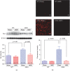
MBP attenuates morphological changes in astrocytes in G93A mice. Mice were orally administered with injection water (vehicle) or MBP, starting at a late symptomatic stage (15 weeks old). Three weeks after the start of the treatment, the lumbar spinal cords were analysed by western blot and the histopathology was imaged. (a) Photographs depict a representative western blot of GFAP, an astrocyte marker, in the lumbar spinal cord of male G93A mice and WT mice. Equal amounts of cell lysates (10 μg) were analysed, with β-actin as an internal marker. The graph shows the relative density of each band on the blots estimated quantitatively using Scion imaging software. Quantitative data are expressed as the ratio of the band intensity of GFAP to the band intensity of β-actin. Each value represents the mean ± SD. Statistical significance was determined by using one-way ANOVA followed by Tukey's post hoc test (n = 6-7). (b) Photographs show representative confocal images of immunofluorescence staining for GFAP in the lumbar spinal cord sections from the indicated groups of mice at 18 weeks old. Representative data from four separate experiments are presented. Scale bar indicates 20 μm. The graph shows semiquantitative analysis of changes in GFAP immunoreactivity in motor neurons. The fluorescence intensity of GFAP immunoreactivity was analysed quantitatively using Scion imaging software. Values represent the mean ± SEM. Statistical significance was determined by using one-way ANOVA followed by Tukey's post hoc test (n = 4).
Figure 6
Figure 6
MBP attenuates morphological changes in microglia in G93A mice. Mice were orally with administered injection water (vehicle) or MBP, starting at a late symptomatic stage (15 weeks old). Three weeks after the start of the treatment, the lumbar spinal cords were analysed by western blot and histopathology was analysed. (a) Photographs show representative western blots of Iba-1, a microglia marker, in the lumbar spinal cord of male G93A mice and WT mice. Equal amounts of cell lysates (10 μg) were analysed, with β-actin as an internal marker. The graph shows the relative density of bands on the blots estimated quantitatively using Scion imaging software. Quantitative data are expressed as the ratio of the band intensity of Iba-1 relative to the band intensity of β-actin. Each value represents the mean ± SD. Statistical significance was determined by using one-way ANOVA followed by Tukey's post hoc test (n = 6-7). (b) Photographs show representative confocal images of immunofluorescence staining for Iba-1 in the lumbar spinal cord sections from the indicated groups of mice at 18 weeks old. Representative data from four separate experiments are presented. Scale bar indicates 20 μm. The graph shows semiquantitative analysis of changes in Iba-1 immunoreactivity in motor neurons. The fluorescence intensity of Iba-1 immunoreactivity was analysed quantitatively using Scion imaging software. Values represent the mean ± SEM. Statistical significance was determined by using one-way ANOVA followed by Tukey's post hoc test (n = 4).
Figure 7
Figure 7
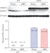
Effect of MBP on SOD1 protein expression in spinal cord tissue of G93A and WT mice. Mice were orally administered with injection water (vehicle) or MBP, starting at a late symptomatic stage (15 weeks old). Three weeks after the start of the treatment, the lumbar spinal cords were analysed by western blot. Photographs show representative western blot of SOD1 in the lumbar spinal cord of male G93A mice and WT mice. The upper band represents human SOD1 (hSOD1; 21 kDa) and the lower band represents mouse SOD1 (mSOD1; 16 kDa). Equal amounts of cell lysates (10 μg) were analysed, with β-actin as an internal marker. The graph shows the relative density of bands on the blots estimated quantitatively using Scion imaging software. Quantitative data are expressed as the ratio of the band intensity of hSOD1 relative to the band intensity of β-actin. Each value represents the mean ± SD. Statistical significance was determined by unpaired Student's t-test (n = 6-7). ND: not detected.

Similar articles

Cited by

References

    1. Rosen D. R., Siddique T., Patterson D., et al. Mutations in Cu/Zn superoxide dismutase gene are associated with familial amyotrophic lateral sclerosis. Nature. 1993;362(6415):59–62. doi: 10.1038/362059a0. - DOI - PubMed
    1. Gurney M. E., Pu H., Chiu A. Y., et al. Motor neuron degeneration in mice that express a human Cu, Zn superoxide dismutase mutation. Science. 1994;264(5166):1772–1775. doi: 10.1126/science.8209258. - DOI - PubMed
    1. Ferraiuolo L., Kirby J., Grierson A. J., Sendtner M., Shaw P. J. Molecular pathways of motor neuron injury in amyotrophic lateral sclerosis. Nature Reviews Neurology. 2011;7(11):616–630. doi: 10.1038/nrneurol.2011.152. - DOI - PubMed
    1. Dachet F., Liu J., Ravits J., Song F. Predicting disease specific spinal motor neurons and glia in sporadic ALS. Neurobiology of Disease. 2019;130, article 104523 doi: 10.1016/j.nbd.2019.104523. - DOI - PubMed
    1. Jiang L. L., Zhu B., Zhao Y., et al. Membralin deficiency dysregulates astrocytic glutamate homeostasis leading to ALS-like impairment. The Journal of Clinical Investigation. 2019;129(8):3103–3120. doi: 10.1172/JCI127695. - DOI - PMC - PubMed

Substances