. 2020 Feb 6:2020:7912763.
doi: 10.1155/2020/7912763. eCollection 2020.

Cordyceps cicadae Mycelia Ameliorate Cisplatin-Induced Acute Kidney Injury by Suppressing the TLR4/NF- κ B/MAPK and Activating the HO-1/Nrf2 and Sirt-1/AMPK Pathways in Mice

Affiliations

Cordyceps cicadae Mycelia Ameliorate Cisplatin-Induced Acute Kidney Injury by Suppressing the TLR4/NF- κ B/MAPK and Activating the HO-1/Nrf2 and Sirt-1/AMPK Pathways in Mice

Jeng-Shyan Deng et al. Oxid Med Cell Longev. .

Abstract

Acute kidney injury (AKI) is a common clinical problem, characterized by a sudden loss of renal function, a high risk of death, and the eventual development of renal fibrosis and renal failure. Cordyceps cicadae is a traditional Chinese medicine with the potential function of kidney protection. We analyze two sputum extracts, a water extract (WCC), and an ethanol extract (ECC), to assess the potential of treating AKI in an animal model of kidney injury induced by cisplatin. A nephrotoxic mouse model was first established by intraperitoneal injection of cisplatin. Subsequently, WCC and ECC were orally administered in these mice. The results show that WCC and ECC significantly alleviated cisplatin-induced AKI renal histological changes, serum creatinine (CRE) and blood urea nitrogen (BUN) production, and the levels of NO, TNF-α, IL-1β, and IL-6. The levels of malondialdehyde (MDA) and glutathione (GSH) were suppressed by administration of WCC and ECC. However, WCC treatment prevented these changes significantly better than ECC treatment. In addition, Western blot data showed that WCC attenuated the cisplatin-induced protein expression of cyclooxygenase-2 (COX-2) and inducible NO synthase (iNOS), as well as inhibiting nuclear factor-kappa B (NF-κB) and mitogen-activated protein kinase (MAPK) activation in the kidney tissues. Furthermore, WCC greatly inhibited the expression of Toll-like receptor 4 (TLR4) and cisplatin-induced NF-κB activation, as well as dramatically increasing the production of antioxidative enzymes (i.e., superoxide dismutase (SOD), glutathione peroxidase (GPx), catalase, nuclear factor erythroid 2-related factor 2 (Nrf2), and heme oxygenase 1 (HO-1)), silent information regulator T1 (Sirt1), and p-AMP-activated protein kinase (AMPK) in the kidney tissues. In addition, we found that WCC increased the expression levels of the autophagy-related proteins LC3B and Beclin-1; proapoptotic proteins, including cleaved caspase-3 and cleaved poly (ADP-ribose) polymerase (PARP) 1; and organic anion transporters 1 (OAT1) and 3 (OAT3) in the kidney tissues. Finally, WCC, ECC, and two bioactive compounds-adenosine and N6-(2-hydroxyethyl) adenosine (HEA)-inhibited the production of nitrite oxide (NO) and intracellular reactive oxygen species (ROS) triggered by lipopolysaccharide- (LPS-) stimulated RAW264.7 macrophages in vitro. Collectively, WCC could provide a potential therapeutic candidate for the prevention of cisplatin-induced kidney injury through the inhibition of oxidative stress and inflammation.


Conflict of interest statement

All authors have no conflicts of interests.

Figures

Figure 1
Figure 1
Protective effects of WCC and ECC on cisplatin-induced kidney damage in AKI mice. Mice were orally administered WCC and ECC daily for 10 days, they received cisplatin (20 mg/kg, i.p.) one hour after WCC and ECC administration on the seventh day, and the mice were euthanized on the eleventh day. The morphological changes in the kidneys (a). Blood urea nitrogen (BUN) levels (b). Serum creatinine (CRE) levels (c). Kidneys stained with H&E (d) and the tubular injury scores (e). Macrophage infiltration in kidney tissues was detected by immunohistochemistry with the F4/80 antibody (f). After cisplatin challenge, kidneys in each group were prepared for histological evaluation. Representative histological section of the kidneys was stained by H&E staining (magnification (400x)) and F4/80 immunohistochemical staining (magnification (200x)). The data are presented as the means ± SEM (n = 5). ###p < 0.001 compared with the sample of the control group. ∗∗p < 0.01 and ∗∗∗p < 0.001 compared with the cisplatin group. Tubular cell necrosis is marked with arrows. The bar indicates 50 μm.
Figure 2
Figure 2
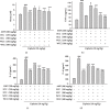
C. cicadae mycelium extract downregulated (a) NO, (b) TNF-α, (c) IL-1β, and (d) IL-6 in serum. Serum was collected. Nitrite concentration in the serum was measured by using the Griess reaction. Serum levels of TNF-α, IL-1β, and IL-6 were determined with commercial ELISA kits. Data are represented as mean ± SEM (n = 5). ###p < 0.001 compared with the sample of the control group. ∗∗p < 0.01 and ∗∗∗p < 0.001 compared with the cisplatin-only group.
Figure 3
Figure 3
The effect of WCC on the oxidative stress in the kidney of cisplatin-treated mice. MDA levels (a). GSH levels (b). Kidney tissue homogenates were evaluated by the MDA and GSH assay. GSH was determined and expressed as μmol/g kidney tissues. Data are represented as mean ± SEM (n = 5). ###p < 0.001 compared with the sample of the control group. ∗∗p < 0.01 and ∗∗∗p < 0.001 compared with the cisplatin-only group.
Figure 4
Figure 4
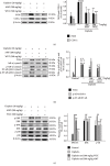
Effects of WCC on cisplatin-induced (a) iNOS, COX-2, (b) TLR4, IκBα, p-IκBα, NF-κB, p-NF-κB, and (c) MAPK phosphorylation signaling expression in kidneys. Protein levels of iNOS, COX-2, TLR4, IκBα, NF-κB, p-IκBα, p-NF-κB, and MAPK phosphorylation protein expression in kidney homogenates were evaluated by Western blot analysis after cisplatin challenge. Densitometric analysis of the relevant bands was performed. Data are represented as mean ± SEM (n = 3). ###p < 0.001 compared with the control group. ∗∗p < 0.01 and ∗∗∗p < 0.001 compared with the cisplatin-only group.
Figure 5
Figure 5
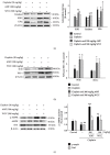
Effects of WCC on (a) cisplatin-induced antioxidative enzyme (catalase, SOD, and GPx), (b) HO-1, Nrf2, (c) AMPK, and Sirt1 protein expression and in the renal tissues. The protein levels of antioxidative enzyme, HO-1, Nrf2, AMPK, and Sirt1 protein expression in kidney homogenates were evaluated by Western blot analysis after cisplatin challenge. Densitometric analysis of the relevant bands was performed. Data are represented as mean ± SEM (n = 3). ###p < 0.001 compared with the control group. ∗∗p < 0.01 and ∗∗∗p < 0.001 compared with the cisplatin-only group.
Figure 6
Figure 6
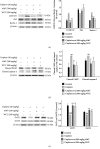
Effects of WCC on (a) cisplatin-induced apoptosis relative enzyme (cleaved caspase-3 and PARP), (b) autophagy relative enzyme (LC3B, p62, and Beclin-1), and (c) renal organic anion transporter (OAT1 and OAT3) protein expression and in the renal tissues. The protein levels of caspase-3 and PARP, LC3B, p62, Beclin-1, OAT1, and OAT3 protein expression in kidney homogenates were evaluated by Western blot analysis after cisplatin challenge. Densitometric analysis of the relevant bands was performed. Data are represented as mean ± SEM (n = 3). ###p < 0.001 compared with the control group. ∗∗p < 0.01 and ∗∗∗p < 0.001 compared with the cisplatin-only group.
Figure 7
Figure 7
HPLC chromatogram of the C. cicadae mycelium water extract (WCC) (A) and ethanolic extract (ECC) (B).
Figure 8
Figure 8
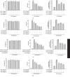
Effect of (a) WCC, (b) ECC, (c) adenosine, and (d) HEA on lipopolysaccharide- (LPS-) induced cell viability and NO and ROS production. Cells were incubated for 24 h with 100 ng/mL of LPS in the absence or presence of samples. Samples were added 1 h before incubation with LPS. The cell viability assay was performed using the MTT assay. Nitrite concentration in the medium was determined using the Griess reagent. Serum-free medium containing H2DCFDA 10 mM was added to cells, followed by 30 min incubation (37°C). 2,7-Dichlorofluorescein fluorescence was evaluated using a fluorescence ELISA reader. Densitometric analysis of the relevant bands was performed. Data are represented as mean ± SEM (n = 3). ###p < 0.001 compared with the control group. ∗∗p < 0.01 and ∗∗∗p < 0.001 compared with the cisplatin-only group.
Figure 9
Figure 9
The schemes of the mechanism for the protective effect of WCC in cisplatin-induced kidney injury.

References

    1. Kim J. S., Kim K. S., Son J. Y., et al. Protective effects of Dendropanax morbifera against cisplatin-induced nephrotoxicity without altering chemotherapeutic efficacy. Antioxidants. 2019;8(8):p. 256. doi: 10.3390/antiox8080256. - DOI - PMC - PubMed
    1. Meng H., Fu G., Shen J., et al. Ameliorative effect of daidzein on cisplatin-induced nephrotoxicity in mice via modulation of inflammation, oxidative stress, and cell death. Oxidative Medicine and Cellular Longevity. 2017;2017:10. doi: 10.1155/2017/3140680.3140680 - DOI - PMC - PubMed
    1. Huang T. H., Wu T. H., Guo Y. H., Li T. L., Chan Y. L., Wu C. J. The concurrent treatment of Scutellaria baicalensis Georgi enhances the therapeutic efficacy of cisplatin but also attenuates chemotherapy-induced cachexia and acute kidney injury. Journal of Ethnopharmacology. 2019;243, article 112075 doi: 10.1016/j.jep.2019.112075. - DOI - PubMed
    1. Qi Z., Li Z., Li W., et al. Pseudoginsengenin DQ exhibits therapeutic effects in cisplatin-induced acute kidney injury via sirt1/NF-κB and caspase signaling pathway without compromising its antitumor activity in mice. Molecules. 2018;23(11):p. 3038. doi: 10.3390/molecules23113038. - DOI - PMC - PubMed
    1. Chen X., Wei W., Li Y., Huang J., Ci X. Hesperetin relieves cisplatin-induced acute kidney injury by mitigating oxidative stress, inflammation and apoptosis. Chemico-Biological Interactions. 2019;308:269–278. doi: 10.1016/j.cbi.2019.05.040. - DOI - PubMed

MeSH terms