Case Reports
. 2020 Jan 9:2020:2586730.
doi: 10.1155/2020/2586730. eCollection 2020.

Subaortic Membrane Papillary Fibroelastoma

Affiliations
Case Reports

Subaortic Membrane Papillary Fibroelastoma

Robyn Bryde et al. Case Rep Cardiol. .

Abstract

A 61-year-old male presented for an annual exam and received a transthoracic echocardiogram (TTE) which revealed a mobile mass arising from a subaortic membrane. Further investigations with a transesophageal echocardiogram (TEE) and cardiac computerized tomography angiography (CTA) confirmed the presence of a mobile 9 mm × 3 mm mass on a subaortic membrane. Cardiothoracic surgery was performed with an open operation removing the mass and subaortic membrane. Upon visual inspection, the mass was likened to a sea anemone and immunohistochemical staining performed pathologically confirmed the diagnosis of cardiac papillary fibroelastoma. This case represents the first reported example of a cardiac papillary fibroelastoma (PFE) arising from a subaortic membrane. Although PFEs are benign cardiac tumors, proper identification and consideration for excision of these lesions may be indicated to prevent thromboembolic complications.


Conflict of interest statement

The authors declare that they have no conflicts of interest.

Figures

Figure 1
Figure 1
(a) TTE imaging of a subaortic membrane and mass (arrow) on the anterior wall of the LVOT. (b) TEE imaging showing the mass (arrow) in close proximity to the aortic valve. (c) CTA showing a subaortic membrane (arrowhead) with an area of extension, likely representing a mass (arrow) in (d).
Figure 2
Figure 2
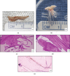
Gross specimens of the resected subaortic membrane (a) and mass (b) which has the classic frond-like appearance after being submerged in water. Histopathology with H&E stain revealing multiple papillae (c) which are avascular and rich in collagen (d) and CD34 staining highlighting the dark single layer of epithelial cells lining the mass.

References

    1. Gowda R. M., Khan I. A., Nair C. K., Mehta N. J., Vasavada B. C., Sacchi T. J. Cardiac papillary fibroelastoma: a comprehensive analysis of 725 cases. American Heart Journal. 2003;146(3):404–410. doi: 10.1016/S0002-8703(03)00249-7. - DOI - PubMed
    1. Val-Bernal J. F., Mayorga M., Garijo M. F., Val D., Nistal J. F. Cardiac papillary fibroelastoma: retrospective clinicopathologic study of 17 tumors with resection at a single institution and literature review. Pathology - Research and Practice. 2013;209(4):208–214. doi: 10.1016/j.prp.2013.02.001. - DOI - PubMed
    1. ElBardissi A. W., Dearani J. A., Daly R. C., et al. Survival after resection of primary cardiac tumors: a 48-year experience. Circulation. 2008;118(14_Supplement_1):S7–S15. doi: 10.1161/CIRCULATIONAHA.107.783126. - DOI - PubMed
    1. Kurup A. N., Tazelaar H. D., Edwards W. D., et al. Iatrogenic cardiac papillary fibroelastoma: a study of 12 cases (1990 to 2000) Human Pathology. 2002;33(12):1165–1169. doi: 10.1053/hupa.2002.130105. - DOI - PubMed
    1. Mariscalco G., Bruno V. D., Borsani P., Dominici C., Sala A. Papillary fibroelastoma: insight to a primary cardiac valve tumor. Journal of Cardiac Surgery. 2010;25(2):198–205. doi: 10.1111/j.1540-8191.2009.00993.x. - DOI - PubMed

Publication types

LinkOut - more resources