Review
. 2020 May 7;12(17):9306-9326.
doi: 10.1039/c9nr10963c. Epub 2020 Feb 24.

Functional nanoarrays for investigating stem cell fate and function

Affiliations
Review

Functional nanoarrays for investigating stem cell fate and function

Jin-Ho Lee et al. Nanoscale. .

Abstract

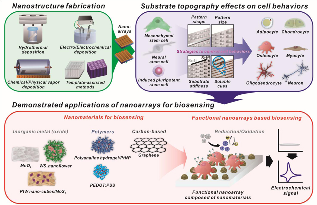
Stem cells show excellent potential in the field of tissue engineering and regenerative medicine based on their excellent capability to not only self-renew but also differentiate into a specialized cell type of interest. However, the lack of a non-destructive monitoring system makes it challenging to identify and characterize differentiated cells before their transplantation without compromising cell viability. Thus, the development of a non-destructive monitoring method for analyzing cell function is highly desired and can significantly benefit stem cell-based therapies. Recently, nanomaterial-based scaffolds (e.g., nanoarrays) have made possible considerable advances in controlling the differentiation of stem cells and characterization of the differentiation status sensitively in real time. This review provides a selective overview of the recent progress in the synthesis methods of nanoarrays and their applications in controlling stem cell fate and monitoring live cell functions electrochemically. We believe that the topics discussed in this review can provide brief and concise guidelines for the development of novel nanoarrays and promote the interest in live cell study applications. A method which can not only control but also monitor stem cell fate and function will be a promising technology that can accelerate stem cell therapies.


Conflict of interest statement

Conflicts of interest

The authors do not have any conflicts of interest to declare.

Figures

Figure. 1
Figure. 1
Schematic illustration of functional nanoarrays for investigating stem cell fate and functions.
Figure. 2
Figure. 2
Fabrication of a nanodot array based on the template-assisted method. (a) Illustration of the fabrication process of an AAO pattern template using ultrathin alumina membranes (UTAM). (b) SEM image of the fabricated template. (c) Digital image of the fabricated template floating on water. (d) Transfer of the fabricated template. (e) SEM image of fabricated nickel nanodot arrays using a template. (f) Plain and oblique view an SEM image of transferred nanodot arrays. (g, h) Fabrication of gold nanodot arrays using a template-assisted method on an FTO substrate (g) and an ITO substrate (h). Scale bars in inset images of (e)-(h) are 500 nm. Reprinted with permission from ref. .
Figure. 3
Figure. 3
Enhanced osteogenic differentiation of MSC using a nanotopographic substrate. (a) SEM images of (i) nanotopographic substrate and (ii) cultured MSC on the substrate. (b) Schematic illustration of the experimental design for osteogenic differentiation of MSC using nanotopographic substrate. (c) ALP staining of osteogenic differentiated MSC on a smooth and nanorough substrate. (d) Quantifications of osteogenic and adipogenic differentiation of MSC on the nanotopographic substrate using ALP staining and oil-red lipid staining. (e) Quantification of osteogenic differentiation for MSC on the nanotopographic substrate in conditioned media. Reprinted with permission from ref. .
Figure. 4
Figure. 4
Neurogenesis of hiPSCs on nanoarrays (nanogratings and nanopillars). (a) SEM images of nanoarrays. Gap size and width size of nanogratings were synchronized as 500 nm (A, D) and 1000 nm (B, E). The diameter of the nanopillar was 500 nm, which was 1.9 times the diameter in center-to-center spacing (C, F). The heights of the nanoarrays were 560 nm (A, B, C) and 150 nm (D, E, F). Scale bars in inset images are 1 μm. (b) Expression of neuronal markers after differentiation on nanoarrays. Scale bars are 100 μm. Reprinted with permission from ref. .
Figure. 5
Figure. 5
Non-destructive, live-cell monitoring technique by H2O2 monitoring using MnO2 nanosheets on a glass carbon electrode (GCE). (a) Schematic illustration of H2O2 detection from SP2/0 cells. (b-d) Characterization of MnO2 nanosheets. (e) Amperometric i-t curves of the response of GCE with MnO2 nanosheets (curve a) and without MnO2 nanosheets (curve d) for the reduction of H2O2 released from SP2/0 cells. Amperometric i-t curves of the response of GCE with MnO2 nanosheets (curve b) and without MnO2 nanosheets (curve e) in the absence of SP2/0 cells. Amperometric i-t curves of the response of GCE with MnO2 nanosheets (curve c) in the presence of SP2/0 cells and catalase. Reprinted with permission from ref. .
Figure. 6
Figure. 6
Non-destructive, live-cell monitoring methods using electrochemical sensing (a-e) and electrophysiological sensing (d-g). (a) Schematic diagram of electrochemical signal change during osteogenic differentiation of MSC on the nanoarray composed of gold and reduced graphene oxide. (b) Cyclic voltammogram of cultured MSC on the nanoarray from time-dependent monitoring (Day 0 to Day 21). (c) The cathodic peak currents of MSC cultured nanoarray from day 1 to day 21. (d) Tested graphene microelectrodes for heart tissue recording. A flexible chip was crumbled to mechanical deformation, then soldered and encapsulated. (e) Picture of HL-1 cells seeded on graphene microelectrodes. (f) Time trace recordings of HL-1 cells on 11 different channels of graphene microelectrodes. (g) The variety of recorded action potential shapes from different HL-1. (a)-(e) are reprinted with permission from ref. . © 2018 WILEY‐VCH Verlag GmbH & Co. KGaA, Weinheim, and (d)-(g) are reprinted with permission from ref. .

References

    1. Ascherio A and Schwarzschild MA, Lancet Neurol, 2016, 15, 1257–1272. - PubMed
    1. Trounson A and DeWitt ND, Nat Rev Mol Cell Biol, 2016, 17, 194–200. - PubMed
    1. Fox IJ, Daley GQ, Goldman SA, Huard J, Kamp TJ and Trucco M, Science, 2014, 345, 1247391. - PMC - PubMed
    1. Knoepfler PS, Adv Drug Deliv Rev, 2015, 82–83, 192–196. - PMC - PubMed
    1. Sterneckert JL, Reinhardt P and Scholer HR, Nat Rev Genet, 2014, 15, 625–639. - PubMed