. 2020 Apr;97(4):407-414.
doi: 10.1002/cyto.a.23987. Epub 2020 Feb 24.

Label-Free Leukemia Monitoring by Computer Vision

Affiliations

Label-Free Leukemia Monitoring by Computer Vision

Minh Doan et al. Cytometry A. 2020 Apr.

Abstract

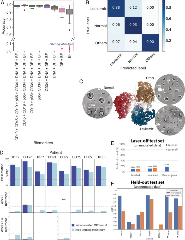
Acute lymphoblastic leukemia (ALL) is the most common childhood cancer. While there are a number of well-recognized prognostic biomarkers at diagnosis, the most powerful independent prognostic factor is the response of the leukemia to induction chemotherapy (Campana and Pui: Blood 129 (2017) 1913-1918). Given the potential for machine learning to improve precision medicine, we tested its capacity to monitor disease in children undergoing ALL treatment. Diagnostic and on-treatment bone marrow samples were labeled with an ALL-discriminating antibody combination and analyzed by imaging flow cytometry. Ignoring the fluorescent markers and using only features extracted from bright-field and dark-field cell images, a deep learning model was able to identify ALL cells at an accuracy of >88%. This antibody-free, single cell method is cheap, quick, and could be adapted to a simple, laser-free cytometer to allow automated, point-of-care testing to detect slow early responders. Adaptation to other types of leukemia is feasible, which would revolutionize residual disease monitoring. © 2020 The Authors. Cytometry Part A published by Wiley Periodicals, Inc. on behalf of International Society for Advancement of Cytometry.

Keywords: computer vision; deep learning; imaging flow cytometry; label-free; leukemia; machine learning; neural networks.


Figures

Figure 1
Figure 1
Sample partitioning strategy for training, validation, inference, and avoiding overfitting. Samples were split for training (including validation), testing (Test 1–3) and hold‐out (Test 4). Training/validation set contained pooled data of 19 entries from 15 patients. Samples were collected and measured at the time of presentation (abbreviated as “pres”) and after round(s) of treatments (noted as days after treatment). Test set 1 contained manually gated ground‐truth populations for leukemic blasts, normal lymphocytes and other cell types (Fig. 2A‐C). Test set 2, which contains DAPI‐positive, in‐focus single white blood cells, was designed to validate whether the learned algorithms were able to derive a correct residual disease (MRD) readout, that is, percentage of leukemic cells within the total number of white cells in the bone marrow sample (Fig. 2D). Note: Although some training data and Test set 2 were generated from the same patients, the training sets use a small number of individually annotated healthy/leukemic cells, while Test set 2 presents a large number of unannotated cells. Test set 3 (>200,000 single cells in total) was conducted with stained/unstained samples in a condition with or without laser illumination, confirming that the performance of the trained neural network was not dependent on the presence of bleed‐through fluorescence or lasers (Fig. 2E). Test set 4 was kept held‐out and only unlocked immediately before submission of the manuscript for the final verification of the success of the machine learning models (Fig. 2F).
Figure 2
Figure 2
Label‐free identification of ALL cells by ResNet50 corresponds well with biomarker‐based analysis. (A) Prediction results on each of 11 test data entries from five patients, who have data at the time of presentation and during treatment (average accuracy from single‐cell classification) (Test 1). The first column reports average accuracy across samples using all channels; subsequent columns correspond to incremental dropping of the next channel. BF, bright‐field; DF, dark‐field. Boxplots show the median line, first and third quartiles. Whiskers are drawn to double interquartile range (+2IQR). Diamonds represent data events that are outside the low and high whisker ends. (B) Three‐class single‐cell classification using bright‐field and dark‐field channels (Test 1). Confusion matrix for three categories, n = 218,747 ground‐truth cells pooled from 11 test sets. (C) Clustering of 9,025 cells randomly sampled from patient LK157 at the time of presentation based on deep learning features. 3D t‐Distributed Stochastic Neighbor Embedding (t‐SNE)20 presentations based on 2,048 label‐free feature vectors for each cell from the second‐to‐last layer of the trained neural network, ResNet50, are shown. The t‐SNE calculation was stabilized after 400 iterations at perplexity of 30. Colors coded according to true class labels: Leukemic cells (blue), normal B lymphocytes (red), other cell types (yellow). The magnified images display true bright‐field channel, randomly zoomed at each cluster. Example data with overlaid images http://projector.tensorflow.org/?config=raw.githubusercontent.com/minh‐doan/Deeplearning_LabelFree_Leukemia/master/Publish/DL_supervised/Data/Step4/Output/projector_config.pbtxt can be visualized with a web‐based projector. (D) Comparison between human‐curated manual gates and label‐free deep learning in predicting residual disease fraction (Test 2). The prediction on each sample was performed on the DAPI‐positive population of in‐focus, white blood cells, which is a mixture of leukemic blasts, normal B lymphocytes, other mature and immature hematopoietic cells such as granulocytes and monocytes. Red dash‐line is 25%—the threshold of treatment effectiveness of chemotherapy. (E) Summary of residual disease estimated by deep learning using information in bright‐field channel of labeled/unlabeled samples (each with laser on/off settings) (Test 3). Residual disease fraction was calculated as the percentage of predicted leukemic cells over the whole population of unannotated in‐focus single cells. For reference, human‐curated manual residual disease fraction (last blue column) was estimated based on the stained version with laser on. (F) Result of label‐free deep learning residual disease readout (yellow columns) on unannotated in‐focus single cells in held‐out data (Test 4). Blue columns are residual disease readout reported by standard clinical flow cytometric protocol. LK881 and LK919 (asterisks) are relapsed ALL patients. [Color figure can be viewed at wileyonlinelibrary.com]

References

    1. Campana D, Pui C‐H. Minimal residual disease–guided therapy in childhood acute lymphoblastic leukemia. Blood 2017;129:1913–1918. - PMC - PubMed
    1. Vora A, Goulden N, Mitchell C, Hancock J, Hough R, Rowntree C, Moorman AV, Wade R. Augmented post‐remission therapy for a minimal residual disease‐defined high‐risk subgroup of children and young people with clinical standard‐risk and intermediate‐risk acute lymphoblastic leukaemia (UKALL 2003): A randomised controlled trial. Lancet Oncol 2014;15:809–818. - PubMed
    1. Vora A, Goulden N, Wade R, Mitchell C, Hancock J, Hough R, Rowntree C, Richards S. Treatment reduction for children and young adults with low‐risk acute lymphoblastic leukaemia defined by minimal residual disease (UKALL 2003): A randomised controlled trial. Lancet Oncol 2013;14:199–209. - PubMed
    1. Gupta S, Devidas M, Loh ML, Raetz EA, Chen S, Wang C, Brown P, Carroll AJ, Heerema NA, Gastier‐Foster JM, et al. Flow‐cytometric vs. ‐morphologic assessment of remission in childhood acute lymphoblastic leukemia: A report from the Children's oncology group (COG). Leukemia 2018;32:1370–1379. 10.1038/s41375-018-0039-7. - DOI - PMC - PubMed
    1. O'Connor D, Moorman AV, Wade R, Hancock J, Tan RMR, Bartram J, Moppett J, Schwab C, Patrick K, Harrison CJ, et al. Use of minimal residual disease assessment to redefine induction failure in pediatric acute lymphoblastic leukemia. J Clin Oncol 2017;35:660–667. - PubMed

Publication types