

Chest CT Findings in Patients With Coronavirus Disease 2019 and Its Relationship With Clinical Features
- PMID: 32091414
- PMCID: PMC7147284
- DOI: 10.1097/RLI.0000000000000670
Chest CT Findings in Patients With Coronavirus Disease 2019 and Its Relationship With Clinical Features
Abstract
Objectives: The aim of this study was to investigate the chest computed tomography (CT) findings in patients with confirmed coronavirus disease 2019 (COVID-19) and to evaluate its relationship with clinical features.
Materials and methods: Study sample consisted of 80 patients diagnosed as COVID-19 from January to February 2020. The chest CT images and clinical data were reviewed, and the relationship between them was analyzed.
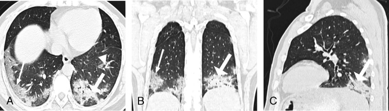
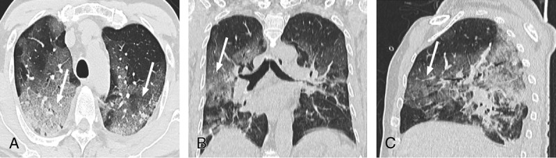
Results: Totally, 80 patients diagnosed with COVID-19 were included. With regards to the clinical manifestations, 58 (73%) of the 80 patients had cough, and 61 (76%) of the 80 patients had high temperature levels. The most frequent CT abnormalities observed were ground glass opacity (73/80 cases, 91%), consolidation (50/80 cases, 63%), and interlobular septal thickening (47/80, 59%). Most of the lesions were multiple, with an average of 12 ± 6 lung segments involved. The most common involved lung segments were the dorsal segment of the right lower lobe (69/80, 86%), the posterior basal segment of the right lower lobe (68/80, 85%), the lateral basal segment of the right lower lobe (64/80, 80%), the dorsal segment of the left lower lobe (61/80, 76%), and the posterior basal segment of the left lower lobe (65/80, 81%). The average pulmonary inflammation index value was (34% ± 20%) for all the patients. Correlation analysis showed that the pulmonary inflammation index value was significantly correlated with the values of lymphocyte count, monocyte count, C-reactive protein, procalcitonin, days from illness onset, and body temperature (P < 0.05).
Conclusions: The common chest CT findings of COVID-19 are multiple ground glass opacity, consolidation, and interlobular septal thickening in both lungs, which are mostly distributed under the pleura. There are significant correlations between the degree of pulmonary inflammation and the main clinical symptoms and laboratory results. Computed tomography plays an important role in the diagnosis and evaluation of this emerging global health emergency.
Conflict of interest statement
Conflicts of interest and sources of funding: none declared.
Figures

References
-
- World Health Organization Novel coronavirus - China. 2020. Available at: https://www.who.int/csr/don/12-january-2020-novel-coronavirus-china/en/.
-
- Chaolin H, Yeming W, Xingwang L, et al. The lancet.Published Online January 24, 2020. Available at: https://doi.org/10.1016/S0140-6736(20)30183-5.
-
- World Health Organization Novel coronavirus - Japan (ex-China). 2020. Available at: http://www.who.int/csr/don/17-january-2020-novel-coronavirusjapan-ex-chi....
-
- World Health Organization Novel coronavirus - Republic of Korea (ex-China). 2020. Available at: http://www.who.int/csr/don/21-january-2020-novelcoronavirus-republic-of-....
Publication types
MeSH terms
LinkOut - more resources
Full Text Sources
Other Literature Sources
Research Materials
