. 2020 Feb 24;16(2):e1008083.
doi: 10.1371/journal.ppat.1008083. eCollection 2020 Feb.

Hinge length contributes to the phagocytic activity of HIV-specific IgG1 and IgG3 antibodies

Affiliations

Hinge length contributes to the phagocytic activity of HIV-specific IgG1 and IgG3 antibodies

Thach H Chu et al. PLoS Pathog. .

Abstract

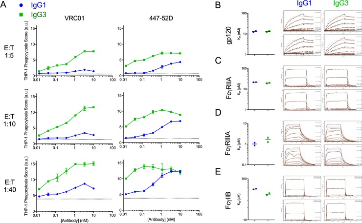
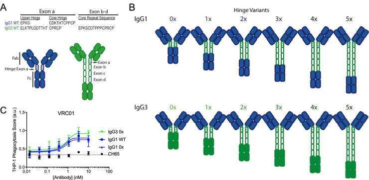
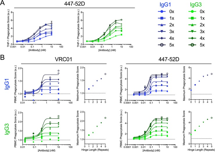
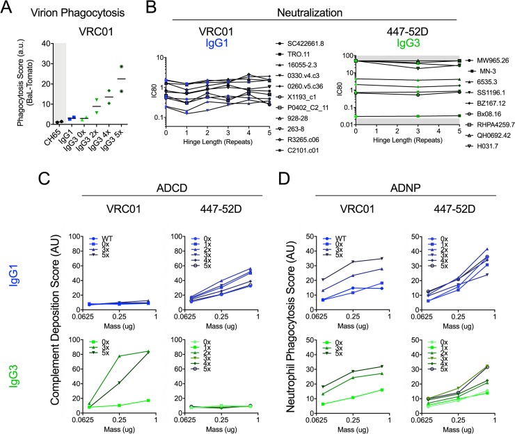
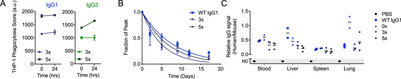
Antibody functions such as neutralization require recognition of antigen by the Fab region, while effector functions are additionally mediated by interactions of the Fc region with soluble factors and cellular receptors. The efficacy of individual antibodies varies based on Fab domain characteristics, such as affinity for antigen and epitope-specificity, and on Fc domain characteristics that include isotype, subclass, and glycosylation profile. Here, a series of HIV-specific antibody subclass and hinge variants were constructed and tested to define those properties associated with differential effector function. In the context of the broadly neutralizing CD4 binding site-specific antibody VRC01 and the variable loop (V3) binding antibody 447-52D, hinge truncation and extension had a considerable impact on the magnitude of phagocytic activity of both IgG1 and IgG3 subclasses. The improvement in phagocytic potency of antibodies with extended hinges could not be attributed to changes in either intrinsic antigen or antibody receptor affinity. This effect was specific to phagocytosis and was generalizable to different phagocytes, at different effector cell to target ratios, for target particles of different size and composition, and occurred across a range of antibody concentrations. Antibody dependent cellular cytotoxicity and neutralization were generally independent of hinge length, and complement deposition displayed variable local optima. In vivo stability testing showed that IgG molecules with altered hinges can exhibit similar biodistribution and pharmacokinetic profiles as IgG1. Overall, these results suggest that when high phagocytic activity is desirable, therapeutic antibodies may benefit from being formatted as human IgG3 or engineered IgG1 forms with elongated hinges.


Conflict of interest statement

The authors have declared that no competing interests exist.

Figures

Fig 1
Fig 1. IgG3 Abs exhibit enhanced ADCP despite similar antigen and FcɣR binding affinities.
A. Phagocytic activity of subclass-switched forms of VRC01 (left) and 447-52D (right) antibodies over a range of effector to target (E:T) ratios in the THP-1 ADCP assay using CH505TF gp140 antigen-conjugated beads as target. Error bars indicate SD of duplicates. Dotted horizontal lines represent phagocytosis values observed in the absence of Ab. B-E. Antigen (BaL gp120) (B), FcɣRIIA (C), and FcɣRIIIA (D), FcɣRIIB (E), binding affinity (left, from duplicate assessments of printed antibody spots, bar indicates average/median) and SPR binding profiles (right, raw data in black, kinetic curve fits in red for the ligand concentration series from a representative antibody spot) for VRC01 in IgG1 and IgG3 forms. AU: arbitrary units.
Fig 2
Fig 2. IgG1 and IgG3 hinge variant panel.
A. Sequences of natural IgG1 and IgG3 hinge exons, noting the upper and core hinge regions encoded by exon a and the core hinge repeat sequences encoded by exons b-d in IgG3. B. Schematic of the panel of hinge swapped and extended IgG1 and IgG3 variants. Domains derived from IgG1 and IgG3 are indicated in blue and green respectively. C. Phagocytic activity of VRC01 WT IgG1 and 0x forms of IgG1 and IgG3 in the THP-1 ADCP assay against CH505TF gp140 antigen-conjugated beads. Error bars indicate mean and SD of duplicates. Dotted horizontal line represent phagocytosis value observed in the absence of Ab. Phagocytic scores for CH65, an IgG1 Ab specific for hemagglutinin are shown as an additional negative control. Connecting lines indicate curve fit models. AU: arbitrary units.
Fig 3
Fig 3. Increasing hinge length increases phagocytic activity.
A. Phagocytic activity of IgG1 (left) and IgG3 (right) forms of 447–52 in the THP-1 ADCP assay against CH505TF gp140 antigen-conjugated beads. Error bars indicate mean and SD of duplicates. B. Phagocytic activity of VRC01 (left) and 447-52D (right) in IgG1 (top) and IgG3 (bottom) in the PBMC ADCP assay against CH505TF gp140 antigen-conjugated beads. Connecting lines indicate curve fit models. Inset scatterplots depict the relationship between hinge length and peak phagocytic score observed from curve fits. Data was collected in duplicate and normalized to a no-Ab control within each plate. Dotted horizontal lines represent phagocytosis values observed in the absence of Ab. AU: arbitrary units.
Fig 4
Fig 4. Impact of hinge modification on additional antibody functions.
A. Uptake of fluorescent virions for IgG1 and IgG3 hinge variant forms of VRC01 and the CH65 control antibody (assessed in duplicate, bar indicates mean). B. Neutralization profiles of IgG1 hinge variants of VRC01 (left) and IgG3 hinge variants of 447-52D (right) on selected sensitive viruses. Titrations were performed in duplicate. Gray region indicates the limit of detection. C-D. Antibody dependent complement deposition (ADCD) (C) and antibody dependent neutrophil phagocytosis (ADNP) (D) activity of VRC01 (left) and 447-52D (right) hinge variants in IgG1 (top) and IgG3 (bottom) forms of gp120 MN antigen-coated beads. Data represent means of duplicates. AU: arbitrary units.
Fig 5
Fig 5. In vitro and in vivo stability tests.
A. Serum stability of 447-52D IgG1 and IgG3 in 3x and 5x forms (n = 2, mean and SD plotted) as defined by phagocytic activity of THP-1 cells against JRFL SOSIP antigen-conjugated beads. AU: arbitrary units. B. Pharmacokinetic assessment of infused WT VRC01 IgG1, 3x, and 5x variants (n = 2–3, mean and SD are plotted along with 1 phase decay curve fit). C. Biodistribution of WT IgG1, 0x, 3x, 5x hinge variants as defined by relative levels of human IgG as compared to mouse IgG detected in each tissue. No human IgG could be detected (ND) in the PBS-infused mouse. Bars indicate means.

References

    1. Burton DR, Williamson RA, Parren PW. Antibody and virus: binding and neutralization. Virology. 2000;270(1):1–3. 10.1006/viro.2000.0239 . - DOI - PubMed
    1. Klasse PJ. Neutralization of Virus Infectivity by Antibodies: Old Problems in New Perspectives. Advances in biology. 2014;2014 10.1155/2014/157895 - DOI - PMC - PubMed
    1. Corti D, Lanzavecchia A. Broadly neutralizing antiviral antibodies. Annual review of immunology. 2013;31:705–42. 10.1146/annurev-immunol-032712-095916 . - DOI - PubMed
    1. Crowe JE Jr. Principles of Broad and Potent Antiviral Human Antibodies: Insights for Vaccine Design. Cell host & microbe. 2017;22(2):193–206. 10.1016/j.chom.2017.07.013 - DOI - PMC - PubMed
    1. Saphire EO, Schendel SL, Fusco ML, Gangavarapu K, Gunn BM, Wec AZ, et al. Systematic Analysis of Monoclonal Antibodies against Ebola Virus GP Defines Features that Contribute to Protection. Cell. 2018;174(4):938–52 e13. 10.1016/j.cell.2018.07.033 - DOI - PMC - PubMed

Publication types