. 2020 Feb 24;16(2):e1008263.
doi: 10.1371/journal.ppat.1008263. eCollection 2020 Feb.

Cryo-EM structure of the Shigella type III needle complex

Affiliations

Cryo-EM structure of the Shigella type III needle complex

Michele Lunelli et al. PLoS Pathog. .

Abstract

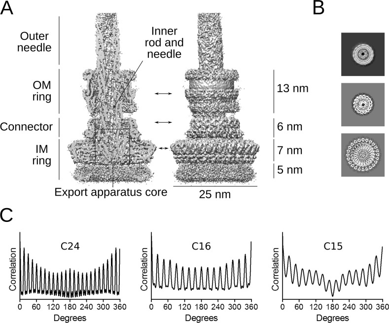
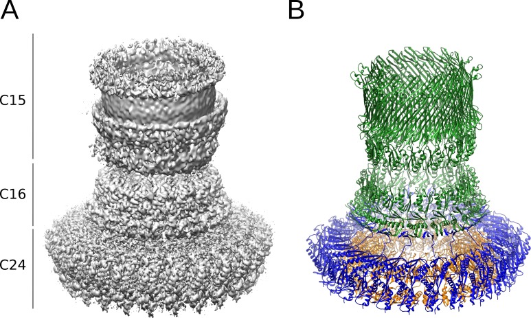
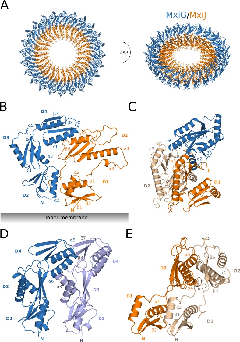
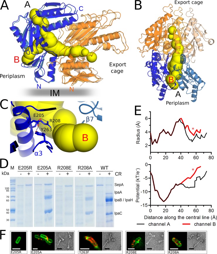
The Type III Secretion Systems (T3SS) needle complex is a conserved syringe-shaped protein translocation nanomachine with a mass of about 3.5 MDa essential for the survival and virulence of many Gram-negative bacterial pathogens. This system is composed of a membrane-embedded basal body and an extracellular needle that deliver effector proteins into host cells. High-resolution structures of the T3SS from different organisms and infection stages are needed to understand the underlying molecular mechanisms of effector translocation. Here, we present the cryo-electron microscopy structure of the isolated Shigella T3SS needle complex. The inner membrane (IM) region of the basal body adopts 24-fold rotational symmetry and forms a channel system that connects the bacterial periplasm with the export apparatus cage. The secretin oligomer adopts a heterogeneous architecture with 16- and 15-fold cyclic symmetry in the periplasmic N-terminal connector and C-terminal outer membrane ring, respectively. Two out of three IM subunits bind the secretin connector via a β-sheet augmentation. The cryo-EM map also reveals the helical architecture of the export apparatus core, the inner rod, the needle and their intervening interfaces.


Conflict of interest statement

The authors have declared that no competing interests exist.

Figures

Fig 1
Fig 1. Needle complex reconstruction and symmetry analysis.
(A) Cross-section and side view of the symmetry-free (C1) reconstruction of the Shigella needle complex. The structural features and their dimensions are indicated. Arrows show the position of the slices shown in panel B. (B) Horizontal slices of the periplasmic IM ring (bottom panel),the connector (middle panel) and the OM ring (upper panel, from non-sharpened map). (C) Rotational autocorrelation analysis of the three slices shown in panel B indicating C24 symmetry for the IM ring (24 peaks spaced by 15 degrees), C16 symmetry for the connector (16 peaks spaced by 22.5 degrees) and C15 symmetry for the OM ring (15 peaks spaced by 24 degrees). A circular mask with inner radius 55 Å and outer radius 76 Å was applied to the OM ring slice to highlight the 15-fold periodicity.
Fig 2
Fig 2. Architecture of the outer regions of the basal body.
(A) Composite map of the highest-resolution maps obtained for the outer regions of the needle complex (left panel), including the focused IM ring map with C24 symmetry, the connector map from the focused IM-connector reconstruction with C8 symmetry and the OM ring map from C1 reconstruction. The cyclic symmetry of the three regions is indicated on the left. (B) Atomic models of the corresponding needle complex proteins built using maps depicted in (A): MxiG (blue), MxiJ (orange) and MxiD (green).
Fig 3
Fig 3. Structure of the periplasmic IM ring.
The periplasmic IM ring model in cartoon representation (MxiG subunits in blue and violet, MxiJ subunits in orange and beige). (A) Top view (left panel) and tilted view (right panel) of the periplasmic IM ring composed by 24 copies of MxiG and MxiJ subunits forming two concentric rings. The inner membrane is located below the ring. (B) Side view of one MxiG and one MxiJ neighboring subunits. Only the periplasmic domains are shown and labeled. Secondary structure elements involved in IM ring interactions are also labeled. The domains adopt RBM αββαβ topology, except the D1 domain of MxiJ that has inverted RBM βαββα topology. (C-E) Interacting subunits of the IM periplasmic ring: one MxiG between two MxiJ subunits (C), a couple of neighboring MxiG (D) and MxiJ subunits (E). Secondary structure elements involved in IM ring interactions are labeled.
Fig 4
Fig 4. Channels inside the periplasmic IM ring of the Shigella needle complex.
(A) Side view of the surface diagram of the channel connecting the export cage with the periplasm (yellow) at the interface between five IM ring subunits. Two MxiG (shades of blue) and three MxiJ (shades of orange) are represented as cartoons. The two channel exits towards the periplasm are labeled A and B. (B) Bottom radial view of the channel and subunits represented in panel (A). The channel runs between two MxiG (shades of blue) and three MxiJ (shades of orange) subunits. (C) Close-up of channel exit B indicates that the conserved MxiG residues E205, R208 and Y263 are located nearby a bottleneck. Hydrogen bonds involving their side chains are shown as dotted lines. (D) SDS-PAGE analysis of secreted proteins from wild-type (WT) and MxiG R208 and E205 Shigella mutant strains upon congo red induction. SepA serves as a T3S-independent loading control. IpaA, IpaB, IpaH and IpaC are T3SS effectors. Protein secretion was induced adding congo red (CR). (E) Channel radii and electrostatic potential profiles of the channels A and B plotted against the distance along the central line, starting from the export cage towards the periplasm. The asterisk indicates the location of the MxiG residues E205, R208 and Y263 near the channel exit B. (F) Representative immunofluorescence images of Shigella mutant strains and TEM images of negatively stained isolated needle complexes. In the immunofluorescence images the bacterial membrane is stained in green, T3SS needles in red. All the mutants except E205R produce the needle complex, which is localized on the membrane and it was successfully isolated. The white scale bar corresponds to 2 μm, the black scale bar of the TEM images to 50 nm.
Fig 5
Fig 5. Structure of the connector and its interface with the IM ring.
(A) Tilted top view of the connector composed of the N-terminal N0 and N1 domains of MxiD (green) and the C-terminal domain of MxiG (blue) in cartoon representation. The ring is formed by 16 subunits of each protein. (B) Close-up of two neighboring couples of MxiD and MxiG subunits. The MxiG subunits interact with the N-terminus of MxiD via β-sheet augmentation, forming a continuous circular β-sheet at the connector base. (C) N0 and N1 domains of MxiD between the C-terminal domain two MxiG subunits (left panel); residues represented as sticks are involved in intermolecular hydrophobic interactions, the position of the amino acid 347 in MxiG is indicated in red. Detailed view of the MxiG-MxiD hydrophobic interface colored according to the Eisenberg scale (white being the most hydrophobic) (right panel); black-labeled residues are at the interface and belong to MxiD (Y38, A40, I71, F75, L77 and A36, which is not visible in this view), MxiG residues are labeled in blue (L350, Y356, M358, W364 and F366). (D) Representative images of immunofluorescence stained Shigella M90T ΔMxiG complemented with truncated Strep-MxiG comprising residues 1 to 346 (MxiG-347-Stop). Green, lipophilic membrane stain; red, anti-Strep-tag antibody. strep-mxiH and strep-mxiG genes are expressed from a plasmid in non-permeabilized bacteria while permeabilized bacteria expressed only strep-mxiG. No needle complex is visible on the surface of the bacteria, while the MxiG mutant is localized in the bacterial membrane. The scale bar corresponds to 2 μm.
Fig 6
Fig 6. Architecture of the OM ring.
(A) Cartoon representation of the vertical cutaway of the MxiD pore in the C1 map. The subunits form a barrel shaped ring system that is anchored to the outer membrane. The OM ring is composed of the RBM N3 domain, the central secretin domain and its membrane associated (MA) subdomain. (B) A vertical slice of the OM ring in the C1 map. The inner and outer β-barrel walls are indicated. The MA subdomain, the periplasmic gate and the N3 domains form constrictions with diameter ~70 Å. The C-terminal S domain of MxiD is likely located on the outer side of the β-barrel in the indicated region. (C) Top view of the OM ring cartoon fitted into the C1 map. The dotted circle represents the outer diameter of the needle filament, which passes through the OM pore.
Fig 7
Fig 7. Architecture of the export apparatus core, inner rod and start of the needle.
Cartoon representation of the subunits forming the export apparatus core, the inner rod and the start of the needle filament, in a vertical section of the outer needle complex. The individual protein components are depicted on the right side. Five SpaP, five SpaQ and one SpaR subunits, arranged in a helical fashion, form the export apparatus core. The subunits connecting the export apparatus with the needle were modeled as polyalanine peptides and adopt three different conformations (see the main text discussion about their possible identity). They share a helix-turn-helix fold and continue the export apparatus helix forming two turns. The upper turns are likely composed of needle subunits MxiH.

References

    1. WHO (2017) The top 10 causes of death. Fact Sheet.
    1. Wang H NM, Allen C, Barber RM, Bhutta ZA, Carter A, Casey DC, Charlson FJ, Chen AZ, Coates MM, Coggeshall M, Dandona L, Dicker DJ, Erskine HE, Ferrari AJ, Fitzmaurice C, Foreman K, Forouzanfar MH, Fraser MS, Fullman N, Gething PW, Goldberg EM, G (2016) Global, regional, and national life expectancy, all-cause mortality, and cause-specific mortality for 249 causes of death, 1980–2015: a systematic analysis for the Global Burden of Disease Study 2015. Lancet 388: 1459–1544. 10.1016/S0140-6736(16)31012-1 - DOI - PMC - PubMed
    1. Ashida H, Mimuro H, Sasakawa C (2015) Shigella manipulates host immune responses by delivering effector proteins with specific roles. Front Immunol 6: 219 10.3389/fimmu.2015.00219 - DOI - PMC - PubMed
    1. Egan F, Barret M, O'Gara F (2014) The SPI-1-like Type III secretion system: more roles than you think. Front Plant Sci 5: 34 10.3389/fpls.2014.00034 - DOI - PMC - PubMed
    1. Blocker A, Jouihri N, Larquet E, Gounon P, Ebel F, et al. (2001) Structure and composition of the Shigella flexneri "needle complex", a part of its type III secreton. Mol Microbiol 39: 652–663. 10.1046/j.1365-2958.2001.02200.x - DOI - PubMed

Publication types

MeSH terms

Substances