Review
. 2020 May 6:49:309-341.
doi: 10.1146/annurev-biophys-121219-081703. Epub 2020 Feb 24.

Insights into the Structure, Function, and Dynamics of the Bacterial Cytokinetic FtsZ-Ring

Affiliations
Review

Insights into the Structure, Function, and Dynamics of the Bacterial Cytokinetic FtsZ-Ring

Ryan McQuillen et al. Annu Rev Biophys. .

Abstract

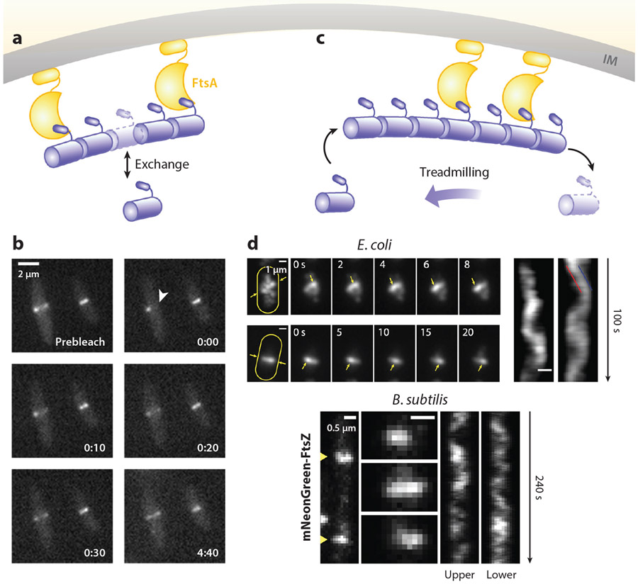
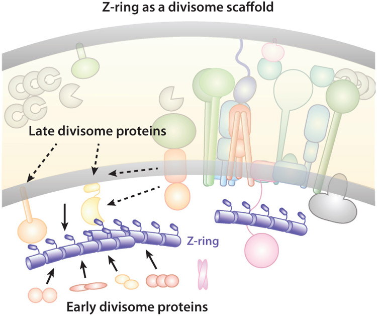
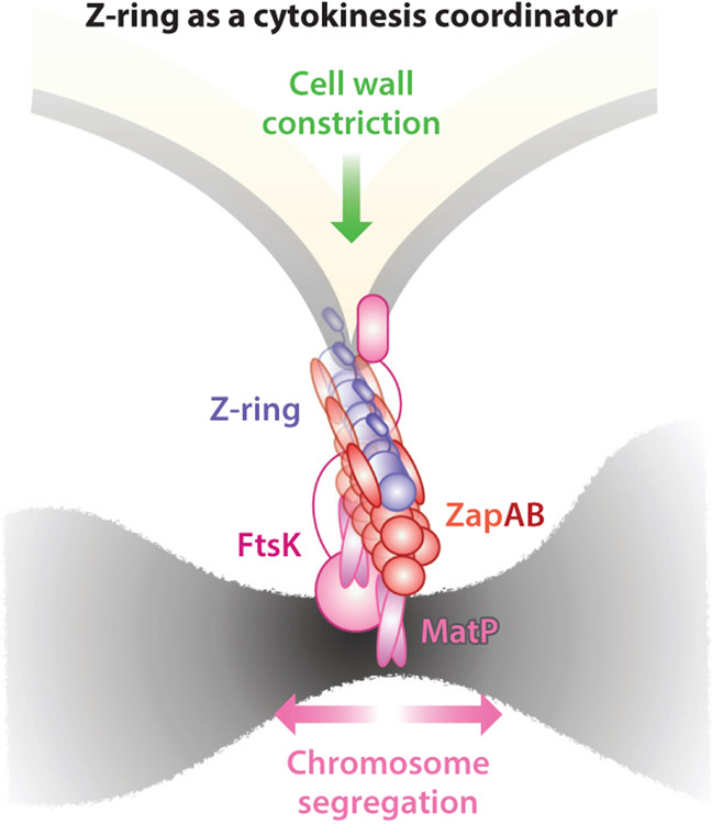
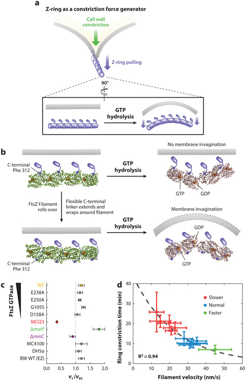
The FtsZ protein is a highly conserved bacterial tubulin homolog. In vivo, the functional form of FtsZ is the polymeric, ring-like structure (Z-ring) assembled at the future division site during cell division. While it is clear that the Z-ring plays an essential role in orchestrating cytokinesis, precisely what its functions are and how these functions are achieved remain elusive. In this article, we review what we have learned during the past decade about the Z-ring's structure, function, and dynamics, with a particular focus on insights generated by recent high-resolution imaging and single-molecule analyses. We suggest that the major function of the Z-ring is to govern nascent cell pole morphogenesis by directing the spatiotemporal distribution of septal cell wall remodeling enzymes through the Z-ring's GTP hydrolysis-dependent treadmilling dynamics. In this role, FtsZ functions in cell division as the counterpart of the cell shape-determining actin homolog MreB in cell elongation.

Keywords: FtsZ; bacterial cell division; cell wall constriction; cytoskeleton dynamics; septum synthesis; single-molecule imaging.


Figures

Figure 1
Figure 1
Uranyl acetate–stained electron microscopy (EM) image of an isolated Escherichia coli sacculus with schematic drawing of the splitting of old (orange) and insertion of new (green) glycan strands in cell wall elongation and constriction. Figure adapted with permission from Reference . Abbreviation: sPG, septal peptidoglycan.
Figure 2
Figure 2
Chromosomal locations and operon organizations of all divisome genes, including the dcw gene cluster, in Escherichia coli. Figure adapted with permission from Reference .
Figure 3
Figure 3
(a) Core and (b) non-core proteins of the Escherichia coli divisome. Whenever possible, verified protein–protein, protein–membrane, and protein–sPG interactions are indicated by the spatial arrangement of respective components. Orange and green lines denote old and new sPG strands, respectively, as in Figure 1. In panel b, core proteins are colored gray in the background for simplicity. Abbreviations: IM, inner membrane; OM, outer membrane; sPG, septal peptidoglycan.
Figure 4
Figure 4
Z-ring structure. (a) Immuno-gold EM images of constricting Escherichia coli cells show labeled FtsZ molecules (scattered black dots) at the leading edge of the invaginating septum (18). (b) Fluorescence images of FtsZ-GFP (green) in live E. coli cells outlined in yellow dashed lines (X. Yang, unpublished data). (c) Cryo-ECT images of FtsZ WT filaments in Caulobacter crescentus (left) (116) and reconstituted FtsZD212A mutant filaments in liposome (right) (212). (d) Three-dimensional fluorescence-based superresolution images of WT FtsZ-rings (left) and FtsZD212A mutant rings in E. coli (116). (e) Current model of the Z-ring depicting the spatial and dimensional features of the Z-ring (not to scale). The Z-ring is most likely composed of a single layer of short FtsZ protofilaments that randomly and heterogeneously associate underneath the inner membrane. Abbreviations: Cryo-ECT, cryo-electron tomography; EM, electron microscopy; FLM, fluorescence microscopy; GFP, green fluorescent protein; SMLM, single-molecule localization microscopy; WT, wild type.
Figure 5
Figure 5
Z-ring dynamics. (a) Schematic of the subunit exchange dynamics of the Z-ring. (b) A FRAP imaging sequence showing the rapid recovery of fluorescence after the photobleaching of half of the Z-ring (white arrowhead) (205). (c) Schematic of the treadmilling dynamics of the Z-ring. (d) Treadmilling dynamics observed in Escherichia coli (top) (239) and Bacillus subtilis (bottom) (20). For E. coli, the maximum intensity projections (left), the montages (middle), and the corresponding kymographs (right) of Z-rings labeled with FtsZ–green fluorescent protein (GFP) in two cells are shown. For B. subtilis, two FtsZ–mNeonGreen-labeled Z-rings (left), montages at 8-s intervals (middle), and the corresponding kymographs (right) of two cells are shown. Abbreviations: FRAP, fluorescence recovery after photobleaching; IM, inner membrane.
Figure 6
Figure 6
Schematic of the Z-ring functioning as a scaffold for the divisome assembly. For clarity, individual divisome proteins are not labeled, but instead are grouped according to their temporal assembly order (early or late). Most early divisome proteins reside in the cytoplasm or inner membrane and interact with FtsZ directly. Most late divisome proteins reside in the periplasm or outer membrane and interact with FtsZ indirectly. Additionally, some of the late divisome proteins’ location patterns do not exactly follow that of the Z-ring in mutants with perturbed Z-ring structures, indicating that the scaffolding function of the Z-ring is likely to mark the future division site and recruit other divisome proteins, but not necessarily to act as a scaffold for the stable assembly of the divisome.
Figure 7
Figure 7
Schematic of the Z-ring functioning as a cytokinesis coordinator. The Z-ring is sandwiched between the inner membrane and chromosome: FtsZ is attached to the inner membrane by FtsA, which makes further contacts with cell wall remodeling enzymes and regulators, and also attached to the chromosome by the ZapA–ZapB–MatP–DNA linkage. The presence of the linkage could coordinate the progression of cell wall constriction and chromosome segregation either mechanically or biochemically.
Figure 8
Figure 8
(a) Schematics of the Z-ring functioning as a constriction force generator through GTP hydrolysis-induced filament bending. (b) A recent crystallographic study (114) shows that GTP hydrolysis causes the FtsZ protofilament to bend, but the bending is toward the membrane (top) if the C-terminal tail of FtsZ (small blue rod) is directly attached to the membrane, which is opposite the direction of membrane invagination. To accommodate the bending conformation, the flexible linker between the globular domain and C-terminal tail of FtsZ has to wrap around the filament to attach to the membrane (dotted outlines, bottom). Note that the models shown are for visualization purposes only and are not based on the molecular dynamics simulations done in Reference . (c) Growth condition–normalized septum closure rate measurements made in wild-type (WT) BW25113 Escherichia coli cells in minimal media compared with different background strains including mutants affecting FtsZ’s GTPase activity (E238A, E250A, G105S and D158A), FtsI’s activity (MC123), chromosome segregation (ΔmatP), and Z-ring stability (ΔminC), in addition to different WT strain backgrounds (MC4100, DH5α) and WT BW25113 cells growing in rich defined growth medium (EZ). The FtsI mutant constricts significantly slower than WT cells, ΔmatP constricts faster, and all other perturbations of the Z-ring did not produce significant changes in septum closure rates (40, 239) (d) Constriction time measured in a series of Bacillus subtilis strains showed a high correlation with Z-ring treadmilling speed (20).
Figure 9
Figure 9
(a) Schematic of the Z-ring functioning as a cell pole shape determinant. The directional treadmilling dynamics of the Z-ring provides a spatial cue to attract sPG synthase molecules to follow the directional movement, thus distributing them evenly along the septum. (b) HADA labeling of Escherichia coli septum synthesis showed that, at a short time scale (10 s), both WT and the D212G mutant had punctate incorporation. At a long time scale (810 s), WT cells showed homogenous incorporation of HADA intensity, corresponding to smooth septum in the scanning electron microscopy image on the right, while the D212G mutant showed incomplete and asymmetric HADA incorporation and deformed septa (yellow arrowheads) (239). (c) SMT of FtsI molecules with maximal intensity projections (left, yellow arrowheads) and corresponding kymographs (right) in the WT (top) and D212G (bottom) FtsZ mutant E. coli cells showed correlated directional movement with FtsZ’s treadmilling speeds (239). Abbreviation: SMT, single-molecule tracking; sPG, septal peptidoglycan; WT, wild-type.

References

    1. Aaron M, Charbon G, Lam H, Schwarz H, Vollmer W, Jacobs-Wagner C. 2007. The tubulin homologue FtsZ contributes to cell elongation by guiding cell wall precursor synthesis in Caulobacter crescentus. Mol. Microbiol 64:938–52 - PubMed
    1. Aarsman ME, Piette A, Fraipont C, Vinkenvleugel TM, Nguyen-Distèche M, den Blaauwen T. 2005. Maturation of the Escherichia coli divisome occurs in two steps. Mol. Microbiol 55:1631–45 - PubMed
    1. Adams DW, Errington J. 2009. Bacterial cell division: assembly, maintenance and disassembly of the Z ring. Nat. Rev. Microbiol 7:642–53 - PubMed
    1. Addinall S, Bi E, Lutkenhaus J. 1996. FtsZ ring formation in fts mutants. J. Bacteriol 178:3877–84 - PMC - PubMed
    1. Addinall S, Lutkenhaus J. 1996. FtsA is localized to the septum in an FtsZ-dependent manner. J. Bacteriol 178:7167–72 - PMC - PubMed

Publication types

MeSH terms

LinkOut - more resources