Review
. 2020 May 6:49:289-308.
doi: 10.1146/annurev-biophys-121219-081550. Epub 2020 Feb 24.

Light Microscopy of Mitochondria at the Nanoscale

Affiliations
Review

Light Microscopy of Mitochondria at the Nanoscale

Stefan Jakobs et al. Annu Rev Biophys. .

Abstract

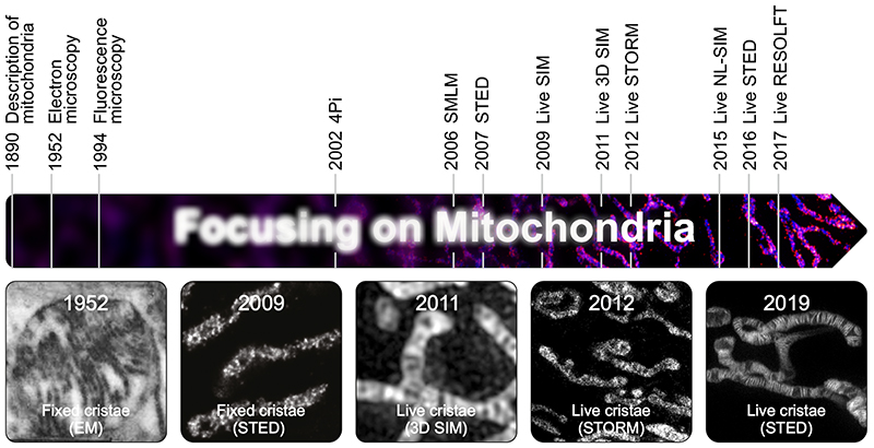
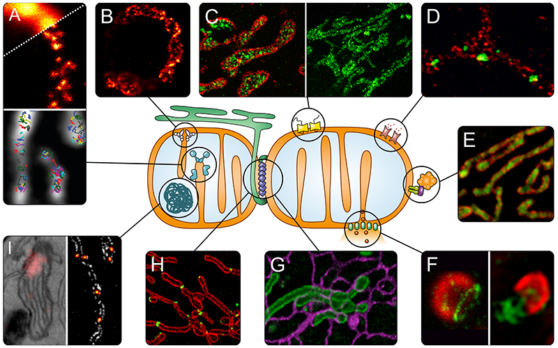
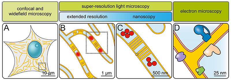
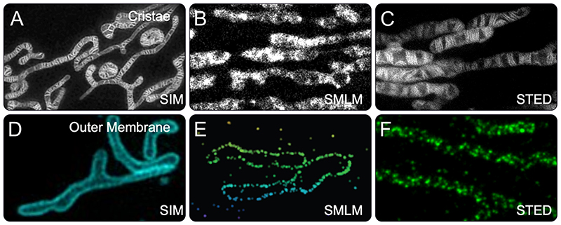
Mitochondria are essential for eukaryotic life. These double-membrane organelles often form highly dynamic tubular networks interacting with many cellular structures. Their highly convoluted contiguous inner membrane compartmentalizes the organelle, which is crucial for mitochondrial function. Since the diameter of the mitochondrial tubules is generally close to the diffraction limit of light microscopy, it is often challenging, if not impossible, to visualize submitochondrial structures or protein distributions using conventional light microscopy. This renders super-resolution microscopy particularly valuable, and attractive, for studying mitochondria. Super-resolution microscopy encompasses a diverse set of approaches that extend resolution, as well as nanoscopy techniques that can even overcome the diffraction limit. In this review, we provide an overview of recent studies using super-resolution microscopy to investigate mitochondria, discuss the strengths and opportunities of the various methods in addressing specific questions in mitochondrial biology, and highlight potential future developments.

Keywords: live-cell microscopy; mitochondria; nanoscopy; super-resolution microscopy.


Figures

Fig. 1
Fig. 1. Mitochondria in the focus of microscopy.
Highlighted are important milestones for visualization of mitochondria. Given are the years when a certain imaging method was first used to visualize mitochondria. The images show (from left to right): One of the first images of cristae recorded by EM (6). First light microscopy image of fixed cristae taken by isoSTED nanoscopy (68). First visualization of mitochondrial inner membrane structures in living cells by 3D SIM (53). First live-cell SMLM of the mitochondrial inner membrane (52). One of the first images of individual mitochondrial cristae recorded in living cells using STED nanoscopy (71).
Fig. 2
Fig. 2. Super-resolution microscopy to address questions of mitochondrial biology.
A) OXPHOS. Upper panel: Distribution of assembly factors recorded with STED nanoscopy (80). Lower panel: Tracking of single OXPHOS subunits using SMLM (77). B) Localization of the MICOS subunit Mic60 in a yeast cell, revealed by STED nanoscopy (89). C) TOM complex in the outer membrane recorded with DNA-PAINT nanoscopy (left, TOM complex in red) (63) and STED nanoscopy (right) (67). D) Differential distributions of the three human hVDAC (mitochondrial porin) isoforms in the outer membrane of human mitochondria (STED nanoscopy, hVDAC in green) (66). E) Sub-mitochondrial localization of PINK1 (green) shown by 3D-SIM (76). F) Apoptosis. Left: Several pro-apoptotic BAX proteins (green) form ring like structures in the outer membrane that may act as pores (STED nanoscopy) (100). Right: During later steps of apoptosis, these large BAX assemblies facilitate the herniation of the inner membrane and release of mtDNA (green) as shown by SIM (102). G) Interactions between mitochondria (green) and the ER (purple) in living cells recorded by STED nanoscopy (70). H) Spatial dynamics of the dynamin-like GTPase DRP1 (green), which is essential for mitochondrial fission, visualized by SIM (109). I) Nucleoids, analyzed with correlative PALM and EM (left panel) (131), and with STED nanoscopy (right panel, nucleoids in fire) (unpublished).
Fig. 3
Fig. 3. Imaging mitochondria across scales.
A) Diffraction-limited microscopy (confocal or widefield microscopy). Overall mitochondrial morphology and network dynamics can be visualized. Only limited information on sub-mitochondrial protein distributions. B) Extended-resolution microscopy (diffraction-limited super-resolution microscopy). Network dynamics, but also inner mitochondrial dynamics. Groups of cristae and, under some conditions, single cristae are visible. Mitochondrial sub-compartments can be analyzed. C) Nanoscopy (diffraction-unlimited super-resolution microscopy). Detailed sub-mitochondrial protein distributions and individual cristae can be resolved. D) Electron microscopy. Precise membrane architecture and lipid bilayers are resolved. With specific approaches, protein distributions and even protein structures can be determined.
Fig. 4
Fig. 4. Cristae and TOM-complexes recorded with different super-resolution microscopies.
A-C) Mitochondrial cristae recorded with Hessian SIM (A) (54), SMLM (STORM) (B) (61) or STED nanoscopy (C) (71) in living human cells. For SIM and STORM, the cristae were labeled with different MitoTracker dyes. For STED nanoscopy, cells expressing a COX8A-Snap-tag fusion protein were labeled with a silicone rhodamine dye. D-F) The mitochondrial outer membrane proteins TOM20 or TOM22 were imaged with SIM (D) (93), SMLM (4PiSMSN) (E) (142) and STED nanoscopy (F) (89) in human cell lines. For 4PiSMSN and STED nanoscopy, fixed cells were decorated with antibodies against TOM20 (4PiSMSN) or TOM22 (STED nanoscopy). SIM was performed on living cells expressing a TOM20-mEmerald fusion protein.

References

    1. Scheffler IE. John Wiley & Sons, Inc; Hoboken, New Jersey, USA: 2008.
    1. Nunnari J, Suomalainen A. Cell. 2012;148:1145–59. - PMC - PubMed
    1. Bereiter-Hahn J, Voth M. Microscopy Research & Technique. 1994;27:198–219. - PubMed
    1. Nunnari J, Marshall WF, Straight A, Murray A, Sedat JW, Walter P. Molecular Biology of the Cell. 1997;8:1233–42. - PMC - PubMed
    1. Westermann B. Nat Rev Mol Cell Biol. 2010;11:872–84. - PubMed

Publication types

LinkOut - more resources