. 2020 Feb 19;18(2):123.
doi: 10.3390/md18020123.

3D Chitin Scaffolds of Marine Demosponge Origin for Biomimetic Mollusk Hemolymph-Associated Biomineralization Ex-Vivo

Affiliations

3D Chitin Scaffolds of Marine Demosponge Origin for Biomimetic Mollusk Hemolymph-Associated Biomineralization Ex-Vivo

Marcin Wysokowski et al. Mar Drugs. .

Abstract

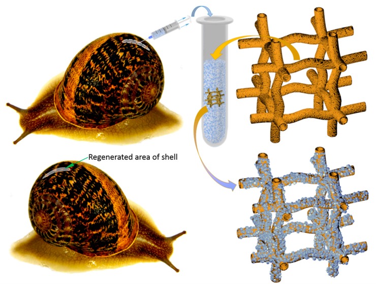
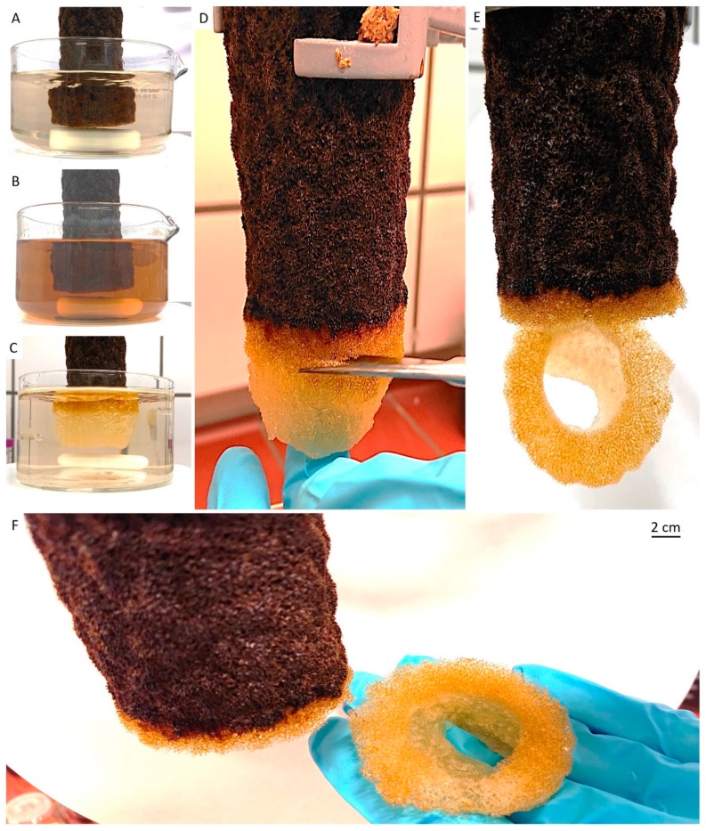
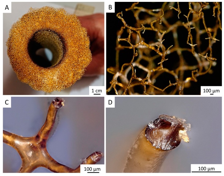
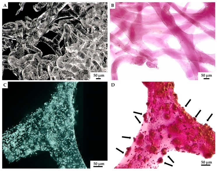
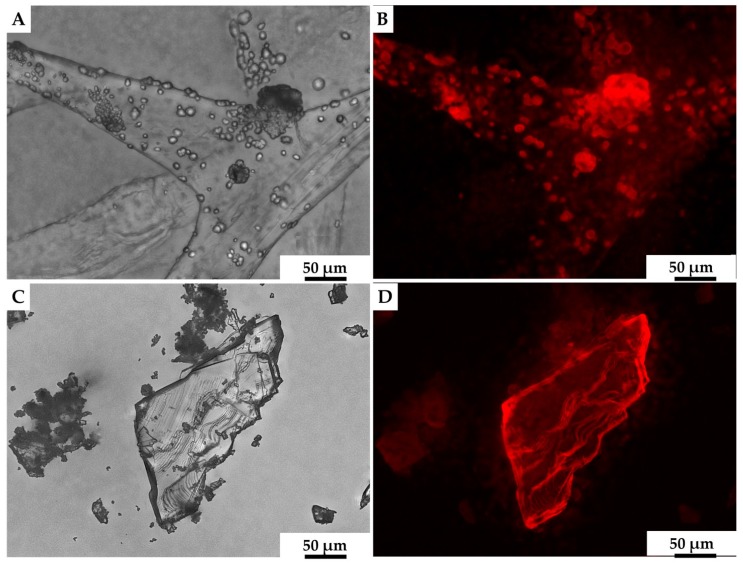
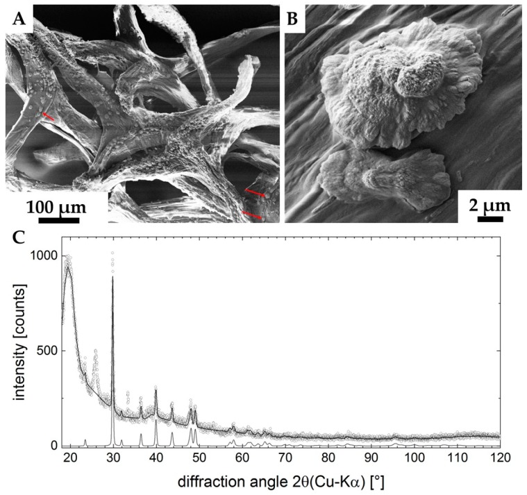
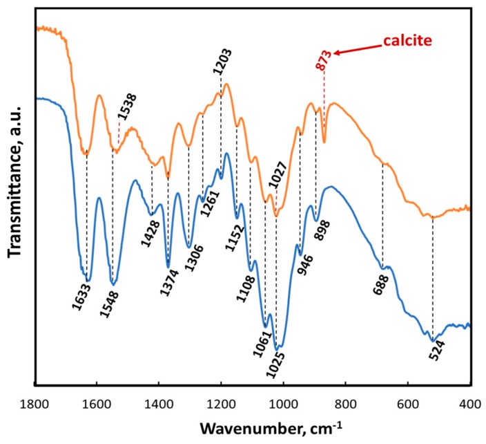
Structure-based tissue engineering requires large-scale 3D cell/tissue manufacture technologies, to produce biologically active scaffolds. Special attention is currently paid to naturally pre-designed scaffolds found in skeletons of marine sponges, which represent a renewable resource of biomaterials. Here, an innovative approach to the production of mineralized scaffolds of natural origin is proposed. For the first time, a method to obtain calcium carbonate deposition ex vivo, using living mollusks hemolymph and a marine-sponge-derived template, is specifically described. For this purpose, the marine sponge Aplysin aarcheri and the terrestrial snail Cornu aspersum were selected as appropriate 3D chitinous scaffold and as hemolymph donor, respectively. The formation of calcium-based phase on the surface of chitinous matrix after its immersion into hemolymph was confirmed by Alizarin Red staining. A direct role of mollusks hemocytes is proposed in the creation of fine-tuned microenvironment necessary for calcification ex vivo. The X-ray diffraction pattern of the sample showed a high CaCO3 amorphous content. Raman spectroscopy evidenced also a crystalline component, with spectra corresponding to biogenic calcite. This study resulted in the development of a new biomimetic product based on ex vivo synthetized ACC and calcite tightly bound to the surface of 3D sponge chitin structure.

Keywords: biomineralization; calcite; chitin; hemocytes; hemolymph; scaffold; sponges.


Conflict of interest statement

The authors declare no conflicts of interest.

Figures

Figure 1
Figure 1
The giant Caribbean stove-pipe sponge Aplysina archeri (Demospongiae, Verongiida: Aplysinidae) produces up to 1.5-m-long skeletal tubes (of inner diameter ≤10 cm) made of mineralized chitin [4]. This image was made by V.N. Ivanenko, on June 12, 2017, in the coastal waters of Curaçao (Playa Marie Pampoen; 12°05’24″N, 68°54’19″W; depth 21 m).
Figure 2
Figure 2
Principal schematic view of non-lethal manual extraction of C. aspersum hemolymph and its application for ex vivo biomineralization, using 3D chitinous scaffolds isolated from sponges with respect to obtain both amorphous and microcrystalline calcium carbonate phases (see also Figure 14).
Figure 3
Figure 3
Step-by-step isolation of selected fragment of 3D tubular chitin scaffold from dried A. archeri segment. In step I, water-soluble salts were removed by pretreatment of the sponge skeleton with distilled water in the beaker placed on a magnetic stirrer (A) for 6 h. Afterward, in step II, the sponge was treated with 2.5 M of NaOH solution during 24 h, for deproteinization and depigmentation (B). In step III, the 3D scaffolds were treated with 20% acetic acid for 24 h, to remove residual calcium and magnesium carbonates, followed by washing with distilled water up to pH 6.8 (C). Steps II and III were repeated twice to obtain tubular scaffolds (D,E), which were cut off from the sponge and used for further studies (F).
Figure 4
Figure 4
Decellularized chitinous skeleton of selected fragment of A. archeri (A) possesses anatomizing 3D microporous architecture (B) due to the presence of naturally mineralized, rigid fibers (C,D). The microstructure of skeletal fibers was characterized by us previously [4].
Figure 5
Figure 5
Alternating treatment of skeletal fibers of A. archeri, using alkali and acid solutions, leads to the visualization of inner channels (arrows) located within fibers (A,B). Such channels are responsible for capillary activity of these constructs with respect to water and other liquids. For details, see [4].
Figure 6
Figure 6
After purification, a translucent tube-like chitinous scaffold derived from A. archeri (see Figure 3) remains stable enough, but flexible after being saturated with water (A). It takes over the shape of respective hard surface, for example, this glass tube. The 3D architecture of the scaffold made of interconnected microtubes is very visible, using digital stereo microscopy (B,C). The porosity of such scaffold ranges between 300 and 800 µm.
Figure 7
Figure 7
Light microscopy imagery. Characteristic aggregates of hemocytes present in isolated hemolymph of C. aspersum snail was observed both without (A) and using eosin and methylene blue staining (B) on the glass slide. The formation of hemocytes-based clumps before (C) and after staining by eosin and methylene blue (D) became visible on the surface of A. archeri chitinous scaffold 24 h after the immersion in hemolymph. Two hemocytes types could be distinguished after staining: most dominant granulocytes (yellow arrow) and single hyalinocytes (violet arrow).
Figure 8
Figure 8
Stereo microscopy imagery of hemocyte clusters formed on the fibers of A. archeri chitinous scaffolds. Round white-colored clusters (A) of disintegrating hemocytes appeared 48 h after chitin immersion into the hemolymph isolated from C. aspersum snail (Figure 2 and Figure 14). Calcium-rich cytoplasmic dense microparticles (arrows) within the disintegrating granulocytes became visible after staining with Alizarin Red S on the chitinous fiber (B), as well as on the surface of the hemocytes cluster (C).
Figure 9
Figure 9
Isolated 3D chitinous scaffolds from A. archeri prior to ex vivo biomineralization with C. aspersum hemolymph (A) became slightly violet after Alizarin Red S staining (B). However, the formation of granular calcium-based deposits on chitin surface (C) after biomineralization ex vivo is quite visible when using digital light microscopy due to the intensive red coloration (D) with the same stain.
Figure 10
Figure 10
Both calcite mineral standard (A) and the mineral phase observed after ex vivo biomineralization of 3D chitin scaffold, using hemolymph of C. aspersum snail (C), possess very similar features with respect to their auto-fluorescence [116] (B,D, respectively).
Figure 11
Figure 11
SEM images (A,B) of the ex vivo mineralized surface of 3D chitinous scaffold confirm the presence of microcrystallites (arrows). X-ray diffraction pattern of the sample is represented in the image C. Measured data are open dots; the refinement is highlighted by the solid line. The diffractogram of calcite-CaCO3 is shown at the bottom, clearly verifying the presence of this phase in the sample. Two unassigned peaks remain in the data (2θ ≈ 27°, 2θ≈ 33°). The large feature at 2θ ≈ 20° indicates a high content of amorphous material in the sample (C).
Figure 12
Figure 12
ATR-FTIR spectra of A. archeri chitin scaffold before (blue line) and after ex vivo biomineralization (orange line) within the region of 1800–400 cm−1.
Figure 13
Figure 13
Raman spectrum of the mineral phase obtained on the surface of 3D chitinous scaffold after ex vivo biomineralization mediated by snail hemolymph.
Figure 14
Figure 14
Nonlethal extraction of the hemolymph from cultivated C. aspersum snail, using syringe (A). Initially, the shell of the snail was disrupted mechanically by using a scalpel, with the aim of obtaining around 5 mm × 5 mm large shell-free surface. After this procedure, the location of the main “blood” vessel becomes visible (B) and accessible to use a needle for the hemolymph extraction (A).

References

    1. Su X., Tan M., Duan B., Cai J., Jiang W., Zhang L. Hierarchical microspheres with macropores fabricated from chitin as 3D cell culture. J. Mater. Chem. B. 2019;7:5190–5198. doi: 10.1039/C9TB01046G. - DOI - PubMed
    1. Morganti P., Coltelli M.B., Santi S. Biobased tissues for innovative cosmetic products: Polybioskin as an EU research project. Glob. J. Nanomedicine. 2018;3:1–6.
    1. Loh Q.L., Choong C. Three-dimensional scaffolds for tissue engineering applications: role of porosity and pore size. Tissue Eng. Part B Rev. 2013;19:485–502. doi: 10.1089/ten.teb.2012.0437. - DOI - PMC - PubMed
    1. Klinger C., Żółtowska-Aksamitowska S., Wysokowski M., Tsurkan M.V., Galli R., Petrenko I., Machałowski T., Ereskovsky A., Martinović R., Muzychka L., et al. Express Method for Isolation of Ready-to-Use 3D Chitin Scaffolds from Aplysina archeri (Aplysineidae: Verongiida) Demosponge. Mar. Drugs. 2019;17:131. doi: 10.3390/md17020131. - DOI - PMC - PubMed
    1. Zhang X., Vecchio K.S. Conversion of natural marine skeletons as scaffolds for bone tissue engineering. Front. Mater. Sci. 2013;7:103–117. doi: 10.1007/s11706-013-0204-x. - DOI