. 2020 Feb 19;12(2):538.
doi: 10.3390/nu12020538.

Targeting Mitochondrial Calcium Uptake with the Natural Flavonol Kaempferol, to Promote Metabolism/Secretion Coupling in Pancreatic β-cells

Affiliations

Targeting Mitochondrial Calcium Uptake with the Natural Flavonol Kaempferol, to Promote Metabolism/Secretion Coupling in Pancreatic β-cells

Flavien Bermont et al. Nutrients. .

Abstract

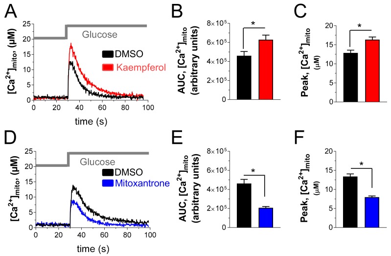
Pancreatic β-cells secrete insulin to lower blood glucose, following a meal. Maintenance of β-cell function is essential to preventing type 2 diabetes. In pancreatic β-cells, mitochondrial matrix calcium is an activating signal for insulin secretion. Recently, the molecular identity of the mitochondrial calcium uniporter (MCU), the transporter that mediates mitochondrial calcium uptake, was revealed. Its role in pancreatic β-cell signal transduction modulation was clarified, opening new perspectives for intervention. Here, we investigated the effects of a mitochondrial Ca2+-targeted nutritional intervention strategy on metabolism/secretion coupling, in a model of pancreatic insulin-secreting cells (INS-1E). Acute treatment of INS-1E cells with the natural plant flavonoid and MCU activator kaempferol, at a low micromolar range, increased mitochondrial calcium rise during glucose stimulation, without affecting the expression level of the MCU and with no cytotoxicity. Enhanced mitochondrial calcium rises potentiated glucose-induced insulin secretion. Conversely, the MCU inhibitor mitoxantrone inhibited mitochondrial Ca2+ uptake and prevented both glucose-induced insulin secretion and kaempferol-potentiated effects. The kaempferol-dependent potentiation of insulin secretion was finally validated in a model of a standardized pancreatic human islet. We conclude that the plant product kaempferol activates metabolism/secretion coupling in insulin-secreting cells by modulating mitochondrial calcium uptake.

Keywords: calcium; insulin; kaempferol; mitochondria; mitoxantrone; polyphenols; β-cell.


Conflict of interest statement

The authors are employees of Nestlé Research, which is part of the Société des Produits Nestlé SA.

Figures

Figure 1
Figure 1
Mitochondrial calcium uniporter (MCU) is expressed in pancreatic INS-1E β-cells and its abundance is not modulated by acute treatment with kaempferol. Detection of MCU by Western blot. (A) Mitochondria were isolated from INS-1E cells, rat brown adipose tissue (BAT) or rat brain and 30 μg of proteins were analyzed. TOM20 was detected as control of mitochondrial fraction. (B) Structure of kaempferol. (C,D) Detection (C) and quantification (D) of the effect of kaempferol on MCU expression, in INS-1E and HeLa cells. Cells were incubated for 30 min with 10 μM kaempferol and then mitochondria were isolated and 30 μg of proteins were analyzed. (C) Representative blot from three independent experiments is shown. (D) The bars of MCU expression indicate optical density of the respective bands of panel C, which are normalized according the corresponding mitochondrial ATP-5A band. They are the mean ± S.E.M of 3 independent experiments (Student’s t-test). NS, not significant.
Figure 2
Figure 2
MCU activator kaempferol and MCU inhibitor mitoxantrone increases and inhibits, respectively, glucose-stimulated mitochondrial Ca2+ rise in INS-1E cells. (A,C) Mitochondrial-targeted mutated aequorin was reconstituted with native coelenterazine in INS-1E cells, as described. Then cells were treated for 30 min with 1% DMSO or 10 μM kaempferol (A), or 50 μM mitoxantrone (D), were placed in the plate reader and stimulated with 16.7 mM glucose, as indicated (Glucose). (B,C,E,F) Statistical evaluation of the effect of kaempferol (B,C) and mitoxantrone (E,F) on the integrated mitochondrial Ca2+ elevation (B,E) and on the amplitude of the mitochondrial Ca2+ signal (C,F), evoked by glucose stimulation. Data are representative (A,D) or the mean ± SEM (B,C,E,F) of 7 independent experiments (Student’s t-test. *, p < 0.05).
Figure 3
Figure 3
Kaempferol potentiates glucose-stimulated insulin secretion, whereas mitoxantrone inhibits glucose-stimulated hormone exocytosis. Static insulin secretion measurements. (A) Insulin release. Control (1% DMSO, black) and kaempferol (10 μM)-treated (red) INS-1E cells were incubated for 30 min in the presence of resting (2.5 mM) and stimulatory (16.7 mM) glucose (Glc) concentrations. Inset, secreted insulin expressed as a percentage of content. Shown is the average of 6 experiments (mean ± SEM; one-way ANOVA test. *, p < 0.05). (B) Effect of 30 min treatment of mitoxantrone (50 μM, blue) and kaempferol (10 μM, red) on glucose-stimulated (16.7 mM glucose) insulin secretion (insulin release), expressed as a fold change of the respective insulin secretion in resting glucose (2.5 mM), set to 1 for each treatment (DMSO; kaempferol; mitoxantrone). Shown is the average of 6 experiments (mean ± SEM; Student’s t-test for the main panel and one-way ANOVA test for the inset. *, p < 0.05). NS, not significant.
Figure 4
Figure 4
Acute kaempferol-dependent mitochondrial Ca2+ rise does not promote cell death. Apoptotic cell death was measured as Annexin-V positive area. (A) Quantification of apoptosis measured at 30 min, 24 h, and 48 h in INS-1E cells treated with 1% DMSO (black), kaempferol at the indicated concentrations (in μM, red), 50 μM mitoxantrone (blue), or 100 nM staurosporine (yellow). Representative results are shown (mean ± SEM of 3 replicates). The experiment was repeated 3 times with comparable results. (B) Representative images of the data quantified in A, at 30 min and at 48 h; 1%, DMSO, 10 µM kaempferol, 50 µM mitoxantrone and 100 nM staurosporine were indicated.
Figure 5
Figure 5
Kaempferol-induced mitochondrial Ca2+ rise and insulin secretion are prevented in mitoxantrone-treated cells. (A) Mitochondrial-targeted mutated aequorin was reconstituted with native coelenterazine in INS-1E cells. Then, the cells were treated for 30 min with 1% DMSO or 10 µM kaempferol in absence (−) or presence (+) of 50 µM mitoxantrone. Finally, the cells were placed in the plate reader and stimulated with 16.7 mM glucose. The bars represent the statistical evaluation of the integrated mitochondrial Ca2+ elevation evoked by glucose stimulation. Data are the mean ± SEM of 10 independent experiments (one-way ANOVA test. *, p < 0.05). (B) Insulin release was quantified as described in Figure 3B in INS-1E cells treated for 30 min with 1% DMSO or 10 μM kaempferol in absence (−) or presence (+) of 50 µM mitoxantrone. Shown is the average of 6 experiments (mean ± SEM; Student’s t-test). (C) Apoptosis measured, as described in Figure 4, at 30 min, in INS-1E cells treated with 1% DMSO (black), or 10 µM kaempferol in absence (−) or presence (+) of 50 μM mitoxantrone. Staurosporine 100 nM is shown as a positive control of cell death (yellow) and it is quantified at 0.5 h and 48 h, as indicated. Representative results are shown (mean ± SEM of 3 replicates. *, p < 0.05). The experiment was repeated 3 times with comparable results.
Figure 6
Figure 6
Kaempferol potentiates glucose-stimulated insulin secretion in human pseudo-islets. (A) Single isolated human pseudo-islets in 96-well plates. (B) Glucose-stimulated insulin secretion of human pseudo-islets exposed to low glucose (2.8 mM, n = 6) or high glucose solution (16.7 mM, n = 12) in combination with either DMSO 0.1% (black) or 10 μM kaempferol (red). Shown is the mean ± SEM. Statistical analysis was performed by ANOVA test (*, p < 0.05).
Figure 7
Figure 7
Proposed mechanism of kaempferol-dependent metabolism/secretion coupling in glucose-stimulated pancreatic β-cells. The glucose-stimulated insulin secretion (A) is potentiated by kaempferol, via mitochondrial calcium rise enhancement (B).

References

    1. Vetere A., Choudhary A., Burns S.M., Wagner B.K. Targeting the pancreatic beta-cell to treat diabetes. Nat. Rev. Drug Discov. 2014;13:278–289. doi: 10.1038/nrd4231. - DOI - PubMed
    1. Wiederkehr A., Szanda G., Akhmedov D., Mataki C., Heizmann C.W., Schoonjans K., Pozzan T., Spat A., Wollheim C.B. Mitochondrial matrix calcium is an activating signal for hormone secretion. Cell Metab. 2011;13:601–611. doi: 10.1016/j.cmet.2011.03.015. - DOI - PubMed
    1. De Marchi U., Thevenet J., Hermant A., Dioum E., Wiederkehr A. Calcium co-regulates oxidative metabolism and ATP synthase-dependent respiration in pancreatic beta cells. J. Biol. Chem. 2014;289:9182–9194. doi: 10.1074/jbc.M113.513184. - DOI - PMC - PubMed
    1. Wiederkehr A., Wollheim C.B. Impact of mitochondrial calcium on the coupling of metabolism to insulin secretion in the pancreatic beta-cell. Cell Calcium. 2008;44:64–76. doi: 10.1016/j.ceca.2007.11.004. - DOI - PubMed
    1. Alam M.R., Groschner L.N., Parichatikanond W., Kuo L., Bondarenko A.I., Rost R., Waldeck-Weiermair M., Malli R., Graier W.F. Mitochondrial Ca2+ uptake 1 (MICU1) and mitochondrial Ca2+ uniporter (MCU) contribute to metabolism-secretion coupling in clonal pancreatic beta-cells. J. Biol. Chem. 2012;287:34445–34454. doi: 10.1074/jbc.M112.392084. - DOI - PMC - PubMed

LinkOut - more resources