Characterization of a Unique Bordetella bronchiseptica vB_BbrP_BB8 Bacteriophage and Its Application as an Antibacterial Agent
- PMID: 32093105
- PMCID: PMC7073063
- DOI: 10.3390/ijms21041403
Characterization of a Unique Bordetella bronchiseptica vB_BbrP_BB8 Bacteriophage and Its Application as an Antibacterial Agent
Abstract
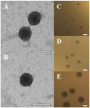
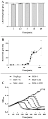
Bordetella bronchiseptica, an emerging zoonotic pathogen, infects a broad range of mammalian hosts. B. bronchiseptica-associated atrophic rhinitis incurs substantial losses to the pig breeding industry. The true burden of human disease caused by B. bronchiseptica is unknown, but it has been postulated that some hypervirulent B. bronchiseptica isolates may be responsible for undiagnosed respiratory infections in humans. B. bronchiseptica was shown to acquire antibiotic resistance genes from other bacterial genera, especially Escherichia coli. Here, we present a new B. bronchiseptica lytic bacteriophage-vB_BbrP_BB8-of the Podoviridae family, which offers a safe alternative to antibiotic treatment of B. bronchiseptica infections. We explored the phage at the level of genome, physiology, morphology, and infection kinetics. Its therapeutic potential was investigated in biofilms and in an in vivo Galleria mellonella model, both of which mimic the natural environment of infection. The BB8 is a unique phage with a genome structure resembling that of T7-like phages. Its latent period is 75 ± 5 min and its burst size is 88 ± 10 phages. The BB8 infection causes complete lysis of B. bronchiseptica cultures irrespective of the MOI used. The phage efficiently removes bacterial biofilm and prevents the lethality induced by B. bronchiseptica in G. mellonella honeycomb moth larvae.
Keywords: Galleria mellonella; animal model; antibiotic resistance; atrophic rhinitis; biofilm; emerging diseases; phage stability; phage therapy; veterinary microbiology; zoonosis.
Conflict of interest statement
The authors declare no conflict of interest. The funders had no role in the design of the study; in the collection, analyses, or interpretation of data; in the writing of the manuscript, or in the decision to publish the results.
Figures


Similar articles
-
The First Siphoviridae Family Bacteriophages Infecting Bordetella bronchiseptica Isolated from Environment.Microb Ecol. 2017 Feb;73(2):368-377. doi: 10.1007/s00248-016-0847-0. Epub 2016 Sep 15. Microb Ecol. 2017. PMID: 27628741
-
Specific Integration of Temperate Phage Decreases the Pathogenicity of Host Bacteria.Front Cell Infect Microbiol. 2020 Feb 4;10:14. doi: 10.3389/fcimb.2020.00014. eCollection 2020. Front Cell Infect Microbiol. 2020. PMID: 32117795 Free PMC article.
-
Specific bacteriophage of Bordetella bronchiseptica regulates B. bronchiseptica-induced microRNA expression profiles to decrease inflammation in swine nasal turbinate cells.Genes Genomics. 2020 Apr;42(4):441-447. doi: 10.1007/s13258-019-00906-7. Epub 2020 Feb 7. Genes Genomics. 2020. PMID: 32034667 Free PMC article.
-
Bordetella bronchiseptica infection of rats and mice.Comp Med. 2003 Feb;53(1):11-20. Comp Med. 2003. PMID: 12625502 Review.
-
Environmental sensing mechanisms in Bordetella.Adv Microb Physiol. 2001;44:141-81. doi: 10.1016/s0065-2911(01)44013-6. Adv Microb Physiol. 2001. PMID: 11407112 Review.
Cited by
-
Antibiotics Act with vB_AbaP_AGC01 Phage against Acinetobacter baumannii in Human Heat-Inactivated Plasma Blood and Galleria mellonella Models.Int J Mol Sci. 2020 Jun 19;21(12):4390. doi: 10.3390/ijms21124390. Int J Mol Sci. 2020. PMID: 32575645 Free PMC article.
-
S2B, a Temperate Bacteriophage That Infects Caulobacter Crescentus Strain CB15.Curr Microbiol. 2022 Feb 12;79(4):98. doi: 10.1007/s00284-022-02799-4. Curr Microbiol. 2022. PMID: 35150327 Free PMC article.
-
Long-Term Effectiveness of Engineered T7 Phages Armed with Silver Nanoparticles Against Escherichia coli Biofilm.Int J Nanomedicine. 2024 Oct 4;19:10097-10105. doi: 10.2147/IJN.S479960. eCollection 2024. Int J Nanomedicine. 2024. PMID: 39381027 Free PMC article.
-
Novel findings in context of molecular diversity and abundance of bacteriophages in wastewater environments of Riyadh, Saudi Arabia.PLoS One. 2022 Aug 18;17(8):e0273343. doi: 10.1371/journal.pone.0273343. eCollection 2022. PLoS One. 2022. PMID: 35980993 Free PMC article.
-
Anti-Bordetella bronchiseptica effects of targeted bacteriophages via microbiome and metabolic mediated mechanisms.Sci Rep. 2023 Dec 8;13(1):21755. doi: 10.1038/s41598-023-49248-1. Sci Rep. 2023. PMID: 38066337 Free PMC article.
References
-
- Datz C. Bordetella Infections in Dogs and Cats: Pathogenesis, Clinical Signs, and Diagnosis. Compend. Contin. Educ. Pract. Vet. N. Am. Ed. 2003;25:896–901.
MeSH terms
Grants and funding
LinkOut - more resources
Full Text Sources

