. 2020 Feb 19;9(2):171.
doi: 10.3390/antiox9020171.

NADPH Oxidase 2 Mediates Myocardial Oxygen Wasting in Obesity

Affiliations

NADPH Oxidase 2 Mediates Myocardial Oxygen Wasting in Obesity

Anne D Hafstad et al. Antioxidants (Basel). .

Abstract

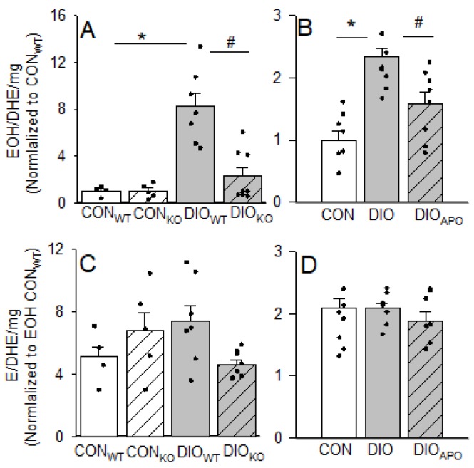
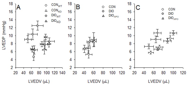
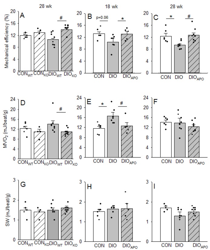
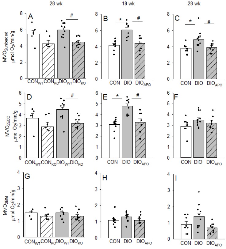
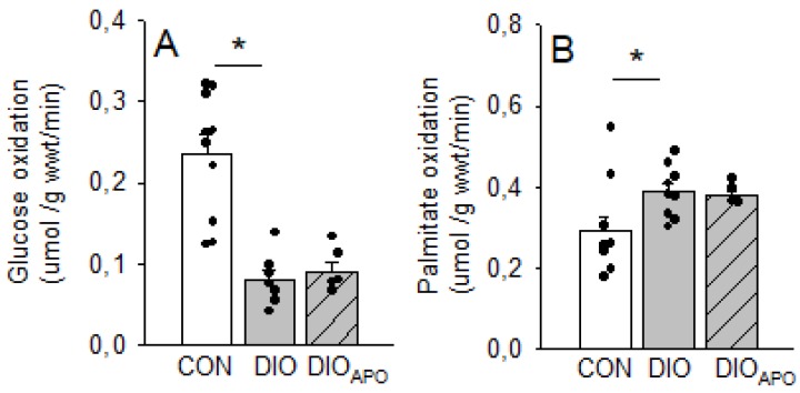
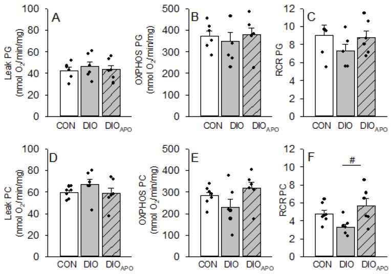
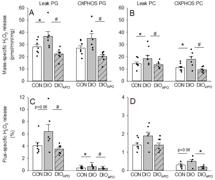
Obesity and diabetes are independent risk factors for cardiovascular diseases, and they are associated with the development of a specific cardiomyopathy with elevated myocardial oxygen consumption (MVO2) and impaired cardiac efficiency. Although the pathophysiology of this cardiomyopathy is multifactorial and complex, reactive oxygen species (ROS) may play an important role. One of the major ROS-generating enzymes in the cardiomyocytes is nicotinamide adenine dinucleotide phosphate (NADPH) oxidase 2 (NOX2), and many potential systemic activators of NOX2 are elevated in obesity and diabetes. We hypothesized that NOX2 activity would influence cardiac energetics and/or the progression of ventricular dysfunction following obesity. Myocardial ROS content and mechanoenergetics were measured in the hearts from diet-induced-obese wild type (DIOWT) and global NOK2 knock-out mice (DIOKO) and in diet-induced obese C57BL/6J mice given normal water (DIO) or water supplemented with the NOX2-inhibitor apocynin (DIOAPO). Mitochondrial function and ROS production were also assessed in DIO and DIOAPO mice. This study demonstrated that ablation and pharmacological inhibition of NOX2 both improved mechanical efficiency and reduced MVO2 for non-mechanical cardiac work. Mitochondrial ROS production was also reduced following NOX2 inhibition, while cardiac mitochondrial function was not markedly altered by apocynin-treatment. Therefore, these results indicate a link between obesity-induced myocardial oxygen wasting, NOX2 activation, and mitochondrial ROS.

Keywords: NADPH-oxidase; ROS; cardiac efficiency; metabolism; myocardial oxygen consumption; obesity.


Conflict of interest statement

“The authors declare no conflict of interest.”

Figures

Figure 1
Figure 1
Reactive oxygen species-products hydroxyethidium (EOH) and ethidium (E) per dihydroethidium (DHE) consumed in cardiac tissue from lean controls (CONWT and CONKO) and obese (DIOWT and DIOKO) wild type and NOX2 KO mice fed a high fat diet for 28 weeks (A,C). Also shown in lean (CON), obese (DIO), and apocynin-treated obese (DIOAPO) C57BL/6J mice fed a western diet for 28 weeks (B,D) n = 4–8 in each group. Values are normalized to EOH in lean controls. Single values and means ± SEM. * p < 0.05 CON vs. DIO within same genotype, # p < 0.05 DIOWT vs. DIOKO and DIO vs. DIOAPO.
Figure 2
Figure 2
Steady state left ventricular end-diastolic volumes (LVEDV) and pressures (LVEDP) at three different workloads (preload: 4,6 and 8 mmHg and afterload: 50mmHg) from lean controls (CONWT and CONKO) and obese (DIOWT and DIOKO) wild type and NOX2 KO mice fed a high fat diet for 28 weeks (A), as well as lean (CON), obese (DIO) and apocynin-treated obese (DIOAPO) C57BL/6J mice fed a western diet for 18 (B) and 28 weeks (C). n = 5–10 per group, the values are mean ± SEM.
Figure 3
Figure 3
Left ventricular mechanical efficiency (AC) expressed as stroke-work (SW, GI) relative to myocardial oxygen consumption (DF) in hearts from lean controls (CONWT and CONKO) and diet-induced obese (DIO) wild type and NOX2 KO mice (DIOWT and DIOKO) fed a high fat diet for 28 weeks (A,D,G) as well as lean controls (CON) and untreated and apocynin-treated obese C57BL/6J mice (DIO and DIOAPO) fed a western diet for 18 weeks (B,E,H) or 28 weeks (C,F,I). Single values and means ± SEM. * p < 0.05 CON vs. DIO within same genotype, # p < 0.05 DIOWT vs. DIOKO and DIO vs. DIOAPO.
Figure 4
Figure 4
Myocardial oxygen consumption in mechanically unloaded hearts (MVO2unloaded, AC) and for processes associated with excitation-contraction coupling (MVO2ECC, DF) and basal metabolism (MVO2BM, GI) that was obtained from lean controls wild-type (CONWT) and NOX2-KO mice (CONKO) and obese mice fed a high fat diet for 28 weeks (DIOWT and DIOKO). (A,D,G). Also shown in lean controls (CON) and untreated and apocynin-treated obese C57BL/6J mice (DIO and DIOAPO) that were fed a western diet for 18 weeks (B,E,H) or 28 weeks (C,F,I). Single values and means ± SEM. * p < 0.05 within the same genotype, # p < 0.05 between genotypes within the same diet.
Figure 5
Figure 5
Glucose (A) and palmitate (B) oxidation rates measured in isolated working hearts from lean controls (CON), diet-induced obese (DIO), and obese apocynin-treated (DIOAPO) C57BL/6J mice fed an obesogenic western diet for 18 weeks. Single values and mean ± SEM, * p < 0.05 vs. DIO.
Figure 6
Figure 6
Oxygen fluxes (leak and maximal mitochondrial respiration capacity, OXPHOS) and respiratory coupling ratio (RCR) in isolated cardiac mitochondria using pyruvate and glutamate (PG, AC) or palmityol-carnitine as substrates (PC, DF). Mitochondria were obtained from lean controls (CON), untreated and apocynin-treated obese C57BL/6J mice (DIO and DIOAPO) fed a western diet for 28 weeks. Single values and mean ± SEM, # p < 0.05 DIO vs. DIOAPO.
Figure 7
Figure 7
Mass-specific and flux-specific H2O2- release from isolated cardiac mitochondria using either pyruvate and glutamate (PG, panel A and C) or palmityol-carnitine as substrates (PC, panel B and D) as substrates. Mitochondria were obtained from lean controls (CON) and untreated and apocynin-treated obese C57BL/6J mice (DIO and DIOAPO) fed a western diet for 28 weeks. Single values and means ± SEM, * p < 0.05 CON versus DIO, # p < 0.05 DIO vs. DIOAPO.

References

    1. Kannel W.B., Hjortland M., Castelli W.P. Role of diabetes in congestive heart failure: The Framingham study. Am. J. Cardiol. 1974;34:29–34. doi: 10.1016/0002-9149(74)90089-7. - DOI - PubMed
    1. Fein F.S., Sonnenblick E.H. Diabetic cardiomyopathy. Cardiovasc. Drugs Ther. 1994;8:65–73. doi: 10.1007/BF00877091. - DOI - PubMed
    1. Anderson E.J., Kypson A.P., Rodriguez E., Anderson C.A., Lehr E.J., Neufer P.D. Substrate-specific derangements in mitochondrial metabolism and redox balance in the atrium of the type 2 diabetic human heart. J. Am. Coll. Cardiol. 2009;54:1891–1898. doi: 10.1016/j.jacc.2009.07.031. - DOI - PMC - PubMed
    1. Giacco F., Brownlee M. Oxidative stress and diabetic complications. Circ. Res. 2010;107:1058–1070. doi: 10.1161/CIRCRESAHA.110.223545. - DOI - PMC - PubMed
    1. Anilkumar N., Weber R., Zhang M., Brewer A., Shah A.M. Nox4 and nox2 NADPH oxidases mediate distinct cellular redox signaling responses to agonist stimulation. Arterioscler. Thromb. Vasc. Biol. 2008;28:1347–1354. doi: 10.1161/ATVBAHA.108.164277. - DOI - PubMed