. 2020 Feb 20;20(4):1157.
doi: 10.3390/s20041157.

Accuracy of CAD/CAM Digital Impressions with Different Intraoral Scanner Parameters

Affiliations

Accuracy of CAD/CAM Digital Impressions with Different Intraoral Scanner Parameters

Asher Chiu et al. Sensors (Basel). .

Abstract

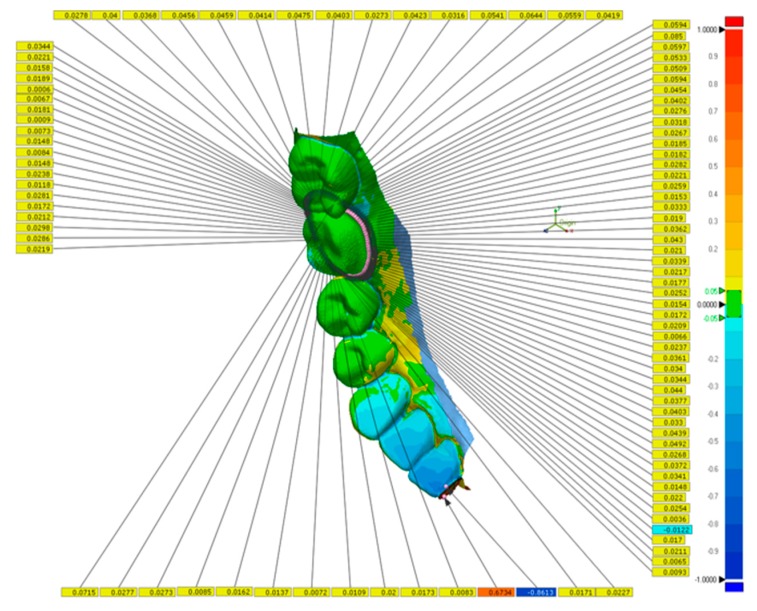
The advancement of intraoral scanners has allowed for more efficient workflow in the dental clinical setting. However, limited data exist regarding the accuracy of the digital impressions produced with various scanner settings and scanning approaches. The purpose of this in vitro study was to compare the accuracy of digital impressions at the crown preparation margin using different scanning resolutions of a specific intraoral scanner system. An all-ceramic crown preparation of a mandibular first molar was constructed in a typodont, and a scan (n = 3) was created with an industrial-grade laboratory scanner (3Shape D2000) as the control. Digital impressions were obtained with an intraoral scanner (3Shape TRIOS 3) under three settings-high resolution (HR), standard resolution (SR), and combined resolution (SHR). Comparative 3D analysis of scans was performed with Geomagic Control X software to measure the discrepancy between intraoral scans and the control scan along the preparation finish line. The scan time and number of images captured per scan were recorded. Statistical analysis was performed by one-way ANOVA, two-way repeated measures ANOVA, Pearson's correlation, and Dunnett's T3 test (α = 0.05). Significant differences were observed for scan time and for number of images captured among scan resolution settings (α < 0.05). The scan time for the SR group was, on average, 34.2 s less than the SHR group and 46.5 s less than the HR group. For discrepancy on the finish line, no significant differences were observed among scanning resolutions (HR: 31.5 ± 5.5 μm, SHR: 33.2 ± 3.7 μm, SR: 33.6 ± 3.1 μm). Significant differences in discrepancy were observed among tooth surfaces, with the distal surface showing the highest discrepancies. In conclusion, the resolution of the intra-oral scanner is primarily defined by the system hardware and optimized for default scans. A software high-resolution mode that obtains more data over a longer time may not necessarily benefit the scan accuracy, while the tooth preparation and surface parameters do affect the accuracy.

Keywords: CAD/CAM; accuracy; digital impression; high resolution; intraoral scanner.


Conflict of interest statement

The authors declare no conflict of interest with regard to the authorship of this manuscript.

Figures

Figure 1
Figure 1
Depiction of the digital interface using the 3Shape TRIOS 3 scanner and software, highlighting the High Resolution (High res) feature.
Figure 2
Figure 2
Superimposition of a master scan obtained from the 3Shape D2000 desktop scanner and intraoral scans from the 3Shape TRIOS 3 scanner using the Geomagic Control X software. The images were aligned using both the Initial Alignment and Best-Fit Alignment functions. The colors depict overlays of multiple scans.
Figure 3
Figure 3
Deviation between master scan and intraoral scan measured at 100 evenly spaced points along the preparation finish line. The specific values of deviation along the finish line are given in yellow legends. The range of deviation across the entire half-arch scan is color graded from −1 mm (blue) to +1 mm (red).
Figure 4
Figure 4
Comparison of total discrepancy, scan time, and number of images captured per scan between scan resolutions: standard resolution (SR), standard resolution with high resolution around the preparation margin (SHR), and high resolution (HR). Statistical analysis was performed using one-way analysis of variance followed by multiple comparisons using Dunnett’s T3 test. Pearson correlations were used to correlate total discrepancy with scan time and number of images captured. Horizontal bars show significant differences (p < 0.05). (A): total discrepancy by scan resolution: no statistically significant difference was observed in the overall comparison (p > 0.05). (B): no correlation was observed between total discrepancy and scan time. (C): a significant difference was observed between scan resolutions in regard to scan time, with the HR group having the longest mean scan time. (D): no correlation was observed between total discrepancy and numbers of images captured per scan. (E): a significant difference was observed between scan resolutions in regard to numbers of images captured per scan, with the HR group showing the highest number. (F): a positive correlation was seen between scan time and number of images captured per scan; the longer the scan time, the more images captured per scan.
Figure 5
Figure 5
Mean discrepancy (μm) along the preparation finish line by tooth surface. Horizontal bars indicate significant differences (p < 0.05). Shared lowercase letters indicate no significant difference in discrepancy between tooth surfaces within groups. Distal surfaces showed significantly higher discrepancies when compared to other tooth surfaces across all three groups.

References

    1. Miyazaki T., Hotta Y., Kunii J., Kuriyama S., Tamaki Y. A review of dental CAD/CAM: Current status and future perspectives from 20 years of experience. Dent. Mater. J. 2009;28:44–56. doi: 10.4012/dmj.28.44. - DOI - PubMed
    1. Persson A., Odén A., Andersson M., Englund G.S. Digitization of simulated clinical dental impressions: Virtual three-dimensional analysis of exactness. Dent. Mater. 2009;25:929–936. doi: 10.1016/j.dental.2009.01.100. - DOI - PubMed
    1. Moörmann W.H., Mörmann W.H. The evolution of the CEREC system. J. Am. Dent. Assoc. 2006;137:7S–13S. doi: 10.14219/jada.archive.2006.0398. - DOI - PubMed
    1. Renne W., Ludlow M., Fryml J., Schurch Z., Mennito A., Kessler R., Lauer A. Evaluation of the accuracy of 7 digital scanners: An in vitro analysis based on 3-dimensional comparisons. J. Prosthet. Dent. 2017;118:36–42. doi: 10.1016/j.prosdent.2016.09.024. - DOI - PubMed
    1. Nedelcu R., Olsson P., Nyström I., Ryden J., Thor A. Accuracy and precision of 3 intraoral scanners and accuracy of conventional impressions: A novel in vivo analysis method. J. Dent. 2018;69:110–118. doi: 10.1016/j.jdent.2017.12.006. - DOI - PubMed

MeSH terms