. 2020 Feb 20;21(4):1433.
doi: 10.3390/ijms21041433.

Efficient Prodrug Activator Gene Therapy by Retroviral Replicating Vectors Prolongs Survival in an Immune-Competent Intracerebral Glioma Model

Affiliations

Efficient Prodrug Activator Gene Therapy by Retroviral Replicating Vectors Prolongs Survival in an Immune-Competent Intracerebral Glioma Model

Shih-Han Chen et al. Int J Mol Sci. .

Abstract

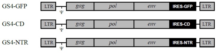
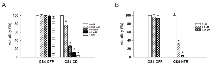
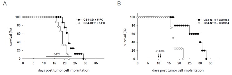
Prodrug activator gene therapy mediated by murine leukemia virus (MLV)-based retroviral replicating vectors (RRV) was previously shown to be highly effective in killing glioma cells both in culture and in vivo. To avoid receptor interference and enable dual vector co-infection with MLV-RRV, we have developed another RRV based on gibbon ape leukemia virus (GALV) that also shows robust replicative spread in a wide variety of tumor cells. We evaluated the potential of GALV-based RRV as a cancer therapeutic agent by incorporating yeast cytosine deaminase (CD) and E. coli nitroreductase (NTR) prodrug activator genes into the vector. The expression of CD and NTR genes from GALV-RRV achieved highly efficient delivery of these prodrug activator genes to RG-2 glioma cells, resulting in enhanced cytotoxicity after administering their respective prodrugs 5-fluorocytosine and CB1954 in vitro. In an immune-competent intracerebral RG-2 glioma model, GALV-mediated CD and NTR gene therapy both significantly suppressed tumor growth with CB1954 administration after a single injection of vector supernatant. However, NTR showed greater potency than CD, with control animals receiving GALV-NTR vector alone (i.e., without CB1954 prodrug) showing extensive tumor growth with a median survival time of 17.5 days, while animals receiving GALV-NTR and CB1954 showed significantly prolonged survival with a median survival time of 30 days. In conclusion, GALV-RRV enabled high-efficiency gene transfer and persistent expression of NTR, resulting in efficient cell killing, suppression of tumor growth, and prolonged survival upon CB1954 administration. This validates the use of therapeutic strategies employing this prodrug activator gene to arm GALV-RRV, and opens the door to the possibility of future combination gene therapy with CD-armed MLV-RRV, as the latter vector is currently being evaluated in clinical trials.

Keywords: E. coli nitroreductase gene; brain tumor; gene therapy; prodrug activator; retroviral replicating vector.


Conflict of interest statement

N.K. has consulted for Tocagen Inc. All other authors declare no conflict of interest.

Figures

Figure 1
Figure 1
Structure of gibbon ape leukemia virus (GALV)-retroviral replicating vectors (RRV) carrying transgenes. Each GALV-RRV contains an internal ribosome entry site (IRES)-green fluorescent protein (GFP), IRES-cytosine deaminase (CD) or IRES-E. coli nitroreductase (NTR) gene expression cassette inserted between GALV env and the 3′ untranslated region (UTR). Ψ, packaging signal. LTR, long terminal repeat.
Figure 2
Figure 2
Replicative spread of GS4-GFP in glioma cells in culture and in intracerebral gliomas in vivo. (A) GS4-GFP-transduced RG-2 cells (5%) and uninfected RG-2 cells (95%) were mixed and seeded onto culture plates. At various time points after cell mixing, the cell populations were analyzed for GFP expression. X-axis: days after cell mixture. Y-axis: % of cells expressing GFP. (B) GS4-GFP (2 × 10⁴ TU) was injected into a pre-established intracerebral RG-2 tumor model in Fischer 344 rats. The spread of GS4-GFP in tumors was examined by quantification of GFP expression at 13 (n = 6) and 19 days (n = 3) after viral vector inoculation.
Figure 3
Figure 3
In vitro cytotoxicity achieved by GALV-based RRV plus prodrug treatment. (A) GS4-CD- and GS4-GFP-transduced RG-2 cells were exposed to 5-FC ranging from 0 to 1 mM, and cell viability was determined 3 days later by MTS assay. (B) GS4-NTR- and GS4-GFP-transduced RG-2 cells were exposed to CB1954 ranging from 0 to 0.25 μM, and cell viability was determined 3 days later by MTS assay. *, p < 0.005.
Figure 4
Figure 4
Survival analysis of immune-competent rats bearing intracerebral RG-2 gliomas. (A) GS4-CD or GS4-GFP was stereotactically injected into pre-established intracerebral RG-2 tumors three days after tumor inoculation. Eight days after viral vector inoculation, the rats received intraperitoneal injections of 5-FC (100 mg/kg), once every other day, for a total of 7 treatments. Survival curves were constructed for two treatment groups: GS4-CD plus 5-FC, and GS4-GFP plus 5-FC. (B) GS4-NTR was stereotactically injected into pre-established intracerebral RG-2 tumors three days after tumor inoculation. Seven and eight days after viral vector inoculation, the rats received daily intraperitoneal injections of CB1954 (2.5 mg/kg) or PBS. Survival curves were constructed for two treatment groups GS4-NTR plus CB1954 and GS4-NTR plus PBS.
Figure 5
Figure 5
MicroPET imaging of intracerebral RG-2 glioma-bearing rats. MicroPET imaging of L-[18F] FET in rats was performed using the R4 system, 20 days after intracerebral RG-2 glioma implantation. Representative examples comparing microPET imaging results from non-tumor-bearing rats (A), tumor-bearing rats treated with GS4-NTR but without prodrug administration (B), or tumor-bearing rats with GS4-NTR and CB1954 prodrug treatments (C) are shown, and tumor regions are indicated by arrowheads. The tumor uptake of L-[18F] FET in the GS4-NTR−CB1954 group relative to the GS4-NTR + CB1954 group was 2.015-fold (p < 0.01).

References

    1. Ostrom Q.T., Bauchet L., Davis F.G., Deltour I., Fisher J.L., Langer C.E., Pekmezci M., Schwartzbaum J.A., Turner M.C., Walsh K.M., et al. The epidemiology of glioma in adults: a “state of the science” review. Neuro-oncology. 2014;16:896–913. doi: 10.1093/neuonc/nou087. - DOI - PMC - PubMed
    1. Rainov N.G. A phase III clinical evaluation of herpes simplex virus type 1 thymidine kinase and ganciclovir gene therapy as an adjuvant to surgical resection and radiation in adults with previously untreated glioblastoma multiforme. Hum. Gene Ther. 2000;11:2389–2401. doi: 10.1089/104303400750038499. - DOI - PubMed
    1. Galanis E., Vile R., Russell S.J. Delivery systems intended for in vivo gene therapy of cancer: targeting and replication competent viral vectors. Crit. Rev. Oncol. Hematol. 2001;38:177–192. doi: 10.1016/S1040-8428(01)00103-2. - DOI - PubMed
    1. Kirn D., Martuza R.L., Zwiebel J. Replication-selective virotherapy for cancer: Biological principles, risk management and future directions. Nat. Med. 2001;7:781–787. doi: 10.1038/89901. - DOI - PubMed
    1. Alemany R., Balague C., Curiel D.T. Replicative adenoviruses for cancer therapy. Nat. Biotechnol. 2000;18:723–727. doi: 10.1038/77283. - DOI - PubMed

MeSH terms