. 2020 Feb 24;18(1):18.
doi: 10.1186/s12915-020-0750-5.

Unexpected cell type-dependent effects of autophagy on polyglutamine aggregation revealed by natural genetic variation in C. elegans

Affiliations

Unexpected cell type-dependent effects of autophagy on polyglutamine aggregation revealed by natural genetic variation in C. elegans

J Alexander-Floyd et al. BMC Biol. .

Abstract

Background: Monogenic protein aggregation diseases, in addition to cell selectivity, exhibit clinical variation in the age of onset and progression, driven in part by inter-individual genetic variation. While natural genetic variants may pinpoint plastic networks amenable to intervention, the mechanisms by which they impact individual susceptibility to proteotoxicity are still largely unknown.

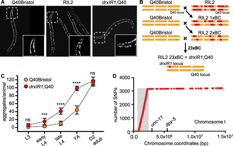
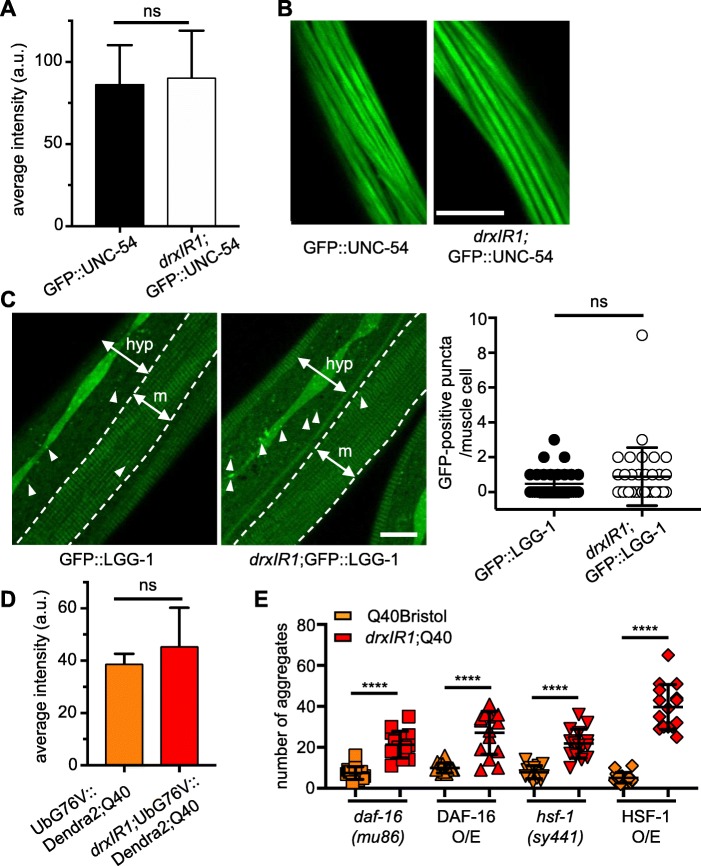
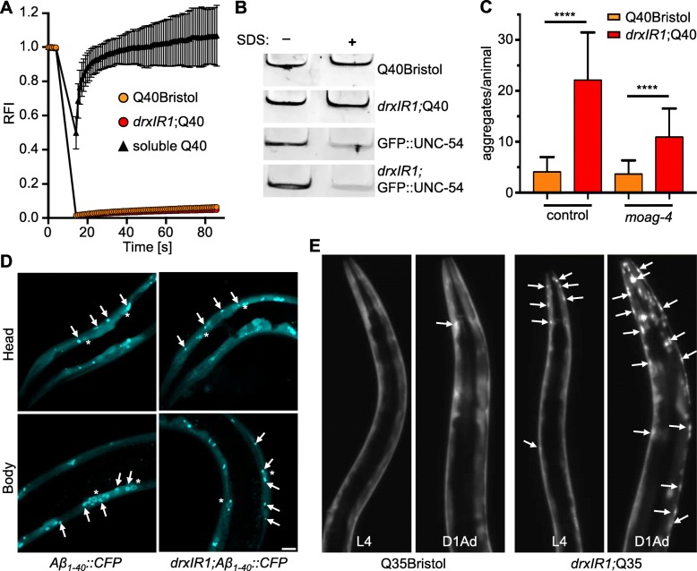
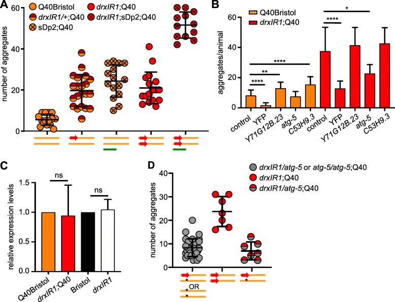
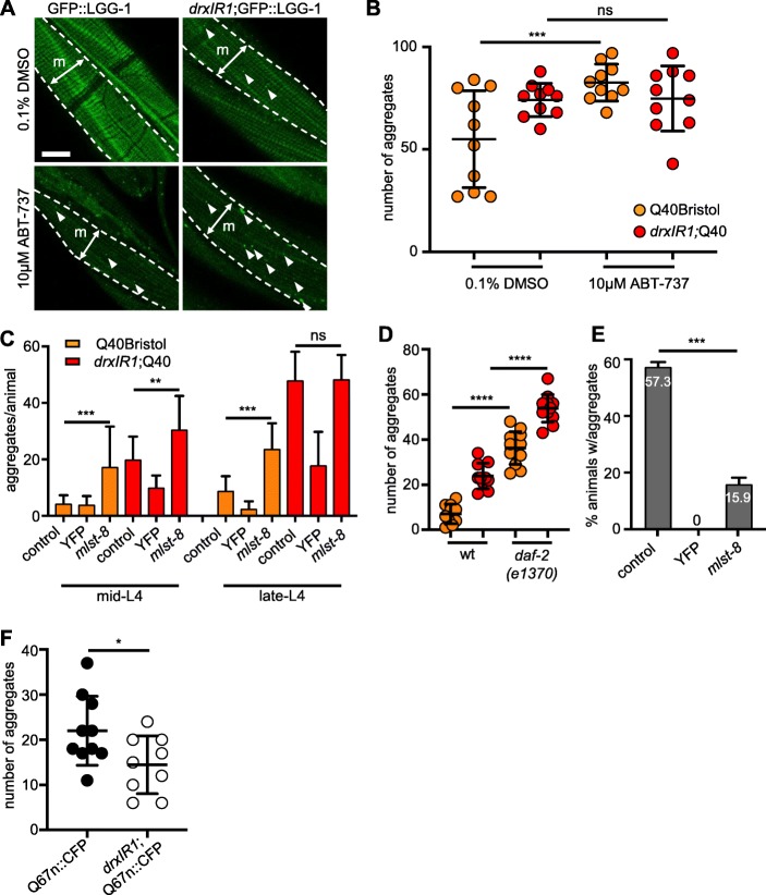
Results: We have previously shown that natural variation modifies polyglutamine (polyQ) aggregation phenotypes in C. elegans muscle cells. Here, we find that a genomic locus from C. elegans wild isolate DR1350 causes two genetically separable aggregation phenotypes, without changing the basal activity of muscle proteostasis pathways known to affect polyQ aggregation. We find that the increased aggregation phenotype was due to regulatory variants in the gene encoding a conserved autophagy protein ATG-5. The atg-5 gene itself conferred dosage-dependent enhancement of aggregation, with the DR1350-derived allele behaving as hypermorph. Surprisingly, increased aggregation in animals carrying the modifier locus was accompanied by enhanced autophagy activation in response to activating treatment. Because autophagy is expected to clear, not increase, protein aggregates, we activated autophagy in three different polyQ models and found a striking tissue-dependent effect: activation of autophagy decreased polyQ aggregation in neurons and intestine, but increased it in the muscle cells.

Conclusions: Our data show that cryptic natural variants in genes encoding proteostasis components, although not causing detectable phenotypes in wild-type individuals, can have profound effects on aggregation-prone proteins. Clinical applications of autophagy activators for aggregation diseases may need to consider the unexpected divergent effects of autophagy in different cell types.

Keywords: Autophagy; Cryptic variation; Natural genetic variation; Polyglutamine; Protein aggregation; Proteostasis; Regulatory variation.


Conflict of interest statement

The authors declare that they have no competing interests.

Figures

Fig. 1.
Fig. 1.
drxIR1 locus causes increased polyQ40 aggregation. a Late-L4 RIL2 and drxlR1;Q40 animals have increased aggregation compared to Q40Bristol animals. Insets show polyQ40 aggregation in the head muscles. b The scheme for generation of the drxIR1;Q40 strain through rounds of serial backcrossing/selection. RIL2 strain was backcrossed (BC) into the Q40Bristol strain 23 times. DR1350-derived variants (red) that are retained through the crossing-selection scheme likely contribute to the RIL2 polyQ phenotype. c The drxIR1;Q40 animals exhibit a faster accumulation of polyQ aggregates compared to Q40Bristol at all development stages, until both strains reach maximum at day 2 of adulthood. L3, L4, YA, and D2 adult indicate third and fourth larval stage, young adult, and day 2 adult stage, respectively. Data are mean ± SD, 10 to 20 animals per data point. Data were analyzed by ANOVA followed by Bonferroni’s multiple comparisons test, ****P < 0.0001, ***P = 0.0004. Orange: Q40Bristol background, red: drxIR1;Q40. Same color scheme is used in all figures. d Distribution of DR1350-derived SNPs and any de novo mutations on chromosome I that distinguish drxIR1;Q40 from Q40Bristol and Hawaiian strains. Gray-shaded area to the left of unc-11 shows a locus with over 3000 unique SNPs in drxIR1;Q40 strain
Fig. 2.
Fig. 2.
Basal protein homeostasis of muscle cells is unaffected in animals carrying the drxIR1 interval. a Expression of GFP::UNC-54 fusion protein from unc-54 promoter is similar between the Bristol and drxIR1 L4 animals. Data are mean ± SD of GFP fluorescence intensity, 16–20 muscle cells per genotype, unpaired t test, two-tailed. b Myofilament assembly is normal in drxIR1 animals. Confocal images of muscle cells. Scale bar, 10 μm. c Muscle cells have very few GFP::LGG-1-positive puncta (arrowheads) in both Bristol and drxIR1 L4 animals. One muscle quadrant is shown between punctate lines. m, muscle; hyp, hypodermis. An increased number of GFP::LGG-1-positive puncta is seen in the hypodermis of drxIR1. Scale bar is 10 μm. Right panel, quantification of GFP::LGG-1 puncta in the muscle cells. Data are mean ± SD, 30 to 40 cells (8 to 10 animals) per genotype, unpaired t test, two-tailed; each symbol represents individual cell. d No difference in the average intensity of the proteasome reporter fluorescence in Q40Bristol and drxIR1;Q40 animals. Data are mean ± SD, 4–5 animals, unpaired t test, two-tailed. e The increased aggregation phenotype in animals carrying the drxIR1 interval does not depend on DAF-16 or HSF-1. Each symbol represents an individual animal, 15 mid-L4 animals per genotype. O/E, overexpression. Means ± SD are overlaid. Data were analyzed by ANOVA followed by Bonferroni’s multiple comparisons test, ****P < 0.0001
Fig. 3.
Fig. 3.
Variants in drxIR1 interval do not alter the biophysical properties of polyQ aggregates. a FRAP analysis. The soluble Q40::YFP protein recovered rapidly (triangles), while aggregated protein (circles) in both Q40Bristol and drxIR1;Q40 backgrounds does not recover. Data are mean ± SD. b PolyQ40 aggregates in native extract from drxIR1;Q40 animals remain resistant to 5% SDS. Aggregated proteins fail to enter the native gel, remaining in the wells (shown). Native extracts containing the fibrillar GFP::UNC-54 protein were used as controls. c The increased aggregation phenotype in animals carrying the drxlR1 interval does not depend on the amyloid-specific modifier moag-4 (mid-L4 animals; YA animals are shown in Suppl. Fig. 3B). Data are mean ± SD, three independent experiments. Thirty-eight to 46 animals per condition. Data were analyzed by ANOVA followed by Bonferroni’s multiple comparisons test, ****P < 0.0001. d Aggregation of a different amyloid protein, Aβ1-40::CFP, in unaffected by the drxlR1 locus. Shown are confocal stacks, arrows point to aggregates, and asterisks indicate Aβ1-40::CFP accumulating in the nuclei of the muscle cells. Scale bar, 10 μm. e The shorter polyQ expansion (Q35::YFP) exhibits both the increased susceptibility of the head muscle cells and the accelerated overall aggregation in animals carrying the drxlR1 interval. Shown are stereo micrographs; arrows point to some of the aggregates. D1Ad, day 1 adults
Fig. 4.
Fig. 4.
Hypermorphic variants in the autophagy gene atg-5 are responsible for the increased polyQ aggregation in the body-wall muscles. a PolyQ aggregation in the body-wall muscles is sensitive to the dosage of the drxlR1 interval, with DR1350-derived interval acting as a hypermorph relative to the Bristol-derived interval. Each symbol represents an individual mid-L4 animal; overlaid are means ± SD. Schematic under the graph represents the genetic composition of chromosome I: Bristol background (orange bar), DR1350-derived drxlR1 interval (red arrow), and the free duplication sDp2 (green bar). b RNAi of three candidate genes affects polyQ40 aggregation. atg-5 RNAi suppresses the increased polyQ aggregation in the muscle cells of drxlR1 but not in Q40Bristol animals. RNAi against YFP downregulates expression of Q40::YFP protein. Data are mean ± SD, 3 independent experiments, 9 to 15 animals per experiment per genotype. Data were analyzed by ANOVA followed by Bonferroni’s multiple comparisons test, ****P < 0.0001, **P = 0.0029, *P = 0.0125. c Relative expression of atg-5 mRNA is unaffected by the DR1350-derived drxIR1 interval. Three independent experiments, statistics as in b. d atg-5(bp484) loss-of-function allele reverses increased aggregation caused by one copy of the DR1350-derived drxIR1 interval. Schematic under the graph as in a, star: atg-5 mutation. Animals were scored at mid-L4 as in a, compare drxIR1/+;Q40 animals (red/orange symbols) in a with drxIR1/atg-5;Q40 animals (red/gray symbols) in d. Gray symbols represent animals that were assumed (but not confirmed) to be heterozygous for the drxIR1 interval, because they did not show the RIL2-like phenotype head muscle phenotype and because atg-5/atg-5 animals exhibit strong developmental delay. Heterozygosity of drxIR1/atg-5;Q40 animals (red/gray symbols) was confirmed by singling them out and scoring segregation of the RIL2-like phenotype among their progeny. Each symbol represents an individual animals, overlaid are means ± SD
Fig. 5.
Fig. 5.
Activation of autophagy has divergent effects on polyQ40 aggregate clearance in different tissues. a Animals carrying the drxIR1 interval accumulate more GFP::LGG-1-positive puncta (arrowheads) in the body-wall muscle cells upon treatment with autophagy-activating drug ABT-737. Animals were treated with 0.1% DMSO (vehicle control) or 10 μM ABT-737 for 24 h. Shown are confocal projections; one muscle quadrant (m) is indicated between punctate lines. Scale bar, 10 μm. b Autophagy-activating drug ABT-737 increases polyQ40 aggregation in the body-wall muscle cells in the wild-type background (Q40Bristol). Aggregation was scored in adult animals, 1 day post-L4 (see the “Methods” section). Aggregation in the drxIR1;Q40 animals is already at maximum under these conditions. Each symbol indicates an individual animal; overlaid are means ± SD. Data were analyzed by ANOVA followed by Bonferroni’s multiple comparisons test, ***P = 0.0006. c Activation of autophagy with mlst-8 RNAi increases aggregation in the body-wall muscles of Q40Bristol mid- or late-L4 animals, and of drxIR1;Q40 mid-L4 animals. Data are mean ± SD, 3 independent experiments, 9 to 13 animals per experiment per treatment. Control RNAi was mec-4. Data were analyzed by ANOVA followed by Bonferroni’s multiple comparisons test, ***P = 0.0007, **P = 0.0082. d Introduction of the daf-2(e1370) allele increases polyQ40 aggregation in the body-wall muscles in both Q40Bristol and drxIR1;Q40 animals. Aggregation was scored at mid-L4. Each symbol indicates an individual animal; overlaid are means ± SD. Colors as in b. Data were analyzed by ANOVA with Bonferroni’s multiple comparisons test, ****P < 0.0001. e Activation of autophagy with mlst-8 RNAi strongly suppresses polyQ aggregation in the intestinal cells. Percent of animals with Q44::YFP aggregates in the intestine of day 4 adult were scored, as in refs. [76, 77], for each indicated RNAi treatment. Control RNAi was mec-4. Data are mean ± SD. Data were analyzed by ANOVA followed by Bonferroni’s multiple comparisons test, ***P = 0.0003. f The drxIR1 interval decreases accumulation of polyQ67 aggregates in the neurites of head neurons. Aggregation was scored in day 1 adults over the dendritic area in the head, as shown in Additional file 4: Figure S4. Each symbol indicates an individual animal; overlaid are means ± SD. Data were analyzed by an unpaired t test, two-tailed, *P = 0.0332

References

    1. Ross CA, Poirier MA. Protein aggregation and neurodegenerative disease. Nat Med. 2004;10(Suppl):S10–S17. doi: 10.1038/nm1066. - DOI - PubMed
    1. David DC, Ollikainen N, Trinidad JC, Cary MP, Burlingame AL, Kenyon C. Widespread protein aggregation as an inherent part of aging in C. elegans. PLoS Biol. 2010;8(8):e1000450. doi: 10.1371/journal.pbio.1000450. - DOI - PMC - PubMed
    1. Carrell RW, Lomas DA. Conformational disease. Lancet. 1997;350(9071):134–138. doi: 10.1016/S0140-6736(97)02073-4. - DOI - PubMed
    1. Bucciantini M, Giannoni E, Chiti F, Baroni F, Formigli L, Zurdo J, Taddei N, Ramponi G, Dobson CM, Stefani M. Inherent toxicity of aggregates implies a common mechanism for protein misfolding diseases. Nature. 2002;416(6880):507–511. doi: 10.1038/416507a. - DOI - PubMed
    1. Zoghbi HY, Orr HT. Glutamine repeats and neurodegeneration. Annu Rev Neurosci. 2000;23:217–247. doi: 10.1146/annurev.neuro.23.1.217. - DOI - PubMed

Publication types

LinkOut - more resources