. 2020 May;58(3):155-168.
doi: 10.1016/j.resinv.2019.12.005. Epub 2020 Feb 21.

Inhibitory effects of glycopyrronium, formoterol, and budesonide on coronavirus HCoV-229E replication and cytokine production by primary cultures of human nasal and tracheal epithelial cells

Affiliations

Inhibitory effects of glycopyrronium, formoterol, and budesonide on coronavirus HCoV-229E replication and cytokine production by primary cultures of human nasal and tracheal epithelial cells

Mutsuo Yamaya et al. Respir Investig. 2020 May.

Abstract

Background: Coronavirus 229E (HCoV-229E), one of the causes of the common cold, exacerbates chronic obstructive pulmonary disease (COPD) and bronchial asthma. Long-acting muscarinic antagonists and β2-agonists and inhaled corticosteroids inhibit the exacerbation of COPD and bronchial asthma caused by infection with viruses, including HCoV-229E. However, the effects of these drugs on HCoV-229E replication and infection-induced inflammation in the human airway are unknown.

Methods: Primary human nasal (HNE) and tracheal (HTE) epithelial cell cultures were infected with HCoV-229E.

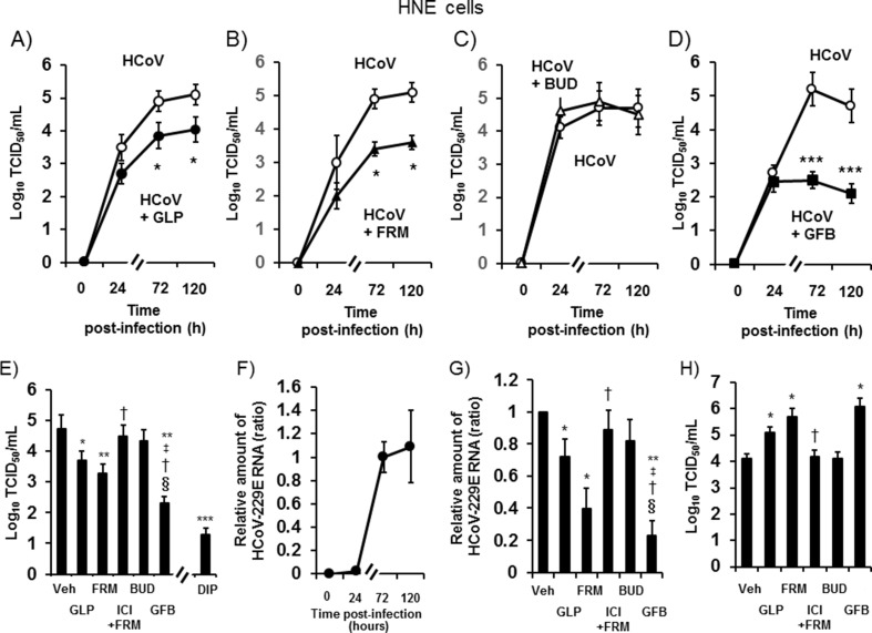
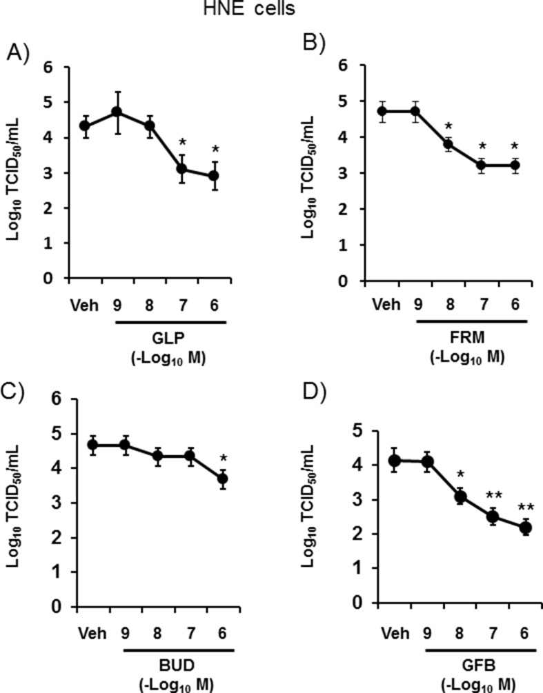
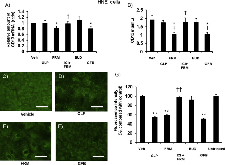
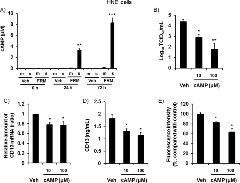
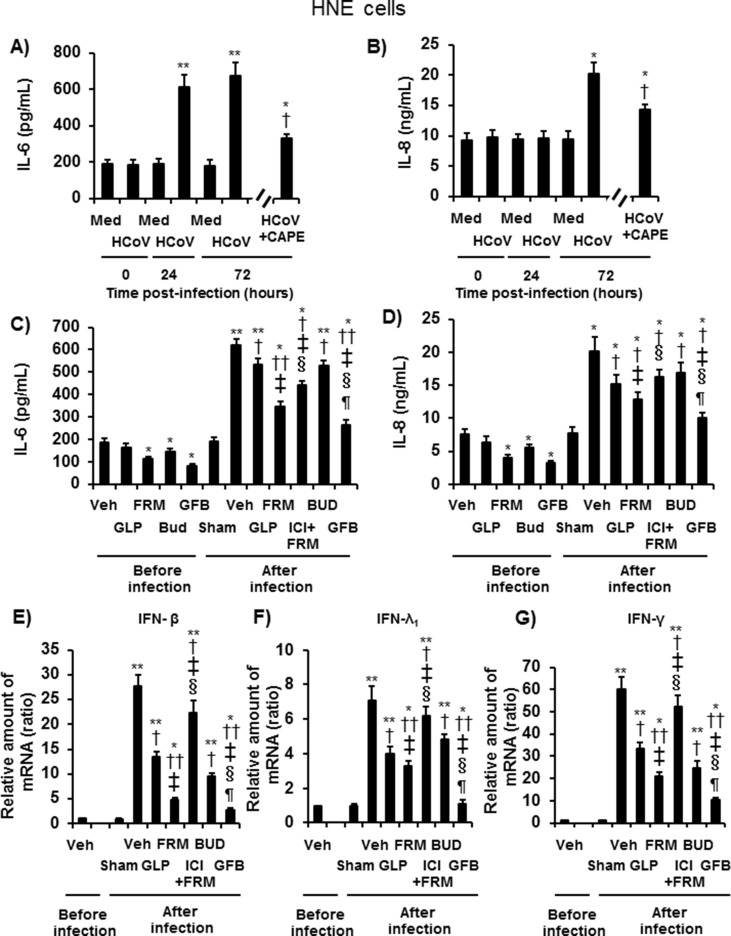
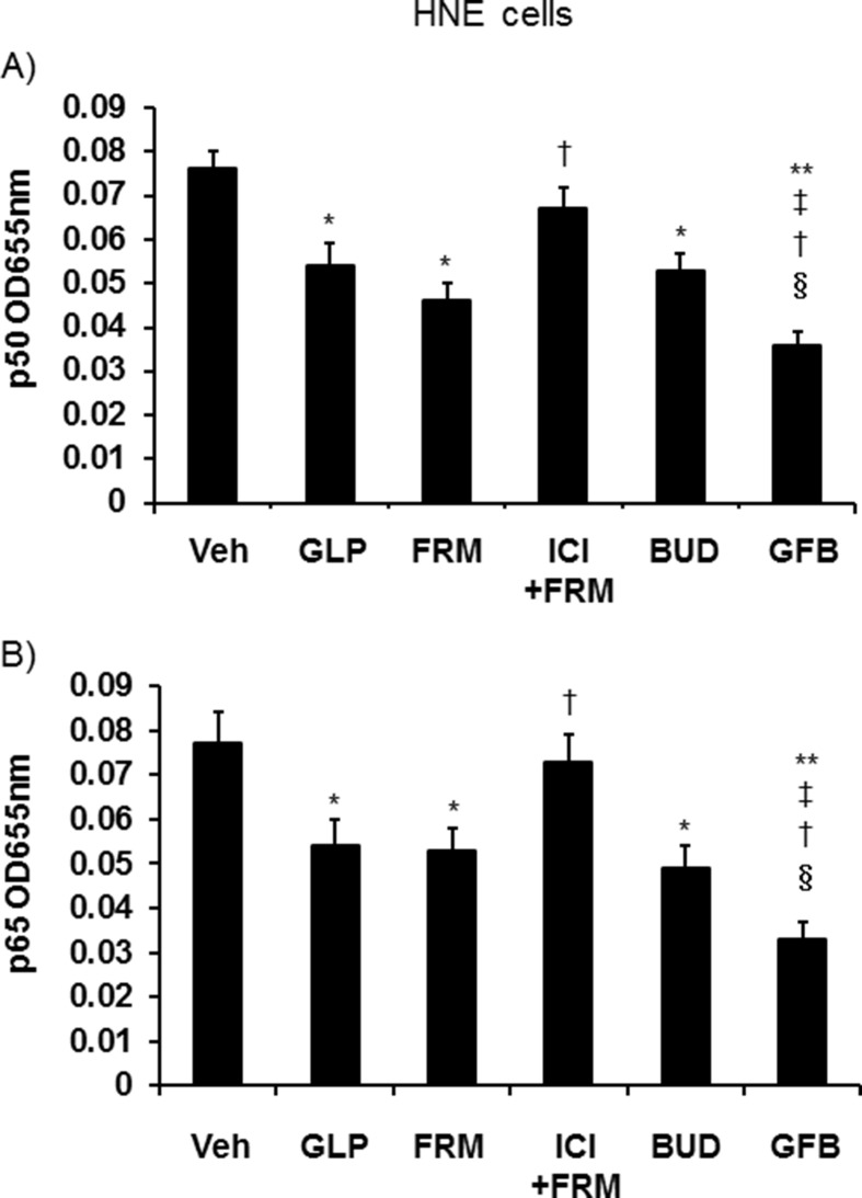
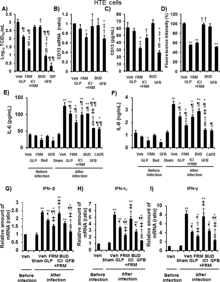
Results: Pretreatment of HNE and HTE cells with glycopyrronium or formoterol decreased viral RNA levels and/or titers, the expression of the HCoV-229E receptor CD13, the number and fluorescence intensity of acidic endosomes where HCoV-229E RNA enters the cytoplasm, and the infection-induced production of cytokines, including IL-6, IL-8, and IFN-β. Treatment of the cells with the CD13 inhibitor 2'2'-dipyridyl decreased viral titers. Pretreatment of the cells with a combination of three drugs (glycopyrronium, formoterol, and budesonide) exerted additive inhibitory effects on viral titers and cytokine production. Pretreatment of HNE cells with glycopyrronium or formoterol reduced the susceptibility to infection, and pretreatment with the three drugs inhibited activation of nuclear factor-kappa B p50 and p65 proteins. Pretreatment with formoterol increased cAMP levels and treatment with cAMP decreased viral titers, CD13 expression, and the fluorescence intensity of acidic endosomes.

Conclusions: These findings suggest that glycopyrronium, formoterol, and a combination of glycopyrronium, formoterol, and budesonide inhibit HCoV-229E replication partly by inhibiting receptor expression and/or endosomal function and that these drugs modulate infection-induced inflammation in the airway.

Keywords: Airway epithelial cells; CD13; HCoV-229E; Long-acting muscarinic antagonist; Long-acting β(2) agonist.


Conflict of interest statement

Declaration of Competing Interest This work was supported by a research support grant from AstraZeneca KK (NCR-17-12892). Glycopyrronium, formoterol, and budesonide were obtained from AstraZeneca PLC.

Figures

Fig. 1
Fig. 1
A–D: The time course of HCoV-229E release into ASL from HNE cells pretreated with glycopyrronium (HCoV + GLP) (A, closed circles), formoterol (HCoV + FRM) (B, closed triangles), budesonide (HCoV + BUD) (C, open triangles), a combination of glycopyrronium, formoterol and budesonide (HCoV + GFB) (D, closed squares), or vehicle (HCoV) (open circles, 0.001% dimethyl sulfoxide: DMSO) at different times after viral infection. The viral titers are expressed as the log10 TCID50 (50% tissue culture infective dose)/mL. E: Viral titers in ASL collected between 24 h and 72 h after infection of HNE cells pretreated with glycopyrronium (GLP), formoterol (FRM), the selective β2-adrenergic receptor antagonist ICI 118,551 (1 μM) plus formoterol (100 nM) (ICI + FRM), budesonide (BUD), a combination of these three drugs (GFB), the CD13 inhibitor 2′2′-dipyridyl (2.5 mM) (DIP), or vehicle (Veh). The cells were pretreated with drugs starting at 72 h before infection and lasting until the end of the experiments. The cells were pretreated with 2′2′-dipyridyl starting at 1 h before infection. F: The time course of HCoV-229E RNA replication in HNE cells measured at different times after infection. G: HCoV-229E RNA replication in HNE cells pretreated with glycopyrronium (GLP), formoterol (FRM), ICI 118,551 plus formoterol (ICI + FRM), budesonide (BUD), a combination of the three drugs (GFB), or vehicle (Veh) at 72 h after infection. H: The minimum dose of virus necessary to cause infection in HNE cells treated with glycopyrronium (GLP), formoterol (FRM), ICI 118,551 plus formoterol (ICI + FRM), budesonide (BUD), a combination of the three drugs (GFB), or vehicle (Veh). A-H: The concentrations of glycopyrronium, formoterol, and budesonide were 100 nM. The results are presented as the mean ± SEM of five (A-D, F–H) or seven (E) subjects. Significant differences compared with cells pretreated with vehicle are indicated by *p < 0.05, **p < 0.01, and ***p < 0.001. Significant differences compared with cells treated with glycopyrronium, formoterol, and budesonide are indicated by p < 0.05, p < 0.05, and §p < 0.05, respectively.
Fig. 2
Fig. 2
Concentration-dependent effects of pretreatment with glycopyrronium (A) (GLP), formoterol (B) (FRM), budesonide (C) (BUD), a combination of these three drugs (D) (GFB), or vehicle (Veh) on HCoV-229E release into ASL collected between 24 h and 72 h after infection. The results are presented as the mean ± SEM of five subjects. Significant differences compared with cells pretreated with vehicle are indicated by *p < 0.05 and **p < 0.01.
Fig. 3
Fig. 3
A and B: CD13 mRNA (A) and protein concentrations in the ASL (B) of uninfected HNE cells pretreated with glycopyrronium (GLP), formoterol (FRM), ICI 118,551 (1 μM) plus formoterol (100 nM) (ICI + FRM), budesonide (BUD), a combination of these three drugs (GFB), or vehicle (Veh) for 72 h. C–F: Distribution of acidic endosomes exhibiting green fluorescence in HNE cells at 72 h after pretreatment with glycopyrronium (GLP) (D), formoterol (FRM) (E), a combination of the three drugs (GFB) (F), or vehicle (C). Data are representative of four different experiments. Scale bar = 100 μm. G: The fluorescence intensity of acidic endosomes in HNE cells at 72 h after pretreatment with glycopyrronium (GLP), formoterol (FRM), ICI 118,551 plus formoterol (ICI + FRM), budesonide (BUD), a combination of the three drugs (GFB), or vehicle (Veh), and in untreated cells (Untreated). The mean value of the fluorescence intensity of the vehicle-treated cells was set to 100%. A–G: The concentrations of glycopyrronium, formoterol, and budesonide were 100 nM. The results are presented as the mean ± SEM of five (A and B) or eight (G) subjects. Significant differences compared with cells pretreated with vehicle are indicated by *p < 0.05 and **p < 0.01 (A, B, and G). Significant differences compared with cells pretreated with formoterol are indicated by p < 0.05 and ††p < 0.01 (A, B, and G). Significant differences compared with cells pretreated with glycopyrronium are indicated by p < 0.05 (B).
Fig. 4
Fig. 4
A: Time course of the cAMP concentration in ASL (m) or basolateral supernatant (s) of uninfected HNE cells treated with formoterol (FRM, 100 nM) or vehicle (Veh) collected before and at 24 h and 72 h after pretreatment. B: Viral titers in ASL from HNE cell cultures pretreated with cAMP (10 μM or 100 μM) or vehicle (Veh) collected between 24 h and 72 h after infection. C and D: The CD13 mRNA (C) and protein concentrations in the ASL (D) of uninfected HNE cells pretreated with cAMP (10 μM or 100 μM) or vehicle (Veh) for 72 h. E: The fluorescence intensity of acidic endosomes in HNE cells at 72 h after pretreatment with cAMP (10 μM or 100 μM) or vehicle (Veh). The mean value of the fluorescence intensity of the vehicle-treated cells was set to 100%. A–E: The results are presented as the mean ± SEM of four (A, C-E) or five (B) subjects. Significant differences compared with cells pretreated with vehicle are indicated by *p < 0.05, **p < 0.01, and ***p < 0.001.
Fig. 5
Fig. 5
A and B: Time course of the release of IL-6 (A) and IL-8 (B) into the ASL of HNE cells before (time 0) and after infection with HCoV-229E in the presence or absence of the NF-κB inhibitor CAPE or sham treatment (medium) (Med). HNE cells were pretreated with CAPE (25 μg/mL) starting at 2 h before infection and lasting until the end of the experiments. The results are reported as the mean ± SEM of cells from five different subjects. Significant differences compared with sham (medium)-infected cells are indicated by *p < 0.05 and **p < 0.01. Significant differences compared with cells infected with HCoV-229E alone (HCoV) at 72 h after infection are indicated by p < 0.05. C and D: The release of IL-6 (C) and IL-8 (D) into the ASL of HNE cells pretreated with glycopyrronium (GLP), formoterol (FRM), budesonide (BUD), a combination of the three drugs (GFB), ICI 118,551 plus formoterol (ICI + FRM), or vehicle collected before and between 24 h and 72 h after infection with HCoV-229E or after sham infection (Sham). E–G: The mRNA expression of IFN-β (E), IFN-λ1 (F), or IFN-γ (G) in HNE cells pretreated with glycopyrronium (GLP), formoterol (FRM), budesonide (BUD), a combination of the three drugs (GFB), ICI plus formoterol (ICI + FRM), or vehicle (Veh) collected before and at 72 h after infection with HCoV-229E or sham infection (Sham). C–G: The results are reported as the mean ± SEM of cells from five different subjects. Significant differences compared with the values from cells treated with vehicle (Veh) before infection are indicated by *p < 0.05 and **p < 0.01. Significant differences compared with the values from cells infected with HCoV-229E alone in the presence of vehicle (Veh) are indicated by p < 0.05 and ††p < 0.01. Significant differences compared with the values from cells pretreated with glycopyrronium, formoterol, and budesonide after infection are indicated by p < 0.05, §p < 0.05, and p < 0.05, respectively.
Fig. 6
Fig. 6
Levels of p50 (A) and p65 (B) in nuclear extracts of uninfected HNE cells treated with glycopyrronium (GLP), formoterol (FRM), ICI 118,551 plus formoterol (ICI + FRM), budesonide (BUD), and a combination of glycopyrronium, formoterol, and budesonide (GFB), or vehicle (Veh) for 72 h. HNE cells were cultured in 24-well dishes. The results are expressed as the OD and the mean ± SEM of five samples. Significant differences compared with the values from vehicle-treated cells (Veh) are indicated by *p < 0.05 and **p < 0.01. Significant differences compared with the values from cells treated with glycopyrronium, formoterol, and budesonide are indicated by p < 0.05, p < 0.05, and §p < 0.05, respectively.
Fig. 7
Fig. 7
A: The viral titers in ASL collected between 24 h and 72 h after the infection of HTE cells pretreated with glycopyrronium (GLP), formoterol (FRM), the selective β2-adrenergic receptor antagonist ICI 118,551 (1 μM) plus formoterol (100 nM) (ICI + FRM), budesonide (BUD), a combination of these three drugs (GFB), the CD13 inhibitor 2′2′-dipyridyl (2.5 mM) (DIP), or vehicle (Veh). The cells were pretreated with drugs starting at 72 h before infection and lasting until the end of the experiments. The cells were pretreated with 2′2′-dipyridyl starting at 1 h before infection. B–D: CD13 mRNA (B) and protein concentrations in the ASL (C) of uninfected HTE cells and the fluorescence intensity of acidic endosomes in HTE cells (D) after pretreatment with glycopyrronium (GLP), formoterol (FRM), ICI 118,551 plus formoterol (ICI + FRM), budesonide (BUD), a combination of these three drugs (GFB), or vehicle (Veh) for 72 h. The mean value of the fluorescence intensity of the vehicle-treated cells was set to 100%. E and F: The release of IL-6 (E) and IL-8 (F) into the ASL of HTE cells pretreated with glycopyrronium (GLP), formoterol (FRM), ICI 118,551 plus formoterol (ICI + FRM), budesonide (BUD), a combination of three drugs (GFB), or vehicle (Veh) collected before and between 24 h and 72 h after infection with HCoV-229E or sham infection (Sham). G–I: The mRNA expression of IFN-β (G), IFN-λ1 (H), or IFN-γ (I) in HTE cells pretreated with glycopyrronium (GLP), formoterol (FRM), ICI plus formoterol (ICI + FRM), budesonide (BUD), a combination of the three drugs (GFB), or vehicle (Veh) extracted before and at 72 h after infection with HCoV-229E or after sham infection (Sham). A–I: The concentrations of glycopyrronium, formoterol, and budesonide were 100 nM. The results are reported as the mean ± SEM for cells of five (A-C, E-I) or seven (D) different subjects. A: Significant differences compared with the values of HTE cells after infection that were treated with vehicle, glycopyrronium, formoterol, or budesonide are indicated by p < 0.05 and ¶¶p < 0.01, p < 0.05, p < 0.05, or §p < 0.01, respectively. B–D: Significant differences compared with the values of uninfected HTE cells treated with vehicle, glycopyrronium, formoterol, or budesonide are indicated by *p < 0.05 and **p < 0.05, p < 0.05, p < 0.05 and ††p < 0.01, or §p < 0.01, respectively. E–I: Significant differences compared with the values from uninfected vehicle-treated HTE cells are indicated by *p < 0.05 and **p < 0.01. Significant differences compared with the values of HTE cells after infection treated with vehicle, glycopyrronium, formoterol, or budesonide are indicated by p < 0.05 and ¶¶p < 0.01, p < 0.05, p < 0.05, or §p < 0.01, respectively.

References

    1. El-Sahly H.M., Atmar R.L., Glezen W.P., Greenberg S.B. Spectrum of clinical illness in hospitalized patients with “common cold” virus infections. Clin Infect Dis. 2000;31:96–100. - PMC - PubMed
    1. Shirato K., Kawase M., Watanabe O., Hirokawa C., Matsuyama S., Nishimura H. Differences in neutralizing antigenicity between laboratory and clinical isolates of HCoV-229E isolated in Japan in 2004- 2008 depend on the S1 region sequence of the spike protein. J Gen Virol. 2012;93:1908–1917. - PubMed
    1. Nicholson K.G., Kent J., Ireland D.C. Respiratory viruses and exacerbations of asthma in adults. BMJ. 1993;307:982–986. - PMC - PubMed
    1. Seemungal T., Harper-Owen R., Bhowmik A., Moric I., Sanderson G., Message S. Respiratory viruses, symptoms, and inflammatory markers in acute exacerbations and stable chronic obstructive pulmonary disease. Am J Respir Crit Care Med. 2001;164:1618–1623. - PubMed
    1. Yeager C.L., Ashmum R.A., Williams R.K., Cardellichio C.B., Shapiro L.H., Look A.T. Human aminopeptidase N is a receptor for human coronavirus 229E. Nature. 1992;357:420–422. - PMC - PubMed

MeSH terms