. 2020 Mar 10;117(10):5339-5350.
doi: 10.1073/pnas.1901720117. Epub 2020 Feb 24.

The aging skin microenvironment dictates stem cell behavior

Affiliations

The aging skin microenvironment dictates stem cell behavior

Yejing Ge et al. Proc Natl Acad Sci U S A. .

Abstract

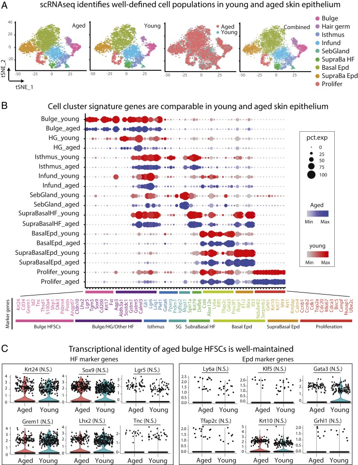
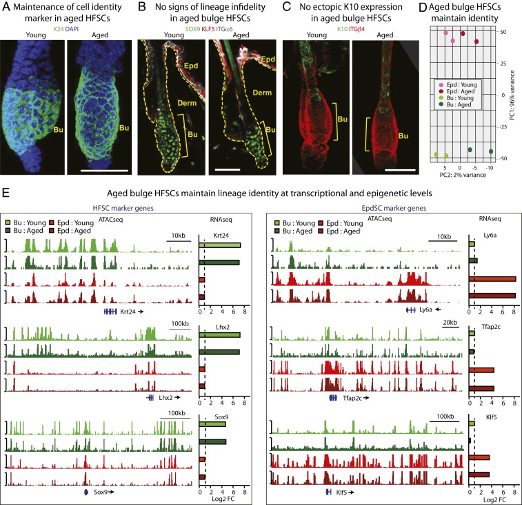
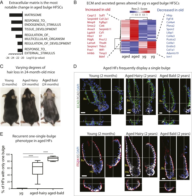
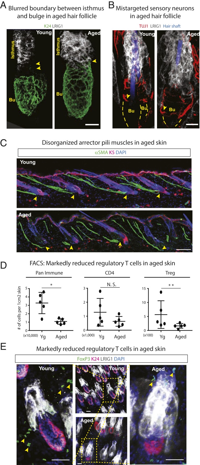
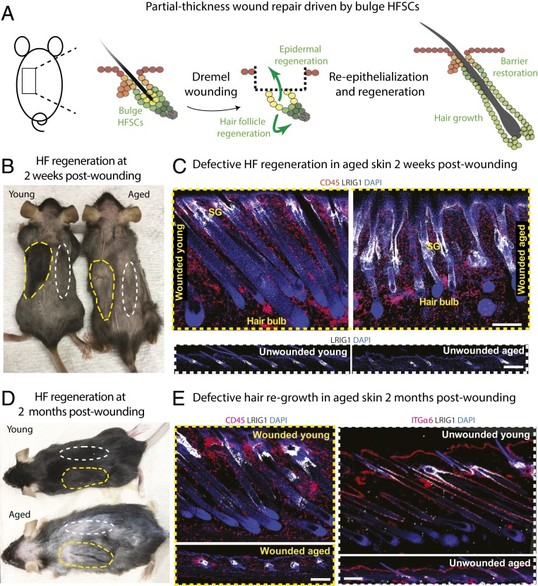
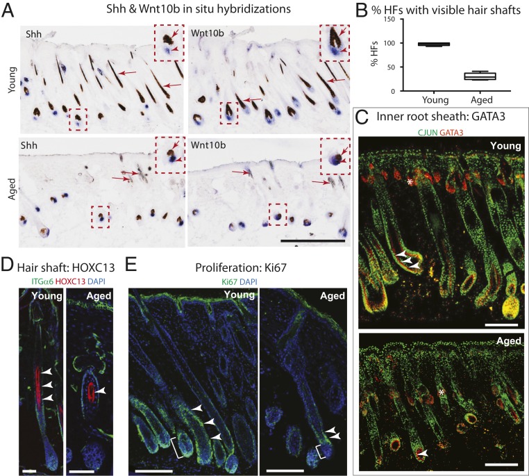
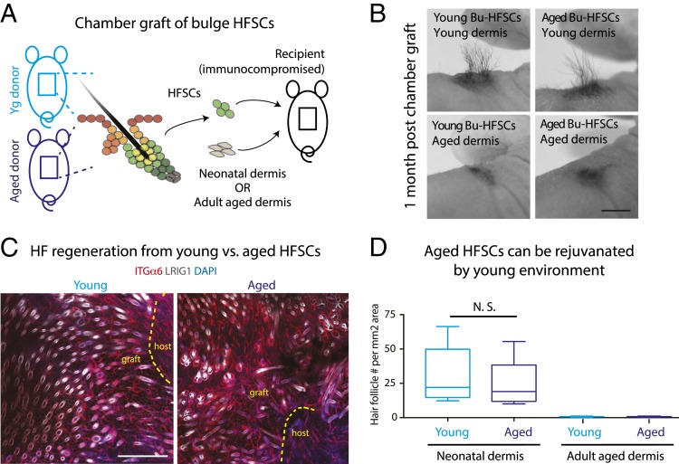
Aging manifests with architectural alteration and functional decline of multiple organs throughout an organism. In mammals, aged skin is accompanied by a marked reduction in hair cycling and appearance of bald patches, leading researchers to propose that hair follicle stem cells (HFSCs) are either lost, differentiate, or change to an epidermal fate during aging. Here, we employed single-cell RNA-sequencing to interrogate aging-related changes in the HFSCs. Surprisingly, although numbers declined, aging HFSCs were present, maintained their identity, and showed no overt signs of shifting to an epidermal fate. However, they did exhibit prevalent transcriptional changes particularly in extracellular matrix genes, and this was accompanied by profound structural perturbations in the aging SC niche. Moreover, marked age-related changes occurred in many nonepithelial cell types, including resident immune cells, sensory neurons, and arrector pili muscles. Each of these SC niche components has been shown to influence HF regeneration. When we performed skin injuries that are known to mobilize young HFSCs to exit their niche and regenerate HFs, we discovered that aged skin is defective at doing so. Interestingly, however, in transplantation assays in vivo, aged HFSCs regenerated HFs when supported with young dermis, while young HFSCs failed to regenerate HFs when combined with aged dermis. Together, our findings highlight the importance of SC:niche interactions and favor a model where youthfulness of the niche microenvironment plays a dominant role in dictating the properties of its SCs and tissue health and fitness.

Keywords: aging; hair follicle; lineage identity; skin; stem cells.


Conflict of interest statement

The authors declare no competing interest.

Figures

Fig. 1.
Fig. 1.
scRNA-seq analysis suggest that bulge HFSCs in the aged skin maintain their lineage identity. scRNA-seq was performed on purified epithelial cells from the back skins of young (2 mo) and aged (24 mo) mice (Materials and Methods). (A) tSNE plots show similar patterns of cell clustering for young and aged skins. (B) Split dot-plots show that each cell cluster stratifies according to known lineage and identity markers. Each circle is a gene, with size representing percentage of cells expressing this gene and color representing expression level (see annotation to the right, blue = aged, red = young). The x axis lists gene names and cell identity; the y axis lists cluster assignments. (C) Violin plots of HF and epidermal (Epd) marker genes expressed by young and aged bulge HFSCs. These cells were defined by clustering and signature expression. Note that there are no detectable changes in the expression of marker genes. N.S., not significant.
Fig. 2.
Fig. 2.
During the hair cycle resting stage, aged bulge HFSCs maintain their lineage identity. (AC) Immunofluorescence of HFSC markers K24 (A) and SOX9 (B), and EpdSC marker KLF5 (B), and Epd differentiation marker K10 (C). Bu, bulge; Derm, dermis; Epd, epidermis; ITGα6, INTEGRIN α6; ITGβ4, INTEGRIN β4. At least five independent biological replicates were performed; shown are representative images. (Scale bars, 30 µm.) (D) PCA on bulk RNA-seq of aged versus young bulge HFSCs (green) and EpdSCs (red) further suggest that aged bulge HFSCs do not convert to an EpdSC-like fate. Data are from two biologically independent replicates. (E) ATAC-seq tracks show accessible chromatin regions of genes transcribed in: HFSCs (Krt24, Lhx2, Sox9) and EpdSCs (Ly6a [=Sca1], Tfap2c [=AP2g], Klf5), and how their patterns differ in young versus aged bulge HFSCs (green) and EpdSCs (red). Bulk RNA-seq log2 fold-change values (dashed line shows the baseline) are plotted next to ATAC-seq tracks, showing specific expression of HFSC and EpdSC identity genes, respectively.
Fig. 3.
Fig. 3.
Age-related changes in the HFSC transcriptome and bulge structure point to ECM perturbations in the niche. (A) GO term analysis on bulk RNA-seq. Note that the top changed category between young and aged HFSCs is matrisome (ECM genes). Note also marked changes in endogenous and exogenous stimuli. (B) Heatmap showing hierarchical clustering of ECM genes and secreted factors that are significantly changed between aged and young HFSCs. Red, increased expression; blue, decreased expression. (C) Examples of variability in hair loss phenotype displayed by aged (24-mo-old) C57BL/6 mice. (D) Whole-mount immunofluorescence of telogen-phase HFs labeled for E-CADHERIN (ECAD) and P-CADHERIN (PCAD) (Upper), and K5 and INTEGRIN β4 (ITGβ4) (Lower). Images of vertical and horizontal planes (crossbars) are shown at the right and bottom of each main frame, respectively. Note that young HFs display the expected two-bulge structure, but aged HFs show only a single bulge, regardless of whether hairy or bald. Bu, bulge; HG, hair germ. At least five independent biological replicates were analyzed; shown are representative images. (Scale bars, 10 μm.) (E) Quantifications of data shown in D. n = 5. Data are presented as mean ± SEM. Paired t test was performed, ****P < 0.0001.
Fig. 4.
Fig. 4.
Multifaceted changes in the aged skin microenvironment. (A and B) Whole-mount immunofluorescence of telogen-phase young and aged HFs immunolabeled for HFSC marker K24, isthmus marker LRIG1, and sensory neuronal marker TUJ1. Arrowheads in A point to the spatial separation between the bulge (Bu) and isthmus. In young HFs, this zone harbors the four sensory neurons that wrap around the HFs. In aged HFs, this zone is missing. Arrowheads in B show that sensory neurons have relocalized to the bulge in aged HFs. (Scale bars, 20 μm.) (C) Immunofluorescence of α-smooth muscle actin (αSMA) in young and aged skin reveals that APMs lose their anchorage (arrowheads) to the bulge in aged skin. (Scale bar, 50 μm.) (D) FACS analysis of skin resident immune cell populations showing age-related reductions in CD45+ pan-immune populations, no significant change in the overall cohort of CD4+ effector T cells, but specific reductions in CD4+FOXP3+CD25+ Tregs. n = 5. Data are presented as mean ± SEM. Paired t test was performed, *P < 0.05, **P < 0.01, N.S., not significant. (E) Whole-mount immunofluorescence of telogen-phase young and aged HFs immunolabeled for HFSC marker K24, isthmus marker LRIG1, and Treg marker FOXP3. Arrowheads in E point to the FoxP3+ Treg cells in the young and aged skin. (Scale bars, 30 μm.) For AC and E, at least five independent biological replicates were analyzed. Shown are representative images.
Fig. 5.
Fig. 5.
Age-related deficiency in hair coat recovery following wounding. (A) Partial-thickness wound repair model used to challenge telogen-phase bulge HFSCs to re-epithelialize the epidermis and initiate HF regeneration. A dremel tool was used to remove the skin directly above the HF bulges. (B and D) Photos of mice at either 2 wk (B) or 2 mo (D) after shaving and dremel wounding of telogen-phase back skin. Yellow dashed lines denote wounded areas; white dashed lines denote unwounded areas used for controls. (C and E) Immunofluorescence for LRIG1 (white), which marks the HF isthmus, where sebaceous glands (SG) reside, and CD45 (red), a pan-immune cell marker. Notes: At 2 wk, the wounded skin of young mice has turned black, reflecting melanin pigment in full anagen-phase HFs. Unwounded regions and aged wounded regions remain pale, reflecting telogen-phase HFs. The unwounded sample was taken from the 2-wk or 2-mo postwounding mice, respectively, from the unwounded side of the backskin. Anagen has been induced in both young and aged wounded areas, but downgrowth is delayed/impaired in aged HFs, which appear immature. By 2 mo, the unwounded skin of young mice has entered its normal anagen, and the hair coat has regrown. However, aged wounded areas appear to be in telogen at 2 mo postwounding, despite having activated hair cycling at 2 wk. At least five independent biological replicates were performed for each time point; shown are representative images. (Scale bars, 50 μm.) SG, sebaceous glands, which attach at the isthmus.
Fig. 6.
Fig. 6.
Defective HF regeneration following wounding in aged skin. (A) In situ hybridizations of skin sections processed at 7 d postdremel-wounding of young and aged mice. Shh and Wnt10b (blue, in situ hybridization signals) are expressed by HFSC progenitors that form at the start of the hair cycle and persist in the hair bulb through anagen (arrowheads in Insets). Arrows point to hair shaft cells, which take up brown melanosome pigment as they differentiate to form the hair shaft. (Scale bar, 200 μm.) (B) Quantifications showing markedly reduced visible (pigmented) hair shafts in wound-stimulated regions of aged versus young mice. (C and D) Immunofluorescence for inner root sheath marker GATA3 (C, arrowheads) and hair shaft marker HOXC13 (D, arrowheads) revealing their reduced numbers if wound-induced HFs in aged compared to young mice (2 wk postdremel-wounding). Notes: AP-1 (CJUN) in green marks the entire skin epithelium during wound repair. An asterisk denotes nonspecific binding. (E) Immunofluorescence for proliferation marker Ki67 shows reduced proliferation of short-lived HFSC progenitors within the lower outer root sheath (arrowheads) and the hair bulb (brackets) of wound-induced HFs in aged compared to young mice (2 wk postdremel-wounding). For CE, at least five independent biological replicates were analyzed; shown are representative images. (Scale bars, 50 μm.)
Fig. 7.
Fig. 7.
Tissue microenvironment overrides stem cell intrinsic differences and rejuvenates aged HFSCs. (A) Schematics illustrating chamber graft assays, where a small area of skin is removed from the recipient Nude mouse back skin and grafted with bulge HFSCs mixed with dermal cells within a domed chamber (chamber graft; see Materials and Methods). This method is used to analyze HF regeneration from HFSCs in vivo (39). (BD) Photos (B) whole-mount immunofluorescence microscopy (C), and quantifications (D) of grafted young or aged donor HFSCs, combined with either neonatal dermal cells (B and D) or aged dermal cells (D). Note that both aged and young HFSCs produced visible hairs and HFs when grafted with neonatal dermal cells, but failed to do so with aged dermal cells. Immunolabeling of LRIG1 marks HF isthmus in white, and that of INTEGRIN α6 (ITGα6) outlines HFs (also blood vessels) in red. Dashed lines delineate graft and host boundaries. At least five independent biological replicates were performed; shown are representative images. (Scale bars, 1 cm in B; 100 μm in C.) Paired t tests were performed for quantifications (D). Data are presented as mean ± SEM. n = 5. N.S., not significant.

References

    1. Till J. E., McCulloch E. A., A direct measurement of the radiation sensitivity of normal mouse bone marrow cells. Radiat. Res. 14, 213–222 (1961). - PubMed
    1. Becker A. J., McCulloch E. A., Till J. E., Cytological demonstration of the clonal nature of spleen colonies derived from transplanted mouse marrow cells. Nature 197, 452–454 (1963). - PubMed
    1. Sharpless N. E., DePinho R. A., How stem cells age and why this makes us grow old. Nat. Rev. Mol. Cell Biol. 8, 703–713 (2007). - PubMed
    1. Rossi D. J., Jamieson C. H., Weissman I. L., Stems cells and the pathways to aging and cancer. Cell 132, 681–696 (2008). - PubMed
    1. Signer R. A., Morrison S. J., Mechanisms that regulate stem cell aging and life span. Cell Stem Cell 12, 152–165 (2013). - PMC - PubMed

Publication types

MeSH terms