. 2020 Apr 1;40(14):2960-2974.
doi: 10.1523/JNEUROSCI.2402-19.2020. Epub 2020 Feb 24.

Microglial Depletion with CSF1R Inhibitor During Chronic Phase of Experimental Traumatic Brain Injury Reduces Neurodegeneration and Neurological Deficits

Affiliations

Microglial Depletion with CSF1R Inhibitor During Chronic Phase of Experimental Traumatic Brain Injury Reduces Neurodegeneration and Neurological Deficits

Rebecca J Henry et al. J Neurosci. .

Abstract

Chronic neuroinflammation with sustained microglial activation occurs following severe traumatic brain injury (TBI) and is believed to contribute to subsequent neurodegeneration and neurological deficits. Microglia, the primary innate immune cells in brain, are dependent on colony stimulating factor 1 receptor (CSF1R) signaling for their survival. In this preclinical study, we examined the effects of delayed depletion of chronically activated microglia on functional recovery and neurodegeneration up to 3 months postinjury. A CSF1R inhibitor, Plexxikon (PLX) 5622, was administered to adult male C57BL/6J mice at 1 month after controlled cortical impact to remove chronically activated microglia, and the inhibitor was withdrawn 1-week later to allow for microglial repopulation. Following TBI, the repopulated microglia displayed a ramified morphology similar to that of Sham uninjured mice, whereas microglia in vehicle-treated TBI mice showed the typical chronic posttraumatic hypertrophic morphology. PLX5622 treatment limited TBI-associated neuropathological changes at 3 months postinjury; these included a smaller cortical lesion, reduced hippocampal neuron cell death, and decreased NOX2- and NLRP3 inflammasome-associated neuroinflammation. Furthermore, delayed depletion of chronically activated microglia after TBI led to widespread changes in the cortical transcriptome and altered gene pathways involved in neuroinflammation, oxidative stress, and neuroplasticity. Using a variety of complementary neurobehavioral tests, PLX5622-treated TBI mice also had improved long-term motor and cognitive function recovery through 3 months postinjury. Together, these studies demonstrate that chronic phase removal of neurotoxic microglia after TBI using CSF1R inhibitors markedly reduce chronic neuroinflammation and associated neurodegeneration, as well as related motor and cognitive deficits.SIGNIFICANCE STATEMENT Traumatic brain injury (TBI) is a debilitating neurological disorder that can seriously impact the patient's quality of life. Microglial-mediated neuroinflammation is induced after severe TBI and contributes to neurological deficits and on-going neurodegenerative processes. Here, we investigated the effect of breaking the neurotoxic neuroinflammatory loop at 1-month after controlled cortical impact in mice by pharmacological removal of chronically activated microglia using a colony stimulating factor 1 receptor (CSF1R) inhibitor, Plexxikon 5622. Overall, we show that short-term elimination of microglia during the chronic phase of TBI followed by repopulation results in long-term improvements in neurological function, suppression of neuroinflammatory and oxidative stress pathways, and a reduction in persistent neurodegenerative processes. These studies are clinically relevant and support new concepts that the therapeutic window for TBI may be far longer than traditionally believed if chronic and evolving microglial-mediated neuroinflammation can be inhibited or regulated in a precise manner.

Keywords: CSF1R; functional recovery; microglia; neurodegeneration; neuroinflammation; traumatic brain injury.


Figures

Figure 1.
Figure 1.
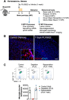
Experimental timeline and in vivo characterization of the CSF1R inhibitor, PLX5622. A, Experimental timeline: adult male C57BL/6 mice underwent CCI/Sham surgery. At 4 weeks after CCI/Sham surgery mice were placed on PLX5622 (1200 ppm) or normal chow (Veh) for 1 week, following which mice were returned to normal chow for the remainder of the study. One cohort of mice were killed at 8 WPI and samples were collected for flow cytometry and nanostring analysis. A separate cohort of mice underwent a battery of neurobehavioral tasks (BW, Y maze, NOR, MWM) through 12 WPI, and samples were collected for histological analysis (Stereology). B, Representative immunofluorescence images of P2Y12+ microglia demonstrated that oral administration of PLX5622 (1200 ppm) in chow for 7 d leads to an almost complete depletion of microglia (P2Y12+, red) in the mouse brain. Scale bar, 200 μm. C, Flow cytometry analysis of microglia (CD11b+/CD45int) demonstrated that 7-day PLX5622 treatment resulted in ∼95% depletion of microglia counts in the mouse brain. Returning animals to normal chow feed resulted in repopulation of microglia in mouse brain within 7 d of removing PLX5622 chow. Data expressed as mean ± SEM (n = 3/group).
Figure 2.
Figure 2.
Delayed depletion of microglia starting at 4 weeks postinjury improves long-term motor function recovery. A, Delayed depletion of microglia using the CSF1R inhibitor, PLX5622, significantly reduced long-term TBI-induced fine-motor impairments (number of foot faults in a BW task) at 9, 10, and 12 WPI. B, At 12 WPI, TBI+Veh-treated animals spent less time on the accelerating rod when compared with Sham counterparts. ****p < 0.0001 vs Sham; +p < 0.05, ++p < 0.01 vs TBI + Veh. Data expressed as mean ± SEM (n = 7–20/group).
Figure 3.
Figure 3.
Delayed depletion of microglia starting at 4 weeks postinjury improves cognitive function recovery. A, At 10 WPI, TBI significantly decreased percentage spontaneous alterations in the Y-maze task in TBI+Veh-treated animals, when compared with Sham. TBI+PLX5622-treated animals had increased percentage spontaneous alterations, equal to levels in Sham. B, At 11 WPI, TBI significantly decreased percentage time spent with the novel object in a novel object task in TBI+Veh-treated animals, when compared with Sham. TBI+PLX5622-treated animals spent increased time with the novel objects, similar to levels of Sham. C, In the MWM task at 12 WPI, TBI+Veh- and TBI+PLX5622-treated animals required significantly increased time to locate the hidden submerged escape platform on acquisition days 2–4, when compared with Sham. There was no significant difference in escape latencies between TBI+Veh- and TBI+PLX5622-treated animals. D, In the probe trial, TBI+Veh-treated animals spent significantly less time in the escape quadrant, when compared with Sham. TBI+PLX5622-treated animals spent significantly more time in the escape quadrant, when compared with TBI+Veh-treated animals. E, TBI+Veh-treated and TBI+PLX5622-treated animals had similar swim speeds to that of Sham animals during the acquisition days of the MWM. F, Escape strategy used during the probe trial was assessed, and the percentage composition for each strategy (spatial, systematic, and looping) demonstrated that TBI+Veh-treated animals used increased looping strategies, and decreased systematic and spatial search strategies when compared with Sham and TBI+PLX5622-treated animals. *p < 0.05, **p < 0.01, ***p < 0.001, ****p < 0.0001 vs Sham; +p < 0.05 vs TBI+Veh. Data expressed as mean ± SEM (n = 7–20/group).
Figure 4.
Figure 4.
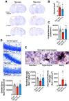
Delayed depletion of microglia starting at 4 weeks postinjury reduces chronic neurodegeneration and alters microglial morphology. A, Representative images of the cortical lesion in TBI+Veh- and TBI+PLX5622-treated animals at 3 months postinjury. B, Stereological analysis demonstrated that TBI+PLX5622-treated animals had a decreased lesion volume, when compared with TBI+Veh-treated animals. C, Stereological analysis demonstrated that TBI significantly increased neuron loss in ipsilateral cortex, when compared with Sham. PLX5622 treatment attenuated cortical neuron loss such that TBI+PLX5622-treated animals had increased numbers of surviving neurons in cortex compared with TBI+Veh-treated animals. D, Representative images of cresyl violet stained neurons in the dentate gyrus (DG) of hippocampus. Stereological analysis demonstrated that TBI increased DG neuron loss, when compared with Sham. In contrast, TBI+PLX5622-treated animals had increased numbers of surviving neurons in DG compared with TBI+Veh-treated animals. E, Representative images of Iba1+ microglia displaying ramified, hypertrophic, and bushy cellular morphologies. Stereological analysis demonstrated that TBI+Veh-treated animals had decreased numbers of ramified and increased hypertrophic/bushy Iba1+ microglia in injured cortex, when compared with Sham. In contrast, TBI+PLX5622-treated animals had increased ramified and decreased hypertrophic/bushy Iba1+ microglia when compared with TBI+Veh-treated animals. *p < 0.05, **p < 0.01 vs Sham; +p < 0.05, ++p < 0.01 vs TBI+Veh. Data expressed as mean ± SEM (n = 8–12/group).
Figure 5.
Figure 5.
Delayed depletion of microglia during chronic TBI alters neuroinflammation, oxidative stress, apoptosis, and neuroplasticity pathways in the cortex. A Nanostring Neuropathology Panel was used to assess cortical transcriptional patterns at 2 months postinjury. A, A PLSDA model was generated to classify each of the 4 treatment groups: Group 1 (Sham+Veh), Group 2 (TBI+Veh), Group 3 (Sham+PLX5622), and Group 4 (TBI+PLX5622). Principal components (PC) 1 and 2 identified key genes changed by TBI and PLX5622 treatment, respectively. B, Hierarchical clustering of differentially expressed genes was performed on different functional annotations. TBI induced a large microglial-related gene cluster (MgC1) associated with inflammation and immune function, representing the inflammatory state induced by TBI. C, Analysis of oxidative stress genes identified 2 clusters of genes altered by PLX5622 treatment. The first cluster (OxC1) included genes that were increased after TBI, and were significantly reduced by TBI+PLX5622 treatment. The other cluster (OxC2) was significantly increased in TBI+PLX5622 animals when compared with the TBI+Veh animals. D, Clustering analysis of apoptosis-related genes showed that TBI increased expression of the mitochondrial membrane permeator, Bax, which was decreased by PLX5622 treatment. E, Clustering analysis of neuroplasticity genes showed that the most highly expressed neuroplasticity genes in Sham+Veh were downregulated by TBI, including in TBI+PLX5622-treated animals. This indicated that PLX5622 treatment did not return neuroplasticity gene expression to a preinjured state. F, Analysis of transcript counts (Violin plots and -log10 adjusted P value assessment) for selected genes related to NLRP3 inflammasome (Il1r1, Casp1, Il1b P2rx7, Nlrp3) and NADPH oxidase (Cybb, Ncf1) pathways demonstrated that key genes in both inflammatory pathways were upregulated by TBI, and subsequently reduced by PLX5622 treatment. (n = 8–9/group). (Fig. 5-1).
Figure 6.
Figure 6.
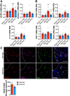
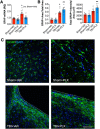
Delayed depletion of microglia during chronic TBI decreases NLRP3 inflammasome and NOX2 expression in the injured cortex. A, Analysis of ipsilateral cortical tissue at 2 months postinjury demonstrated that TBI increased Nlrp3, Casp1, and Il1b mRNA. The TBI-induced increase in Nlrp3 mRNA was significantly reduced by PLX5622 treatment. B, TBI increased NADPH oxidase subunits, Cybb, Cyba, Ncf1, and Ncf4 mRNA in injured cortex. The TBI-induced increase in Cybb mRNA was significantly reduced by PLX5622 treatment. C, Immunofluorescence analysis of reactive microglia (NOX2+, red; CD68+, cyan; Iba1+, green) in the injured cortex at 3 months postinjury demonstrated that delayed depletion of microglia decreased the number of NOX2+ reactive microglia when compared with levels in TBI+Veh-treated animals. The dotted line represents the lesion boundary. Scale bar, 100 μm. **p < 0.01, ***p < 0.001 vs Sham animals; +p < 0.05, ++p < 0.01 vs TBI+Veh-treated animals. Data expressed as mean ± SEM (n = 6–9/group).
Figure 7.
Figure 7.
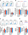
Delayed depletion of microglia during chronic TBI reduces expression of NLRP3 inflammasome components in microglia. A, Flow cytometry analysis and representative dot plots show populations of resident CD45int microglia and infiltrating peripherally derived CD45hi myeloid cells in the ipsilateral cortex at 2 months postinjury. There was no difference in absolute microglial numbers between Sham, TBI+Veh-treated, and TBI+PLX5622-treated groups. TBI increased numbers of infiltrating CD45hi myeloid cells in brain in TBI+Veh-treated animals, but numbers were small compared with numbers of resident microglia. B, Assessment of microglial cell size as measured by forward scatter (FSC), and microglial granularity as measured by side scatter (SSC), demonstrated that TBI increased microglial cell size and granularity when compared with levels in Sham microglia. PLX5622 treatment reduced both markers in microglia. C, Representative dot plots show resident CD45int microglia expressing active caspase-1. TBI increased active caspase-1 in microglia, when compared with levels in Sham. Levels of active caspase-1 were decreased by PLX5622 treatment, but failed to reach statistical significance. D, TBI increased IL-1β production in microglia, when compared with levels in Sham. Levels of microglial IL-1β was decreased in TBI+PLX5622-treated animals. *p < 0.05, ***p < 0.001, vs Sham; ++p < 0.01, +++p < 0.001, ++++p = 0.000 vs TBI+Veh-treated animals. Data expressed as mean ± SEM (n = 5–9/group).
Figure 8.
Figure 8.
Delayed depletion of microglia during chronic TBI does not alter astrocyte reactivity in the cortex. A, Analysis of ipsilateral cortical tissue at 2 months postinjury demonstrated that TBI increased Gfap mRNA. TBI-induced increase in Gfap mRNA was not altered by PLX5622 treatment. B, In situ analysis of astrocytes in the perilesional cortex at 3 months postinjury. TBI increased astrocyte cellular area and GFAP expression when compared with levels in Sham. PLX5622 treatment did not change posttraumatic GFAP cellular distribution or expression. C, Representative images of GFAP (green) in the perilesional cortex at 3 months post injury. The dotted line represents the lesion boundary. Scale bar, 100 μm. *p < 0.05, **p < 0.01 vs Sham. Data expressed as mean ± SEM (n = 5–8/group).

Comment in

References

    1. Adamczak S, Dale G, de Rivero Vaccari JP, Bullock MR, Dietrich WD, Keane RW (2012) Inflammasome proteins in cerebrospinal fluid of brain-injured patients as biomarkers of functional outcome: clinical article. J Neurosurg 117:1119–1125. 10.3171/2012.9.JNS12815 - DOI - PMC - PubMed
    1. Asai H, Ikezu S, Tsunoda S, Medalla M, Luebke J, Haydar T, Wolozin B, Butovsky O, Kügler S, Ikezu T (2015) Depletion of microglia and inhibition of exosome synthesis halt tau propagation. Nat Neurosci 18:1584–1593. 10.1038/nn.4132 - DOI - PMC - PubMed
    1. Babior BM. (1999) NADPH oxidase: an update. Blood 93:1464–1476. - PubMed
    1. Barrett JP, Henry RJ, Villapol S, Stoica BA, Kumar A, Burns MP, Faden AI, Loane DJ (2017) NOX2 deficiency alters macrophage phenotype through an IL-10/STAT3 dependent mechanism: implications for traumatic brain injury. J Neuroinflammation 14:65. 10.1186/s12974-017-0843-4 - DOI - PMC - PubMed
    1. Beckmann N, Giorgetti E, Neuhaus A, Zurbruegg S, Accart N, Smith P, Perdoux J, Perrot L, Nash M, Desrayaud S, Wipfli P, Frieauff W, Shimshek DR (2018) Brain region-specific enhancement of remyelination and prevention of demyelination by the CSF1R kinase inhibitor BLZ945. Acta Neuropathol Commun 6:9. 10.1186/s40478-018-0510-8 - DOI - PMC - PubMed

Publication types

MeSH terms

Substances