. 2020 Apr 3;295(14):4541-4555.
doi: 10.1074/jbc.RA120.012783. Epub 2020 Feb 24.

Mammalian lectin arrays for screening host-microbe interactions

Affiliations

Mammalian lectin arrays for screening host-microbe interactions

Sabine A F Jégouzo et al. J Biol Chem. .

Abstract

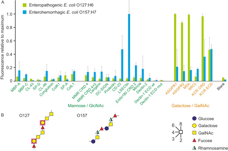
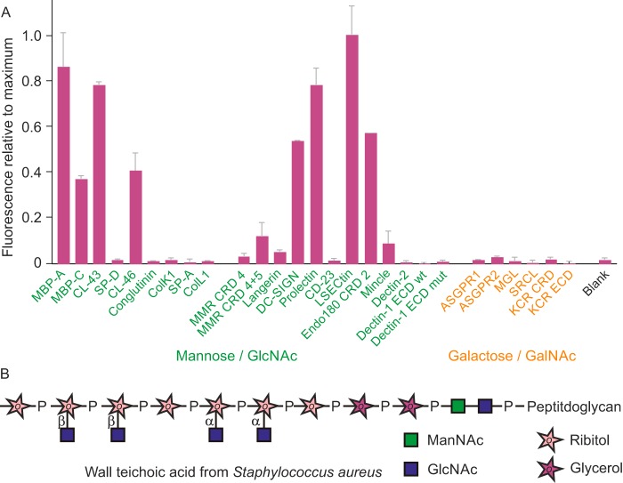
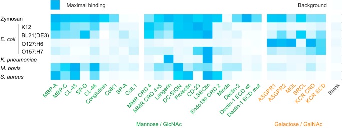
Many members of the C-type lectin family of glycan-binding receptors have been ascribed roles in the recognition of microorganisms and serve as key receptors in the innate immune response to pathogens. Other mammalian receptors have become targets through which pathogens enter target cells. These receptor roles have often been documented with binding studies involving individual pairs of receptors and microorganisms. To provide a systematic overview of interactions between microbes and the large complement of C-type lectins, here we developed a lectin array and suitable protocols for labeling of microbes that could be used to probe this array. The array contains C-type lectins from cow, chosen as a model organism of agricultural interest for which the relevant pathogen-receptor interactions have not been previously investigated in detail. Screening with yeast cells and various strains of both Gram-positive and -negative bacteria revealed distinct binding patterns, which in some cases could be explained by binding to lipopolysaccharides or capsular polysaccharides, but in other cases they suggested the presence of novel glycan targets on many of the microorganisms. These results are consistent with interactions previously ascribed to the receptors, but they also highlight binding to additional sugar targets that have not previously been recognized. Our findings indicate that mammalian lectin arrays represent unique discovery tools for identifying both novel ligands and new receptor functions.

Keywords: array screening; carbohydrate function; carbohydrate-binding protein; glycan-binding receptors; glycobiology; innate immune system; lectin; ligand binding; lipopolysaccharide (LPS); pathogen.


Conflict of interest statement

The authors declare that they have no conflicts of interest with the contents of this article

Figures

Figure 1.
Figure 1.
Cow C-type CRDs. Dendrogram shows a comparison of the CRD sequences for the CRDs from cow C-type lectins included in the lectin array. The aligned sequences are shown in Fig. S1. The lectin groups correspond to the human classification RRID:SCR_018122 (1). Only a single form of DC-SIGN (green) is found in cows, rather than the multiple related genes in humans and mice. Several collectins are ruminant-specific (red).
Figure 2.
Figure 2.
Lectin organization and immobilization strategy. A, domain organization of membrane-bound receptors containing C-type CRDs. Many of the receptors are type 2 transmembrane proteins that often assemble into oligomers (left). The mannose receptor and Endo180 contain multiple C-type lectin-like domains, only some of which are involved in binding sugars (middle). Other receptors interact with other types of polypeptides, such as the FcRγ subunit (right). B, arrangement of domains in collectins. C, C-type CRD from dectin-2 illustrating the location of the N and C termini opposite to the ligand-binding site. Prepared from Protein Data Bank code 5VYB using PyMOL. D, arrangement of lectin fragments on streptavidin-coated wells. In addition to the fragments employed in the initial test array described here, the extracellular portion of the mannose receptor expressed with a C-terminal biotinylation sequence and treated with biotin ligase in vitro is also depicted. E, sequence of biotinylation tag showing site of attachment of biotin to a lysine side chain.
Figure 3.
Figure 3.
Demonstration of saturation of streptavidin-coated wells with biotin-tagged receptors. Examples of screening with zymosan at 5 × 10⁶ particles/ml for three receptors are shown.
Figure 4.
Figure 4.
Validation of lectin array using yeast zymosan. A, structures of polysaccharides in zymosan. B, screening of lectin array with FITC-labeled zymosan extract from S. cerevisiae. Labeled zymosan at a concentration of 5 × 10⁶ particles/ml was used in the screening. The results are plotted relative to the maximal value of 215,000 fluorescence units for binding to dectin-1. In this and subsequent figures, error bars represent standard deviations for duplicate samples. C, binding as a function of zymosan concentration. Zymosan was screened against a receptor panel at three different concentrations.
Figure 5.
Figure 5.
Testing of lectin array with laboratory E. coli strains. A, comparison of different methods for preparing bacterial strain BL21(DE3) prior to screening of the array. Cells expressing GFP and fixed with paraformaldehyde were screened at 1 × 10⁸ cells/ml, and cells labeled with CFDA-SE followed by paraformaldehyde fixation or used directly were screened at 3 × 10⁸ cells/ml. For comparison, results for all three samples were normalized to the SP-D result, which was the highest value for both of the CFDA-SE samples. The values were 40,400, 16,400, and 39,000 fluorescence units, respectively. B, summary of structures of LPS from three laboratory strains of E. coli. C, binding of different strains of E. coli to the lectin array. Strain K12 cells were labeled with FITC (1.5 × 10⁷ cells/ml), and BL21(DE3) (5 × 10⁷ cells/ml) and the ClearColi derivative of BL21(DE3) (3 × 10⁸ cells/ml) were expressing GFP. All results were normalized to the signals for CD23, which was the strongest signal for K12 and BL21(DE3). The signals were 56,000, 49,700, and 46,200 fluorescence units for K12, BL21(DE3), and BL21(DE3) ClearColi, respectively.
Figure 6.
Figure 6.
Comparison of enteropathogenic and enterohemorrhagic E. coli strains binding to the lectin array. A, bacteria expressing GFP were grown to stationary phase and fixed with paraformaldehyde. Enteropathogenic E. coli strain E2348/69 (O127:H6) was screened at 7 × 10⁸ cells/ml and enterohemorrhagic E. coli strain EDL933 (O157:H7) was screened at 5 × 10⁸ cells/ml. Results were normalized to the values for ASGPR1 (41,400 fluorescence units) and LSECtin (51,800 fluorescence units), respectively. B, structures of two repeat units from the O127 and O157 outer polysaccharides of LPS are shown. Purple shading is used to highlight galactose residues that have free 3- and 4-OH groups in the O127 polysaccharide and would be potential targets for galactose-binding C-type CRDs.
Figure 7.
Figure 7.
Effect of capsule on interaction of K. pneumoniae with lectins on the array. A, summary of structures of the K2 capsule and the O1 outer polysaccharide structure of K. pneumoniae. B, lectin array was screened with two strains of K. pneumoniae with the K2:O1 serotype, strains 43816 and B5055, as well a mutant derived from the B5055 strain in which the wzb/c gene is inactivated to prevent capsule formation. Cells expressing GFP were grown to stationary phase and screened against the array at concentrations of 9 × 10⁸, 4 × 10⁸, and 7 × 10⁸ cells/ml, respectively. Results were normalized to the signals for LSECtin for the WT strains (155,000 and 37,300 fluorescence units) and ASGPR1 for the mutant strain (796,000 fluorescence units).
Figure 8.
Figure 8.
S. aureus binding to lectin array. A, heat-killed cells of the Wood 46 strain of S. aureus labeled with FITC were used to screen the array at a concentration of 1.25 × 10⁸ cells/ml. Results were normalized to the signal of 109,000 fluorescence units, which was obtained for LSECtin. B, schematic diagram of wall teichoic acid from S. aureus, showing relative location of α- and β-linked GlcNAc residues. Only β-linked residues are present in this strain.
Figure 9.
Figure 9.
M. bovis binding to lectin array. A, M. bovis bacillus Calmette-Guerin expressing yellow fluorescent protein growing in log phase was further labeled with CFDA-SE, fixed with paraformaldehyde, and used to screen the lectin array at a concentration of 4 × 10⁸ cells/ml. Results were normalized to the value for binding to CL-43, which was 30,000 fluorescence units. B, structure of mannose caps on lipo-arabinomannan in the outer membrane of M. bovis.
Figure 10.
Figure 10.
Comparative heatmap for zymosan and bacteria binding to lectin array. Binding of laboratory and pathogenic strains of E. coli, as well as K. pneumoniae, M. bovis, and S. aureus, are compared with zymosan results using a heatmap. Results for each organism are normalized to the maximum value for that organism as indicated in legends to Figs. 4–9.

References

    1. Drickamer K., and Taylor M. E. (2015) Recent insights into structures and functions of C-type lectins in the immune system. Curr. Opin. Struct. Biol. 34, 26–34 10.1016/j.sbi.2015.06.003 - DOI - PMC - PubMed
    1. Taylor M. E., and Drickamer K. (2019) Mammalian sugar-binding receptors: known functions and unexplored roles. FEBS J. 286, 1800–1814 10.1111/febs.14759 - DOI - PMC - PubMed
    1. Howard M., Farrar C. A., and Sacks S. H. (2018) Structural and functional diversity of collectins and ficolins and their relationship to disease. Semin. Immunopathol. 40, 75–85 10.1007/s00281-017-0642-0 - DOI - PMC - PubMed
    1. Gibson A. J., Coffey T. J., and Werling D. (2013) Of creatures great and small: the advantages of farm animal models in immunology research. Front. Immunol. 4, 124 10.3389/fimmu.2013.00124 - DOI - PMC - PubMed
    1. Guzman E., and Montoya M. (2018) Contributions of farm animals to immunology. Front. Vet. Sci. 5, 307 10.3389/fvets.2018.00307 - DOI - PMC - PubMed

Publication types

MeSH terms

Associated data

LinkOut - more resources