

Development of Potent Forchlorfenuron Analogs and Their Cytotoxic Effect in Cancer Cell Lines
- PMID: 32094384
- PMCID: PMC7039965
- DOI: 10.1038/s41598-020-59824-4
Development of Potent Forchlorfenuron Analogs and Their Cytotoxic Effect in Cancer Cell Lines
Abstract
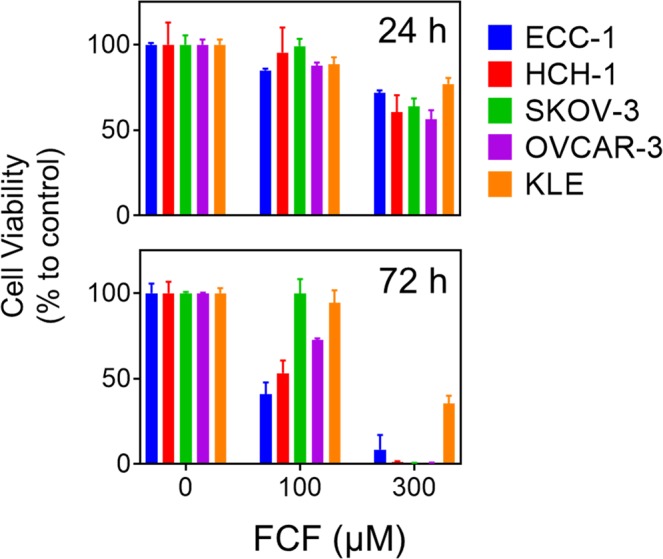
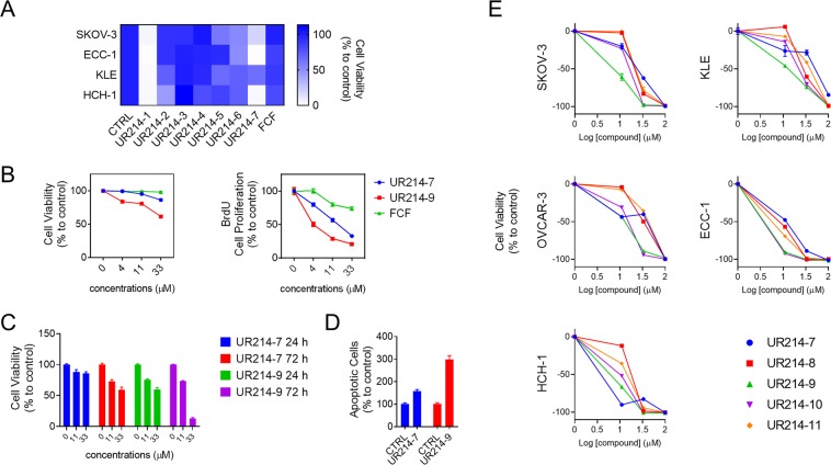
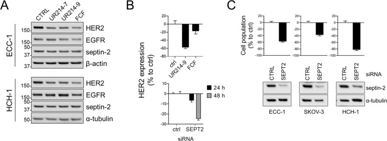
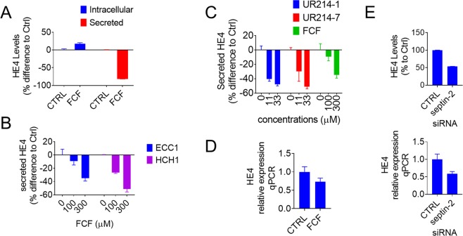
Forchlorfenuron (FCF) is a synthetic plant cytokinin widely used in agriculture to promote fruit size, that paradoxically inhibits proliferation, migration, and invasion in human cancer cell lines. FCF has also been shown to affect HIF-1α and HER2, which are both known to play a crucial role in cancer cell survival. In this study, we have developed potent FCF analogs through structural modification of FCF, coined UR214-1, UR214-7, and UR214-9. Compared to parental FCF, these analogs are more effective in decreasing viability and proliferation in both ovarian and endometrial cancer cell lines. These FCF analogs also suppress HER2 expression at a concentration lower than that of FCF. In addition, we found that treatment with either FCF or its analogs decreases the expression of human epididymis protein 4 (HE4), which is commonly upregulated in ovarian and endometrial cancers. Given the association between cancer behavior and HE4 production in gynecologic cancers, our findings may provide insight useful in the development of new treatment strategies for gynecologic cancers.
Conflict of interest statement
K.K.K., R.K.S., R.T. and R.G.M. are listed on a patent application relating to this study. Other authors declare no competing interests.
Figures
Similar articles
-
Forchlorfenuron and Novel Analogs Cause Cytotoxic Effects in Untreated and Cisplatin-Resistant Malignant Mesothelioma-Derived Cells.Int J Mol Sci. 2022 Apr 2;23(7):3963. doi: 10.3390/ijms23073963. Int J Mol Sci. 2022. PMID: 35409322 Free PMC article.
-
Forchlorfenuron disrupts SEPT9_i1 filaments and inhibits HIF-1.PLoS One. 2013 Aug 19;8(8):e73179. doi: 10.1371/journal.pone.0073179. eCollection 2013. PLoS One. 2013. PMID: 23977378 Free PMC article.
-
A Septin Cytoskeleton-Targeting Small Molecule, Forchlorfenuron, Inhibits Epithelial Migration via Septin-Independent Perturbation of Cellular Signaling.Cells. 2019 Dec 29;9(1):84. doi: 10.3390/cells9010084. Cells. 2019. PMID: 31905721 Free PMC article.
-
Serum HE4 Level as a Biomarker to Predict the Recurrence of Gynecologic Cancers.Curr Drug Targets. 2017;18(10):1158-1164. doi: 10.2174/1389450118666170404154929. Curr Drug Targets. 2017. PMID: 28382859 Review.
-
HE4 tumor marker as a predictive factor for lymphatic metastasis in endometrial cancer.Int J Gynaecol Obstet. 2020 Jun;149(3):265-268. doi: 10.1002/ijgo.13140. Epub 2020 Apr 3. Int J Gynaecol Obstet. 2020. PMID: 32147821 Review.
Cited by
-
Forchlorfenuron and Novel Analogs Cause Cytotoxic Effects in Untreated and Cisplatin-Resistant Malignant Mesothelioma-Derived Cells.Int J Mol Sci. 2022 Apr 2;23(7):3963. doi: 10.3390/ijms23073963. Int J Mol Sci. 2022. PMID: 35409322 Free PMC article.
-
Dysregulation of septin cytoskeletal organization in the trabecular meshwork contributes to ocular hypertension.JCI Insight. 2024 Dec 6;9(23):e179468. doi: 10.1172/jci.insight.179468. JCI Insight. 2024. PMID: 39641270 Free PMC article.
-
Septins in Infections: Focus on Viruses.Pathogens. 2021 Mar 2;10(3):278. doi: 10.3390/pathogens10030278. Pathogens. 2021. PMID: 33801245 Free PMC article. Review.
-
Mechanical Counterbalance of Kinesin and Dynein Motors in a Microtubular Network Regulates Cell Mechanics, 3D Architecture, and Mechanosensing.ACS Nano. 2021 Nov 23;15(11):17528-17548. doi: 10.1021/acsnano.1c04435. Epub 2021 Oct 22. ACS Nano. 2021. PMID: 34677937 Free PMC article.
-
Forchlorfenuron-Induced Mitochondrial Respiration Inhibition and Metabolic Shifts in Endometrial Cancer.Cancers (Basel). 2024 Feb 28;16(5):976. doi: 10.3390/cancers16050976. Cancers (Basel). 2024. PMID: 38473335 Free PMC article.
References
Publication types
MeSH terms
Substances
LinkOut - more resources
Full Text Sources
Other Literature Sources
Research Materials
Miscellaneous
