. 2020 Mar;30(3):229-243.
doi: 10.1038/s41422-020-0287-8. Epub 2020 Feb 24.

Meningeal lymphatic vessels regulate brain tumor drainage and immunity

Affiliations

Meningeal lymphatic vessels regulate brain tumor drainage and immunity

Xueting Hu et al. Cell Res. 2020 Mar.

Abstract

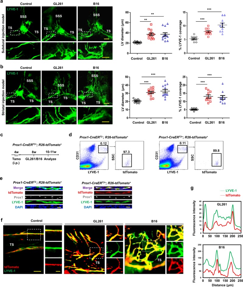
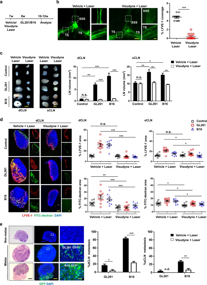
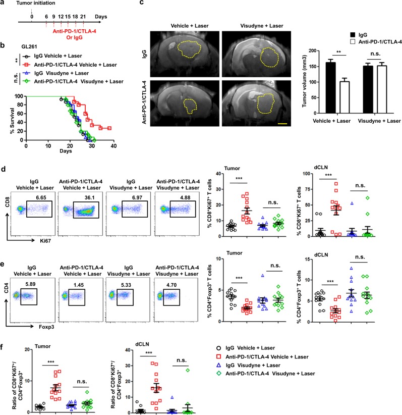
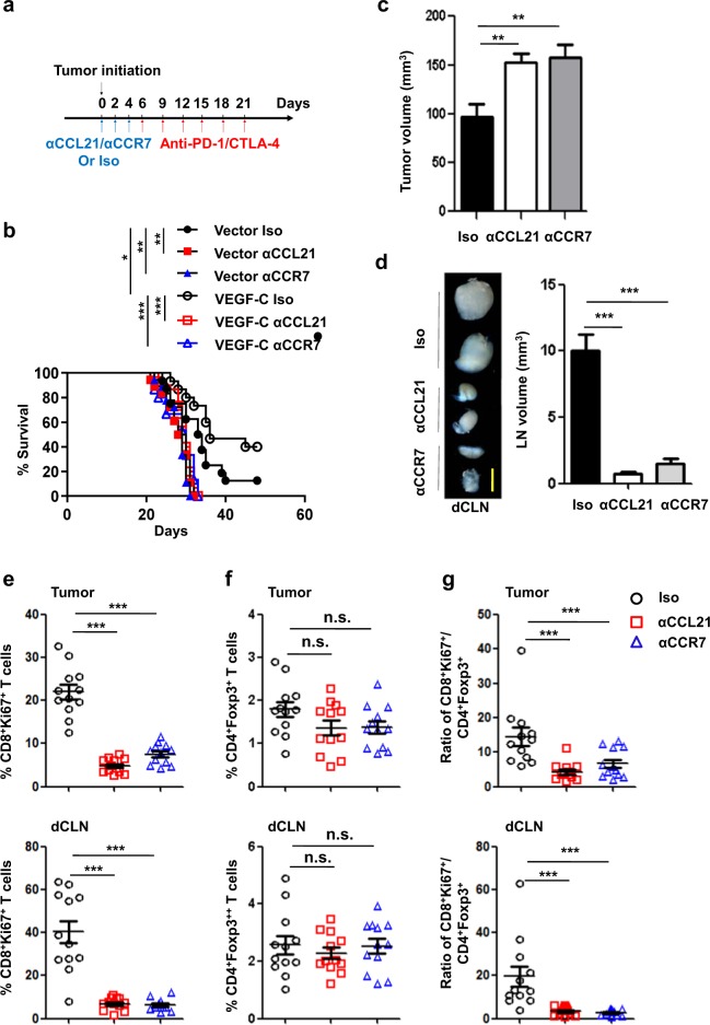
Recent studies have shown that meningeal lymphatic vessels (MLVs), which are located both dorsally and basally beneath the skull, provide a route for draining macromolecules and trafficking immune cells from the central nervous system (CNS) into cervical lymph nodes (CLNs), and thus represent a potential therapeutic target for treating neurodegenerative and neuroinflammatory diseases. However, the roles of MLVs in brain tumor drainage and immunity remain unexplored. Here we show that dorsal MLVs undergo extensive remodeling in mice with intracranial gliomas or metastatic melanomas. RNA-seq analysis of MLV endothelial cells revealed changes in the gene sets involved in lymphatic remodeling, fluid drainage, as well as inflammatory and immunological responses. Disruption of dorsal MLVs alone impaired intratumor fluid drainage and the dissemination of brain tumor cells to deep CLNs (dCLNs). Notably, the dendritic cell (DC) trafficking from intracranial tumor tissues to dCLNs decreased in mice with defective dorsal MLVs, and increased in mice with enhanced dorsal meningeal lymphangiogenesis. Strikingly, disruption of dorsal MLVs alone, without affecting basal MLVs or nasal LVs, significantly reduced the efficacy of combined anti-PD-1/CTLA-4 checkpoint therapy in striatal tumor models. Furthermore, mice bearing tumors overexpressing VEGF-C displayed a better response to anti-PD-1/CTLA-4 combination therapy, and this was abolished by CCL21/CCR7 blockade, suggesting that VEGF-C potentiates checkpoint therapy via the CCL21/CCR7 pathway. Together, the results of our study not only demonstrate the functional aspects of MLVs as classic lymphatic vasculature, but also highlight that they are essential in generating an efficient immune response against brain tumors.


Conflict of interest statement

The authors declare no competing interests.

Figures

Fig. 1
Fig. 1. Brain tumors induce dorsal meningeal lymphangiogenesis.
a, b Left, Representative meningeal LYVE-1 staining 1 week after subdural injection (a) and 2 weeks after striatal injection (b) of GL261 or B16 cells into WT mice (SSS superior sagittal sinus, TS transverse sinus). Right, Quantification of the diameter (n = 12) and percentage area (n = 10) of LYVE-1+ MLVs around the TS. Scale bars, 500 µm in wide-fields; 100 µm in insets. c Schematic diagram of tamoxifen administration and tissue analysis schedule in Prox1-CreERT2+; R26-tdTomato+ mice. d Representative FACS plots and gating scheme of CD31 + LYVE-1+tdTomato+ MLECs isolated from normal Prox1-CreERT2; R26-tdTomato+ and Prox1-CreERT2+; R26-tdTomato+ mice 3 weeks after tamoxifen induction. e Images of Prox1, LYVE-1 staining and tdTomato signals in the TS of meninges from Prox1-CreERT2; R26-tdTomato+ and Prox1-CreERT2+; R26-tdTomato+ mice 3 weeks after tamoxifen induction. Scale bars, 20 µm. f LYVE-1 staining of MLVs around the TS in Prox1-CreERT2+; R26-tdTomato+ mice 2 weeks after subdural injection of GL261 or B16 cells. Scale bars, 100 µm in wide-fields; 50 µm in insets. g Co-localization analysis of tdTomato and LYVE-1 in the insets shown in f. Data are presented as means ± SEM; each symbol represents an individual mouse. **P < 0.01, ***P < 0.001; two-way ANOVA (a, b). Data are from at least three independent experiments (ag).
Fig. 2
Fig. 2. Dorsal meningeal lymphatic vasculature mediates the spread of brain tumor cells to CLNs.
a Protocol: 7-week-old WT mice were treated with Vehicle + Laser or Visudyne + Laser (to ablate dorsal MLVs), and 1 week later GL261 or B16 cells were injected into the striatum. b Left panels, representative meningeal LYVE-1 staining 1 week after mice were treated with Vehicle + Laser or Visudyne + Laser (scale bars, 500 µm in wide-fields; 100 µm in insets). Right panel, quantification of the percentage area of LYVE-1 (n = 12). c Left panels, representative images of dCLNs and sCLNs 2 weeks after striatal injection of GL261 or B16 cells into mice treated with Vehicle + Laser or Visudyne + Laser (scale bars, 1 mm). Right panels, quantification of the volumes of dCLNs and sCLNs (n = 12). d Left panels, representative sections of dCLNs showing DAPI, LYVE-1, and FITC-dextran staining 30 min after injection of FITC-dextran into tumors of mice (same site in control mice) treated with Vehicle + Laser or Visudyne + Laser (scale bars, 300 µm). Right panels, quantification of the fraction of LYVE-1 and FITC-dextran area in dCLNs and sCLNs (n = 10). e Left panels, histology of representative dCLNs containing tumor cell metastases in mice after i.c.m. injection of GL261-GFP+ or B16-GFP+ cells. Fluorescence micrographs of parallel sections reveal strong GFP expression by metastatic tumor cells. Images on the right correspond to the insets (Non-metas, non metastatic; metas, metastatic; scale bars, 500 µm in wide-fields, 50 µm in insets). Right panels, percentages of GFP+ dCLNs and sCLNs (n = 20; n.d. not detected). Data are presented as means ± SEM. *P < 0.05, **P < 0.01, ***P < 0.001, n.s. not significant; two-tailed unpaired Student’s t test (b), or two-way ANOVA (c–e). Data are from at least three independent experiments (ae).
Fig. 3
Fig. 3. Dorsal MLVs are the main route for immune cell entry to draining CLNs.
a Heat map of DEGs (Up, 219; Down, 100; power > 0.4). b, c Gene sets involved in lymphatic remodeling, fluid drainage, as well as inflammatory and immunological responses as shown by the representative upregulated pathways in GL261 tumor-associated and B16 tumor-associated MLECs compared to control MLECs (b), and heat map of DEGs enriched in the antigen processing and presentation pathway (c). d Left panels, treatment scheme and representative flow cytometry dot plots of DC trafficking from GL261 tumors to dCLNs in mice treated with Vehicle + Laser or Visudyne + Laser, determined by the quantity of CD11c+MHCII+FITC+ cells in the dCLNs 24 h after intratumoral injection of FITC-labeled latex beads. Right panel, quantification of Bead+ DCs in the dCLNs of mice treated with Vehicle + Laser or Visudyne + Laser. e Immunoprecipitation of secreted VEGF-C protein (arrow) in conditioned medium from GL261-Vector, GL261-VEGF-C, B16-Vector, and B16-VEGF-C cells. f Left panels, LYVE-1 and CCL21 staining of MLVs in mice bearing Empty and VEGF-C-overexpressing GL261 tumors in the striatum (scale bars, 100 µm in wide-fields; 50 µm in insets). Right panels, quantification of the percentage area of LYVE-1 and CCL21 (n = 10). g Left panels, treatment scheme and representative flow cytometry dot plots of DC trafficking in the dCLNs of mice bearing GL261 tumors overexpressing Vector or VEGF-C. Right panel, quantification of bead+ DCs in dCLNs (n = 10). h Left panels, treatment scheme and representative flow cytometry dot plots of DC trafficking in the dCLNs of GL261 tumor-bearing mice treated with CCL21 (αCCL21)- or IgG (Iso)-blocking antibodies. Right panel, quantification of bead+ DCs in dCLNs (n = 10). Data are presented as means ± SEM. *P < 0.05, **P < 0.01, ***P < 0.001; two-way ANOVA (d, f–h). Data are from at least two (ac) or three (dh) independent experiments.
Fig. 4
Fig. 4. Ablation of dorsal MLVs inhibits anti-brain tumor immune responses.
a Monitoring and treatment scheme of the administration of anti-PD-1/CTLA-4 or IgG controls (days 8, 9, 12, 16, 18, and 21). b Survival of mice treated with Vehicle + Laser or Visudyne + Laser and striatal GL261 tumor injection following the administration of anti-PD-1/CTLA-4 or IgG antibodies (n = 15). c Representative T2-weighted single brain slices (left panels) and quantification (right panel) of tumor volume in MLV-intact and MLV-defective mice bearing striatal GL261 tumors (n = 8). Dashed lines indicate tumor margin. Scale bar, 3 mm. d Representative flow cytometry plots of CD8+Ki67+ T cells in dCLNs (left) and quantification (right) in tumors and dCLNs as percentages of overall CD45+ cells on day 14 after inoculation (n = 12). e Representative flow cytometry plots of CD4+Foxp3+ T cells in dCLNs (left) and quantification (right) in tumors and dCLNs as percentages of overall CD45+cells on day 14 after inoculation (n = 12). f Ratios of CD8+Ki67+ T cells to CD4+Foxp3+ Tregs in tumors and dCLNs. Data are presented as means ± SEM. **P < 0.01, ***P < 0.001, n.s. not significant; long-rank (Mantel–Cox) test (b); two-way ANOVA (c-f). Data are from at least three (a, b, df) or two (c) independent experiments.
Fig. 5
Fig. 5. High level of tumor-derived VEGF-C improves anti-PD-1/CTLA-4 efficacy.
a Survival of mice with striatal Vector- or VEGF-C-overexpressing GL2161 tumors following the administration of anti-PD-1/CTLA-4 or IgG controls (n = 15). b Representative T2-weighted single brain slices from mice with intracranial injection of GL261 cells overexpressing Vector or VEGF-C (n = 6). Scale bar, 3 mm. c Tumor volumes in mice with striatal injection of GL261 cells overexpressing Vector or VEGF-C (n = 6). d, e Quantification of CD8+Ki67+ T cells (d) and CD4+Foxp3+ T cells (e) as percentages of overall CD45+ cells in tumors and in dCLNs on day 14 after inoculation (n = 12 in each). f Ratios of CD8+Ki67+ T cells to Tregs in tumors and in dCLNs. Data are presented as means ± SEM. *P < 0.05, **P < 0.01, ***P < 0.001, n.s. not significant; long-rank (Mantel–Cox) test (a); two-way ANOVA (cf). Data are from at least three (a, df) or two (b, c) independent experiments.
Fig. 6
Fig. 6. Enhancement of immunotherapy by VEGF-C is dependent on CCL21/CCR7 signaling.
a Monitoring and treatment scheme. CCL21/CCR7 were blocked on days 0, 2, and 4 before the administration of anti-PD-1/CTLA-4 antibodies. b Survival of mice with striatal Vector- or VEGF-C-overexpressing GL2161 tumors following the administration of anti-CCL21, anti-CCR7, or IgG (Iso) antibodies combined with anti-PD-1/CTLA-4 antibodies (n = 15). c Tumor volumes in mice with striatal injection of GL261 cells overexpressing VEGF-C following the administration of anti-CCL21, anti-CCR7, or IgG (Iso) antibodies combined with anti-PD-1/CTLA-4 antibodies (n = 8). d Examples (left) and quantification (right) of dCLN volume after GL261 cell injection followed by the administration of anti-CCL21, anti-CCR7, or IgG (Iso) antibodies combined with anti-PD-1/CTLA-4 antibodies (n = 12). Scale bar, 1 mm. e, f Quantification of CD8+Ki67+ T cells (e) and CD4+Foxp3+ T cells (f) as percentages of overall CD45+ cells in tumors and in dCLNs on day 14 after inoculation (n = 12 for each). g Ratios of CD8+Ki67+ T cells to Tregs in tumors and in dCLNs. Data are presented as means ± SEM. *P < 0.05, **P < 0.01, ***P < 0.001, n.s. not significant; long-rank (Mantel–Cox) test (b); two-way ANOVA (cg). Data are from at least two independent experiments (a–g).

Comment in

  • Meningeal lymphatics "drain" brain tumors.
    Kanamori M, Kipnis J. Kanamori M, et al. Cell Res. 2020 Mar;30(3):191-192. doi: 10.1038/s41422-020-0286-9. Cell Res. 2020. PMID: 32111971 Free PMC article. No abstract available.

References

    1. Engelhardt B, Vajkoczy P, Weller RO. The movers and shapers in immune privilege of the CNS. Nat. Immunol. 2017;18:123–131. - PubMed
    1. Shirai Y. On the transplantation of the rat sarcoma in adult heterogeneous animals. Jpn. Med. World. 1921;1:14–15.
    1. Medawar PB. Immunity to homologous grafted skin; the fate of skin homografts transplanted to the brain, to subcutaneous tissue, and to the anterior chamber of the eye. Br. J. Exp. Pathol. 1948;29:58–69. - PMC - PubMed
    1. van Bussel MTJ, Beijnen JH, Brandsma D. Intracranial antitumor responses of nivolumab and ipilimumab: a pharmacodynamic and pharmacokinetic perspective, a scoping systematic review. BMC Cancer. 2019;19:519. - PMC - PubMed
    1. Weller RO, Djuanda E, Yow HY, Carare RO. Lymphatic drainage of the brain and the pathophysiology of neurological disease. Acta Neuropathol. 2009;117:1–14. - PubMed

Publication types