. 2021 Feb;18(2):350-362.
doi: 10.1038/s41423-020-0366-2. Epub 2020 Feb 24.

TRIM34 attenuates colon inflammation and tumorigenesis by sustaining barrier integrity

Affiliations

TRIM34 attenuates colon inflammation and tumorigenesis by sustaining barrier integrity

Qiaoshi Lian et al. Cell Mol Immunol. 2021 Feb.

Abstract

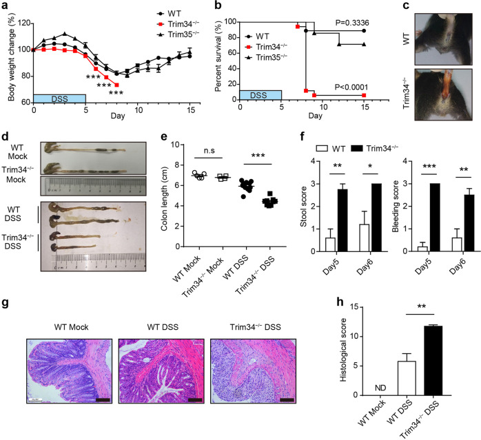
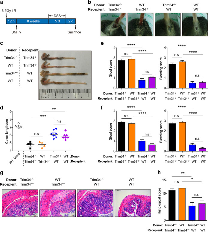
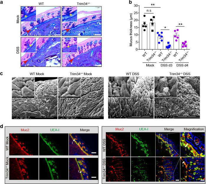
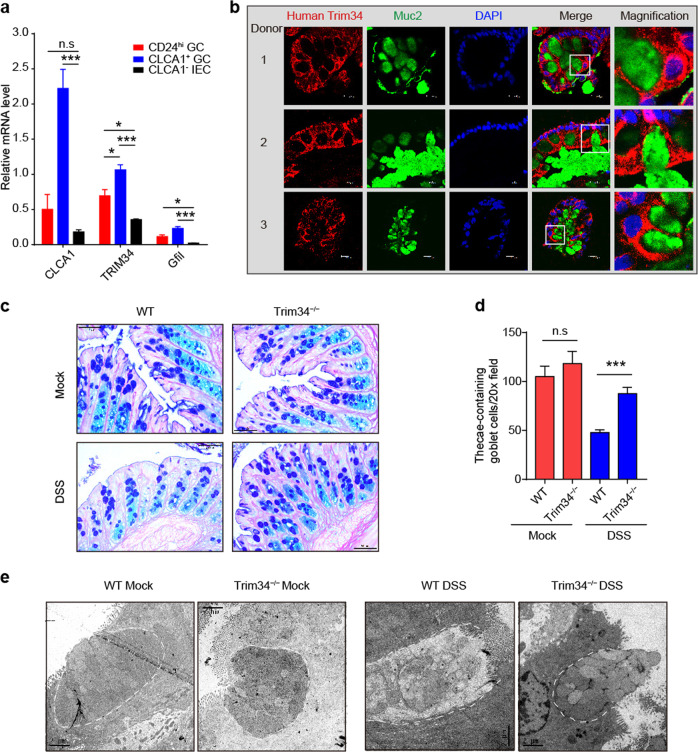
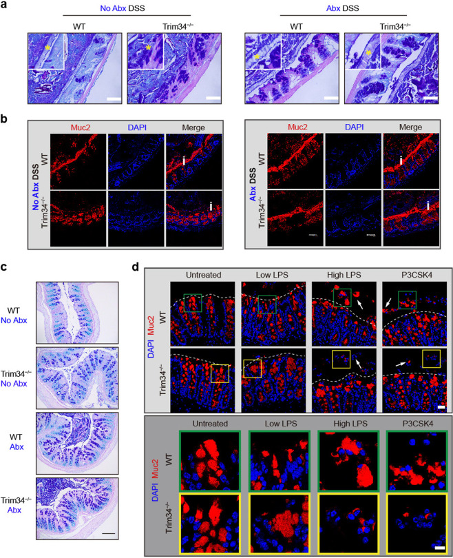
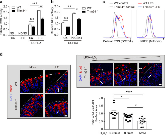
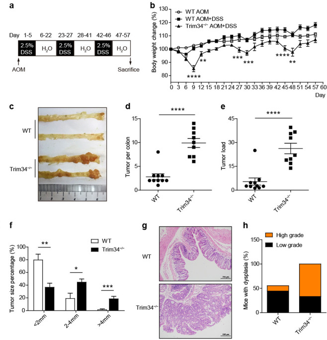
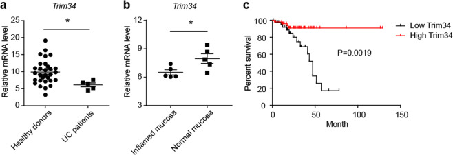
Loss of the colonic inner mucus layer leads to spontaneously severe colitis and colorectal cancer. However, key host factors that may control the generation of the inner mucus layer are rarely reported. Here, we identify a novel function of TRIM34 in goblet cells (GCs) in controlling inner mucus layer generation. Upon DSS treatment, TRIM34 deficiency led to a reduction in Muc2 secretion by GCs and subsequent defects in the inner mucus layer. This outcome rendered TRIM34-deficient mice more susceptible to DSS-induced colitis and colitis-associated colorectal cancer. Mechanistic experiments demonstrated that TRIM34 controlled TLR signaling-induced Nox/Duox-dependent ROS synthesis, thereby promoting the compound exocytosis of Muc2 by colonic GCs that were exposed to bacterial TLR ligands. Clinical analysis revealed that TRIM34 levels in patient samples were correlated with the outcome of ulcerative colitis (UC) and the prognosis of rectal adenocarcinoma. This study indicates that TRIM34 expression in GCs plays an essential role in generating the inner mucus layer and preventing excessive colon inflammation and tumorigenesis.

Keywords: Colon inflammation; Goblet cell; Muc2; TRIM34; Toll-like receptor.


Conflict of interest statement

The authors declare no competing interests.

Figures

Fig. 1
Fig. 1
TRIM34 is required to prevent DSS-induced colitis. a, b Wild-type (n = 7), TRIM34-deficient (n = 7), and TRIM35-deficient (n = 7) mice were treated with 3% DSS as indicated and monitored daily for changes in body weight (a) and survival (b) until the end of the experiment. ce Wild-type (n = 11) and TRIM34-deficient (n = 9) mice were treated with 3% DSS for 5 days. Wild-type mice without DSS challenge (n = 5) and TRIM34-deficient mice without DSS challenge (n = 4) served as controls. c Representative images of diarrhea/bloody stools observed on day 5. d, e Colon length on day 7. fh Wild-type and TRIM34-deficient mice were treated with 2.5% DSS for 5 days (WT, n = 5; KO, n = 4). Untreated wild-type mice served as controls (n = 3). f Clinical stool and bleeding scores were determined on both day 5 and day 6. g Representative images of pathological H&E-stained colon sections collected on day 7. Scale bar, 100 μm. h Histopathological scores for (g). The data shown are representative of three independent experiments and depict the means ± SEM; *P < 0.05, **P < 0.01, and ***P < 0.001; ND not detected; a, e, f, h, unpaired Student’s t test; b log-rank test
Fig. 2
Fig. 2
Nonhematopoietic TRIM34 is responsible for protecting against acute colitis. Chimeric mice were generated by transplanting bone marrow cells from donor mice into lethally irradiated recipient mice (donor mice > recipient mice: WT > WT, n = 7; KO > WT, n = 7; WT > KO, n = 4; KO > KO, n = 4). After 8 weeks of reconstitution, the four groups of mice were treated with 4% DSS for 5 days. a Flow chart of the generation and induction processes. b Representative images of diarrhea and bloody stools on day 5 post-DSS challenge. c Images of the colons from the mice sacrificed on day 7. d Colon length in (c). e, f Stool and bleeding scores on day 5 (e) and day 6 (f). g Representative images of H&E-stained colon sections. Scale bar, 100 μm. h Histopathological scores of (g). The data shown are representative of three independent experiments and depict the means ± SEM; *P < 0.05, **P < 0.01, and ***P < 0.001, ****P < 0.0001; n.s. not significant. df, h One-way ANOVA (Tukey’s multiple-comparisons test)
Fig. 3
Fig. 3
TRIM34 is important for preserving inner mucus layer integrity following challenge with DSS. a Representative images of AB/PAS-stained colonic sections containing stool pellets from mice treated with 3% DSS for 3 days or from untreated mice. Untreated WT (n = 5) and KO (n = 4), DSS-treated WT (n = 6), and KO (n = 5) mice. Asterisks and arrows highlight the inner mucus layers. Scale bar, 50 μm. b Measurement of mucus thickness on day 3 and day 4. c Images obtained by scanning electron microscopy of colons from untreated mice and mice treated with 3% DSS for 3 days. Scale bar, 100 μm. d Colon sections stained for Muc2 (red), fucose (UEA-1, green), and nuclei (DAPI, blue) from untreated mice or mice treated with 3% DSS for 3 days. Scale bar, 50 μm, except for the magnified image, in which the scale bar is 20 μm. The letter i and short line highlight the inner mucus layers. The data shown are representative of two (c) or three (a, b, d) independent experiments and depict the means ± SEM; *P < 0.05 and **P < 0.01; n.s, not significant. b One-way ANOVA (Tukey’s multiple-comparisons test)
Fig. 4
Fig. 4
TRIM34 deficiency attenuates the exocytosis of Muc2-containing granules from goblet cells following treatment with DSS. a Quantitative PCR analysis of CLCA1+ GCs, CD24hi GCs, and CLCA1 IECs. b Human colonic sections obtained from healthy donors were stained for TRIM34 (red), Muc2 (green) and nuclei (DAPI, blue). Scale bar, 10 μm. c Representative images of AB/PAS-stained colonic sections obtained from mice treated with 3% DSS for 3 days or untreated mice. Untreated WT (n = 4) and KO (n = 3), DSS-treated WT (n = 5) and KO (n = 4). Scale bar, 50 μm. d The number of thecae-containing goblet cells shown in (c). e Transmission electron microscopy of colons obtained from untreated mice or mice treated with 3% DSS for 3 days. Scale bar, 2 μm. The discontinuous white line highlights the edge of goblet cells. The data shown are representative of two (e) or three (ad) independent experiments and depict the means ± SEM. *P < 0.05, **P < 0.01, ***P < 0.001. n.s. not significant. a, d, One-way ANOVA (Tukey’s multiple-comparisons test)
Fig. 5
Fig. 5
TRIM34 is critical for the commensal bacterial TLR ligand-induced compound exocytosis of Muc2 from GCs. ac Wild-type and TRIM34-deficient mice were treated with a cocktail of antibiotics for more than 2 weeks to deplete gut commensal bacteria and then challenged with or without 3% DSS for 4 days. Mice not treated with Abx were used as controls; N = 5 per group. a Representative images of AB/PAS-stained colonic sections. Scale bar, 50 μm. Asterisks highlight the inner mucus layers. b Colonic sections stained for Muc2 (red) and nuclei (DAPI, blue). Scale bar, 50 μm. The letter i highlights the inner mucus layer. c AB/PAS staining of colonic sections (no feces). Scale bar, 50 μm. d Freshly isolated colonic explants were unstimulated or stimulated with a low or high concentration of LPS or P3CSK4 for 45 min and then stained for Muc2 (red) and nuclei (DAPI, blue). Scale bar, 25 μm (upper). Scale bar, 10 μm (lower), magnifications from the pictures above. Arrowheads highlight Muc2 granules in crypts. Discontinuous white lines highlight the edges of crypts. The data shown represent three independent experiments
Fig. 6
Fig. 6
TRIM34 controls Muc2 exocytosis by promoting the TLR-induced synthesis of ROS. a, b Relative ROS activity was detected using DCFDA staining in ex vivo colonic epithelial cells that were treated with LPS (a) or P3CSK4 (b). c Bone marrow-derived macrophages were stimulated with LPS (1 μg/ml), and ROS synthesis was then examined with flow cytometry. Total cellular ROS levels were determined using DCFDA (5 μM), and mitochondrial ROS levels were determined using MitoSOX (5 μM). d TRIM34-deficient colonic explants were treated with exogenous H2O2 (50 nM, 500 nM, or 5 μM) during LPS stimulation and then stained for Muc2 (red) and nuclei (DAPI, blue). Quantification of surface area (μm2) based on the surface-markers for Muc2 and DAPI fluorescence using Imaris software with separate samples. The calculation of the ratio of Muc2/DAPI from different sections is shown. Arrowheads highlight either Muc2 granules in or out of crypts. Discontinuous white lines highlight the edges of crypts. The data shown represent three independent experiments and indicate the means ± SEM; *P < 0.05, **P < 0.01, ***P < 0.001, and ****P < 0.0001; ND not detected, n.s. not significant. a, b, d One-way ANOVA (Tukey’s multiple-comparisons test)
Fig. 7
Fig. 7
TRIM34 deficiency renders mice more susceptible to colitis-associated colorectal cancer. Wild-type (n = 10) and TRIM34-deficient (n = 10) mice were treated with AOM and 3 cycles of 2.5% DSS to induce inflammation-driven colorectal cancer. Wild-type mice treated only with AOM were used as controls (n = 5). a Flow chart of the induction procedure. b Body weight changes were monitored every three days. c Representative photos of the colons. d Number of tumors in the colon per mouse. e The sum of the tumor diameters in the colon of each mouse. f The percentage of tumors by different diameter. g Representative H&E-stained colon sections. h Percentages of mice with dysplasia 57 days after AOM injection. Each symbol represents one mouse (d, e). The data shown are representative of two independent experiments and depict the means ± SEM; *P < 0.05, **P < 0.01, ***P < 0.001, and ****P < 0.0001. b, d, e, f Unpaired Student’s t test
Fig. 8
Fig. 8
Colonic TRIM34 expression is correlated with UC and rectal cancer in the clinic. a Relative TRIM34 mRNA abundance was determined using qPCR with colonic mucosal samples obtained from healthy donors and patients diagnosed with UC. b The relative abundance of TRIM34 mRNA in inflamed and normal mucosae in active UC patients. c Overall survival in rectal adenocarcinoma patients (n = 92) based on data stratified by the median normalized amount of TRIM34 mRNA in tumors according to the Colorectal Adenocarcinoma (TCGA, Provisional) data set, which includes tumor RNA-seq data and clinical correlates. *P < 0.05. a Unpaired Student’s t test. b Paired Student’s t test. c Log-rank test

References

    1. Medzhitov R. Origin and physiological roles of inflammation. Nature. 2008;454:428–435. - PubMed
    1. Danese S, Fiocchi C. Ulcerative Colitis. N. Engl. J. Med. 2011;365:1713–1725. - PubMed
    1. Ng SC, et al. Early course of inflammatory bowel disease in a population-based inception cohort study from 8 countries in Asia and Australia. Gastroenterology. 2016;150:86–95. - PubMed
    1. Molodecky NA, et al. Increasing incidence and prevalence of the inflammatory bowel diseases with time, based on systematic review. Gastroenterology. 2012;142:46–54. - PubMed
    1. Ng SC, et al. Incidence and phenotype of inflammatory bowel disease based on results from the Asia-pacific Crohn’s and colitis epidemiology study. Gastroenterology. 2013;145:158–165. - PubMed

Publication types

MeSH terms