Case Reports
. 2020 Feb;13(1):44-51.
doi: 10.14740/gr1262. Epub 2020 Feb 1.

Esophageal Carcinoma Cuniculatum Diagnosed on Mucosal Biopsies Using a Semiquantitative Histologic Schema: Report of Two Esophagectomy-Confirmed Cases

Affiliations
Case Reports

Esophageal Carcinoma Cuniculatum Diagnosed on Mucosal Biopsies Using a Semiquantitative Histologic Schema: Report of Two Esophagectomy-Confirmed Cases

Xiuli Liu et al. Gastroenterology Res. 2020 Feb.

Abstract

Esophageal carcinoma cuniculatum is a rare variant of squamous cell carcinoma characterized by a unique and common histologic pattern including hyperkeratosis, acanthosis, dyskeratosis, deep keratinization, intraepithelial neutrophils, neutrophilic microabscess, focal cytologic atypia, koilocyte-like cells, and keratin-filled cyst/burrows observed in the resection specimens. Preoperative diagnosis can be extremely difficult. A semiquantitative histologic scoring system has been previously proposed for mucosal biopsies, which has been associated with improved diagnostic yield. However, this histologic schema for the diagnosis of carcinoma cuniculatum has not been applied prospectively. Herein, we describe two cases of esophageal carcinoma cuniculatum in patients presenting with progressive dysphagia and esophageal mass. Presurgical endoscopic mucosal biopsies showed features consistent with carcinoma cuniculatum, and a preoperative diagnosis was achieved by applying the aforementioned semiquantitative histologic schema. Both patients underwent neoadjuvant chemoradiation followed by esophagectomy. Both esophagectomy specimens showed residual adventitia-invading carcinoma cuniculatum, negative lymph nodes, marked tumor regression, and an exuberant histiocytic and giant response. To our best knowledge, these represent the first two cases of esophageal carcinoma cuniculatum diagnosed by applying this semiquantitative histologic schema to mucosal biopsies. Large studies are needed to further confirm these preliminary findings and validate this histologic scoring system.

Keywords: Carcinoma cuniculatum; Esophagus; Mucosal biopsy; Squamous cell carcinoma.


Conflict of interest statement

None to declare for all authors.

Figures

Figure 1
Figure 1
(a) Endoscopic finding of an esophageal mass on esophagogastroduodenoscopy (case 1). (b) Mural destruction by the mass on endoscopic ultrasound (EUS) examination. (c) Enlarged hypoechoic nodes, suspicious for metastases on EUS examination. (d) Positron emission tomography (PET) revealed a hypermetabolic mass in the distal esophagus (axial view).
Figure 2
Figure 2
Histologic features of mucosal biopsy from the mass (case 1). Squamous epithelium shows hyperkeratosis and acanthosis ((a) hematoxylin and eosin stain, original magnification × 40), focal atypia ((b) hematoxylin and eosin stain, original magnification × 200), intraepithelial neutrophilic inflammation and microabscesses and dyskeratosis ((c) hematoxylin and eosin stain, original magnification × 200), koilocyte-like cells ((d) hematoxylin and eosin stain, original magnification × 200), deep keratinization ((e) hematoxylin and eosin stain, original magnification × 100), and furrow ((f) hematoxylin and eosin stain, original magnification × 20).
Figure 3
Figure 3
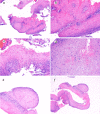
Esophagectomy (case 1) shows mural fibrosis and inflammation ((a) hematoxylin and eosin stain, original magnification × 20), few residual cyst lined with well-differentiated bland squamous epithelium ((b) hematoxylin and eosin stain, original magnification × 100), and histiocytic and giant cell response to keratinous material ((c) hematoxylin and eosin stain, original magnification × 200) and residual squamous carcinomatous cells ((d) hematoxylin and eosin stain, original magnification × 200).
Figure 4
Figure 4
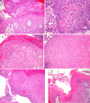
Histologic features of mucosal biopsy from the mass (case 2). Squamous epithelium shows hyperkeratosis and acanthosis ((a) hematoxylin and eosin stain, original magnification × 40), focal atypia ((b) hematoxylin and eosin stain, original magnification × 400), intraepithelial neutrophilic inflammation and microabscesses and dyskeratosis ((c) hematoxylin and eosin stain, original magnification × 200), koilocyte-like cells ((d) hematoxylin and eosin stain, original magnification × 200), deep keratinization ((e) hematoxylin and eosin stain, original magnification × 200), and furrow ((f) hematoxylin and eosin stain, original magnification × 40).
Figure 5
Figure 5
Esophagectomy (case 2) shows histiocytic and giant cell response to keratinous material and residual terminally differentiated squamous carcinomatous cells ((a) and (b) hematoxylin and eosin stain, original magnification × 100 and × 200, respectively).

References

    1. Aird I, Johnson HD, Lennox B, Stansfeld AG. Epithelioma cuniculatum: a variety of squamous carcinoma peculiar to the foot. Br J Surg. 1954;42(173):245–250. doi: 10.1002/bjs.18004217304. - DOI - PubMed
    1. Barreto JE, Velasquez EF, Ayala E. et al. Carcinoma cuniculatum: a distinct variant of penile squamous cell carcinoma. Am J Surg Pathol. 2007;31:71–75. doi: 10.1097/01.pas.0000213401.72569.22. - DOI - PubMed
    1. Landau M, Goldblum JR, DeRoche T, Dumot J, Downs-Kelly E, Rice TW, Xiao SY. et al. Esophageal carcinoma cuniculatum: report of 9 cases. Am J Surg Pathol. 2012;36(1):8–17. doi: 10.1097/PAS.0b013e3182348aa1. - DOI - PubMed
    1. De Petris G, Lewin M, Shoji T. Carcinoma cuniculatum of the esophagus. Ann Diagn Pathol. 2005;9(3):134–138. doi: 10.1016/j.anndiagpath.2005.02.002. - DOI - PubMed
    1. Goh GH, Venkateswaran K, Leow PC, Loh KS, Thamboo TP, Petersson F. Carcinoma cuniculatum of the esophagus and tongue: report of two cases, including TP53 mutational analysis. Head Neck Pathol. 2014;8(3):261–268. doi: 10.1007/s12105-014-0522-z. - DOI - PMC - PubMed

Publication types

LinkOut - more resources