Review
. 2020 Feb 17:9:7.
doi: 10.1186/s40035-020-0185-5. eCollection 2020.

Insights into the pathogenesis of multiple system atrophy: focus on glial cytoplasmic inclusions

Affiliations
Review

Insights into the pathogenesis of multiple system atrophy: focus on glial cytoplasmic inclusions

Seiji Kaji et al. Transl Neurodegener. .

Abstract

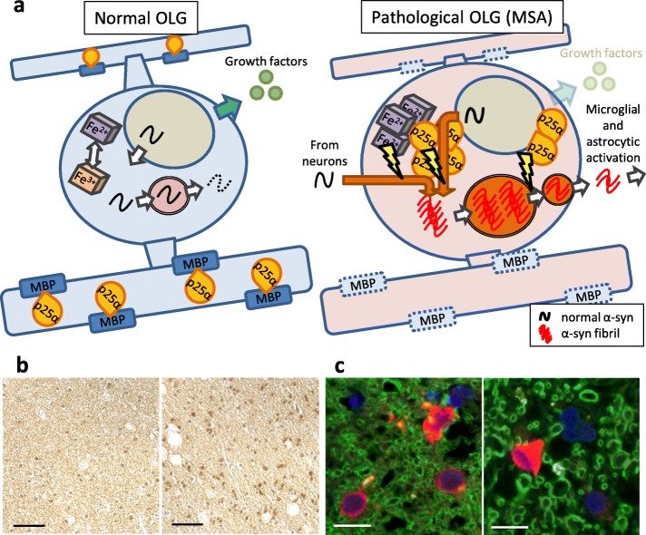
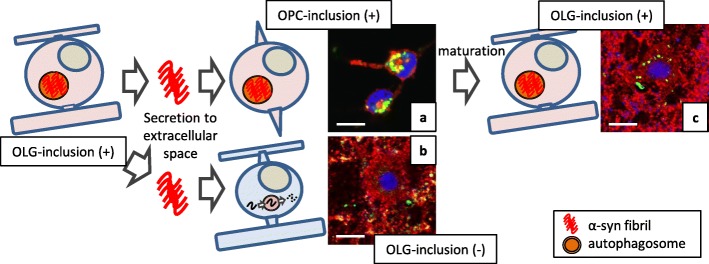
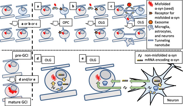
Multiple system atrophy (MSA) is a debilitating and fatal neurodegenerative disorder. The disease severity warrants urgent development of disease-modifying therapy, but the disease pathogenesis is still enigmatic. Neurodegeneration in MSA brains is preceded by the emergence of glial cytoplasmic inclusions (GCIs), which are insoluble α-synuclein accumulations within oligodendrocytes (OLGs). Thus, preventive strategies against GCI formation may suppress disease progression. However, although numerous studies have tried to elucidate the molecular pathogenesis of GCI formation, difficulty remains in understanding the pathological interaction between the two pivotal aspects of GCIs; α-synuclein and OLGs. The difficulty originates from several enigmas: 1) what triggers the initial generation and possible propagation of pathogenic α-synuclein species? 2) what contributes to OLG-specific accumulation of α-synuclein, which is abundantly expressed in neurons but not in OLGs? and 3) how are OLGs and other glial cells affected and contribute to neurodegeneration? The primary pathogenesis of GCIs may involve myelin dysfunction and dyshomeostasis of the oligodendroglial cellular environment such as autophagy and iron metabolism. We have previously reported that oligodendrocyte precursor cells are more prone to develop intracellular inclusions in the presence of extracellular fibrillary α-synuclein. This finding implies a possibility that the propagation of GCI pathology in MSA brains is mediated through the internalization of pathological α-synuclein into oligodendrocyte precursor cells. In this review, in order to discuss the pathogenesis of GCIs, we will focus on the composition of neuronal and oligodendroglial inclusions in synucleinopathies. Furthermore, we will introduce some hypotheses on how α-synuclein pathology spreads among OLGs in MSA brains, in the light of our data from the experiments with primary oligodendrocyte lineage cell culture. While various reports have focused on the mysterious source of α-synuclein in GCIs, insights into the mechanism which regulates the uptake of pathological α-synuclein into oligodendroglial cells may yield the development of the disease-modifying therapy for MSA. The interaction between glial cells and α-synuclein is also highlighted with previous studies of post-mortem human brains, cultured cells, and animal models, which provide comprehensive insight into GCIs and the MSA pathomechanisms.

Keywords: Astrocyte; Glial cytoplasmic inclusion; Microglia; Multiple system atrophy; Neurodegeneration; Oligodendrocyte; Oligodendrocyte precursor cell; Prion; α-Synuclein.


Conflict of interest statement

Competing interestsThe authors declare that they have no competing interests.

Figures

Fig. 1
Fig. 1
Hypothetical overview of GCI pathogenesis and post-mortem analysis of TPPP/p25α translocation. a: Hypothetical overview of a normal OLG (left) and a pathological OLG generating GCI (right). Left: Intracellular homeostasis is maintained by normal expression levels of myelin-associated proteins and their colocalization with TPPP/p25α as well as autophagic degradation of endogenous α-syn and balanced iron metabolism. Right: Aggregation formation is enhanced by decreased expression of myelin-associated proteins, cytosolic translocation of TPPP/p25α, impaired autophagy-lysosomal degradation, and oxidation of ferrous to ferric ions. Secretion of pathological α-syn in response to insufficient degradation leads to microglial and astrocytic activation. OLG dysfunction also causes compromised neuronal support such as reduced production of neurotrophic factors. b: Translocation of TPPP/p25α from myelin to cell bodies in the frontal cortex white matter of a control patient (left) and an MSA patient (right). The scale bar represents 50 μm. c: Localization of TPPP/p25α (red) and its interaction with MBP (green) in the frontal cortex white matter of a control patient (left) and an MSA patient (right). Blue; DAPI. The scale bar represents 10 μm
Fig. 2
Fig. 2
Difference in the response to extracellularly applied pathological α-syn between two oligodendroglial cells. Pathological α-syn with seeding property hypothetically propagates from OLG to OLG. Extracellular α-syn fibrils do not induce inclusions when they are applied to mature OLGs. Seed internalization can occur more drastically during the immature state of OLG differentiation including the precursor state. The mechanism which regulates the uptake of misfolded α-syn may be shared by normal OPCs and pathological OLGs in MSA brains, but not by normal OLGs. a: intercellular localization of α-syn (green)-immunoreactive inclusions in platelet-derived growth factor receptor α (red)-positive primary rat OPCs, which were incubated for 72 h with 1 μM human recombinant α-syn pre-formed fibrils (PFFs). b: MBP (red)-positive primary rat OLGs with extracellular Thioflavin S (green) immunoreactivity, which were incubated for 24 h with 1 μM human recombinant α-syn PFFs after maturation. c: MBP (red)-positive primary rat OLGs containing Thioflavin S (green)-positive intracellular inclusions. OLGs were differentiated from OPCs that were pre-incubated for 24 h with 1 μM human recombinant α-syn PFFs. a-c: Each scale bar represents 10 μm. Blue; DAPI.
Fig. 3
Fig. 3
Hypothetical schema showing OLG-to-OLG propagation and accumulation of pathological α-syn. Pathological α-syn with seeding property hypothetically propagates from OLG to OLG leading to the spreading of GCI pathology. Given that the uptake of extracellularly-applied misfolded α-syn is not usually observed in normal OLGs, the entry of misfolded α-syn into oligodendroglial cells is mediated through an unidentified mechanism (a-c). Once the misfolded α-syn (pre-GCI) enters oligodendroglial cells, the pre-GCI self-assembles through the interaction with non-misfolded α-syn, resulting in the formation of perinuclear fibrillary structure (mature GCIs). The non-misfolded α-syn may be derived from OLGs or neurons (d, e)

References

    1. Watanabe H, Saito Y, Terao S, Ando T, Kachi T, Mukai E, et al. Progression and prognosis in multiple system atrophy: an analysis of 230 Japanese patients. Brain. 2002;125(Pt 5):1070–1083. doi: 10.1093/brain/awf117. - DOI - PubMed
    1. Gilman S, Wenning GK, Low PA, Brooks DJ, Mathias CJ, Trojanowski JQ, et al. Second consensus statement on the diagnosis of multiple system atrophy. Neurology. 2008;71(9):670–676. doi: 10.1212/01.wnl.0000324625.00404.15. - DOI - PMC - PubMed
    1. Mitsui J, Matsukawa T, Sasaki H, Yabe I, Matsushima M, Durr A, et al. Variants associated with Gaucher disease in multiple system atrophy. Ann Clin Trans Neurol. 2015;2(4):417–426. doi: 10.1002/acn3.185. - DOI - PMC - PubMed
    1. Ozawa T, Tada M, Kakita A, Onodera O, Tada M, Ishihara T, et al. The phenotype spectrum of Japanese multiple system atrophy. J Neurol Neurosurg Psychiatry. 2010;81(11):1253–1255. doi: 10.1136/jnnp.2009.182576. - DOI - PubMed
    1. Gai WP, Pountney DL, Power JH, Li QX, Culvenor JG, McLean CA, et al. Alpha-Synuclein fibrils constitute the central core of oligodendroglial inclusion filaments in multiple system atrophy. Exp Neurol. 2003;181(1):68–78. doi: 10.1016/S0014-4886(03)00004-9. - DOI - PubMed

Publication types

MeSH terms

LinkOut - more resources