. 2020 Feb 11:25:e00434.
doi: 10.1016/j.btre.2020.e00434. eCollection 2020 Mar.

Optimization of recombinant Zika virus NS1 protein secretion from HEK293 cells

Affiliations

Optimization of recombinant Zika virus NS1 protein secretion from HEK293 cells

Julieta S Roldán et al. Biotechnol Rep (Amst). .

Abstract

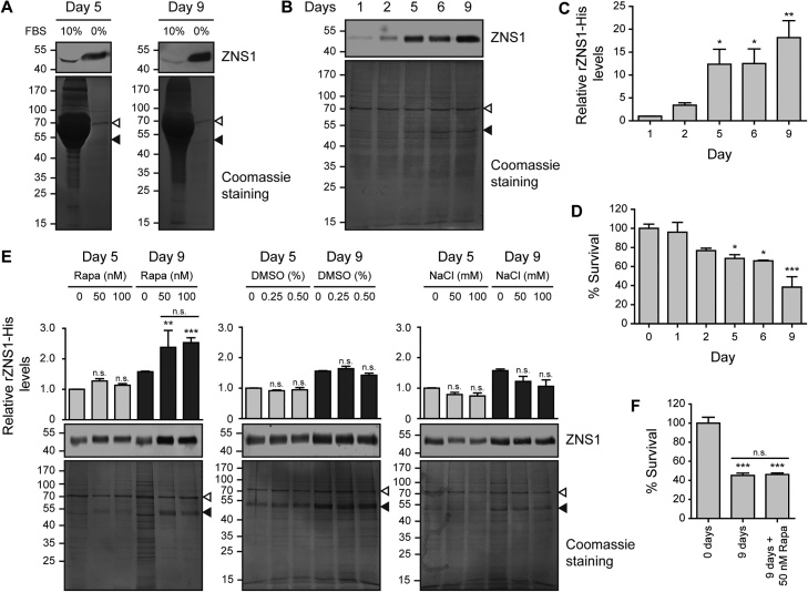
Sensitive, accurate and cost-effective diagnostic tests are urgently needed to detect Zika virus (ZIKV) infection. Nonstructural 1 (NS1) glycoprotein is an excellent diagnostic marker since it is released in a hexameric conformation from infected cells into the patient's bloodstream early in the course of the infection. We established a stable rZNS1-His-expression system in HEK293 cells through lentiviral transduction. A novel optimization approach to enhance rZNS1-His protein secretion in the mammalian expression system was accomplished through 50 nM rapamycin incubation followed by serum-free media incubation for 9 days, reaching protein yields of ∼10 mg/l of culture medium. Purified rZNS1-His hexamer was recognized by anti-NS1 antibodies in ZIKV patient's serum, and showed the ability to induce a humoral response in immunized mice. The obtained recombinant protein is a reliable biological tool that can potentially be applied in the development of diagnostic tests to detect ZIKV in infected patients during the acute phase.

Keywords: Mammalian expression system; NS1; Recombinant protein; Zika virus.


Conflict of interest statement

The authors declare that they have no known competing financial interests or personal relationships that could have appeared to influence the work reported in this paper.

Figures

Fig. 1
Fig. 1
Development of stable rZNS1-His-expressing HEK293 cells. (A) Immunostaining of untransduced (UTD) HEK293 cells, parental, 1E4, 1E4-C9 and 1E4-D11 rZNS1-His HEK293 cells with ZNS1 mouse polyclonal sera. Scale bar, 25 μm. (B) Determination of the percentage of positive rZNS1-His-expressing cells assessed by flow cytometry in UTD, parental, 1E4, 1E4-C9 and 1E4-D11 rZNS1-His HEK293 cells. Only secondary Ab was used as control. (C-D) Western blot analysis of cell extracts (C) and supernatants (D) of UTD, 1E4, 1E4-C9 and 1E4-D11 rZNS1-His HEK293 cells with anti-ZNS1 monoclonal antibody. GAPDH (C) and Coomassie brilliant blue staining (D) were used as loading controls. The position of the molecular mass standards is indicated on the right. Arrowheads in (C) indicate rZNS1-His monomer glycosylation pattern. Data represent the mean ± SEM of at least three independent experiments. P-values were calculated by one-way ANOVA, Bonferroni's: **P < 0.01, ***P < 0.001, n.s. not significant.
Fig. 2
Fig. 2
Optimal conditions for rZNS1-His increased secretion from 1E4-C9 HEK293 cells. (A-C) Western blot analysis with anti-ZNS1 monoclonal antibody of supernatants from 1E4-C9 cells incubated in the presence or absence of fetal bovine serum (FBS) (A) or in serum-free media for the indicated days (B-C). (D) Cell survival assessed by MTT reduction assay in 1E4-C9 cells incubated in serum-free media for the indicated days. (E) Immunoblot analysis with anti-ZNS1 monoclonal antibody of supernatants from 1E4-C9 cells subjected to treatment with 50 nM or 100 nM rapamycin (Rapa), 0.25 % or 0.50 % DMSO, or hypotonic medium (50 mM or 100 mM NaCl), followed by incubation in serum-free media for 5 or 9 days. (F) MTT assay in 1E4-C9 cells treated or not with 50 nM rapamycin and incubated in serum-free media for 9 days. In (A,B,E) Coomassie brilliant blue staining was used as loading control, filled arrowheads show rZNS1-His position whereas hollow arrowheads show the remaining bovine serum albumin contained in the serum-free culture media following a PBS washing step, and the positions of the molecular mass standards are indicated on the left. Data in (C,D,E,F) represent mean ± SEM of at least three independent experiments. P-values were calculated by one-way ANOVA, Dunnett's in (C,D) and Bonferroni's in (E,F): *P < 0.05, **P < 0.01, ***P < 0.001, n.s. not significant.
Fig. 3
Fig. 3
Purification and conformational characterization of secreted rZNS1-His. (A) Nickel affinity chromatography purification of rZNS1-His from 10X dialyzed supernatant of 1E4-C9 cells treated with 50 nM rapamycin and incubated in serum-free media for 9 days. Fractions collected were visualized by Coomassie brilliant blue staining. (B) Western blot analysis with anti-ZNS1 monoclonal antibody of 1E4-C9 supernatant processing steps prior to purification by nickel affinity chromatography. Coomassie brilliant blue staining was used as loading control. (C) Dot blot analysis with ZNS1 mouse polyclonal sera of 1E4-C9 supernatant, and 10 × 1E4-C9 supernatant or permeate fractions obtained by ultrafiltration with a 100 kDa MWCO disc membrane. (D) Immunoblot analysis with anti-ZNS1 monoclonal antibody of purified rZNS1-His subjected or not to heat (5 min 100 °C) and/or 100 mM DTT pretreatment. (E) Gel filtration chromatography of rZNS1-His showing a molecular mass corresponding to a hexameric quaternary structure (upper panel), and Western blot analysis with anti-His tag monoclonal antibody of the eluted fractions corresponding to rZNS1-His peak (lower panel). (F) In vitro cross-linking of rZNS1-His with different concentrations of glutaraldehyde (GA) analyzed by Western blot with anti-His tag monoclonal antibody (left panel) and with Coomassie brilliant blue staining (right panel). In (A,B,E) filled arrowheads show rZNS1-His position whereas in (B) hollow arrowheads show the remaining bovine serum albumin contained in the serum-free culture media following a PBS washing step, and in (A,B,D,E,F), the positions of the molecular mass standards are indicated on the left.
Fig. 4
Fig. 4
rZNS1-His reactivity with ZIKV infected patients sera and immunogenicity in a mouse model. (A) iELISA of rZNS1-His with serum samples obtained from patients with a positive diagnosis of ZIKV. Healthy individuals sera were used as negative control. Each point of the curve represents mean ± SEM of three sample replicates. (B) Dot Blot analysis with the indicated serum samples of rZNS1-His or ZIKV stock obtained from C6/36 cells infected with ZIKV PRVABC59 Puerto Rico strain. Mock-infected C6/36 cells supernatant was loaded as negative control. (C) iELISA showing humoral responses of mice immunized with rZNS1-His. Non-immunized mouse sera was used as a negative control. Each point of the curve represents mean ± SD of two sample replicates.
None
None
None

References

    1. Dick G.W., Kitchen S.F., Haddow A.J. Zika virus. I. Isolations and serological specificity. Trans. R. Soc. Trop. Med. Hyg. 1952;46:509–520. - PubMed
    1. Fauci A.S., Morens D.M. Zika Virus in the Americas--Yet another arbovirus threat. N. Engl. J. Med. 2016;374:601–604. - PubMed
    1. Duffy M.R., Chen T.H., Hancock W.T., Powers A.M., Kool J.L. Zika virus outbreak on Yap Island, Federated States of Micronesia. N. Engl. J. Med. 2009;360:2536–2543. - PubMed
    1. Oehler E., Watrin L., Larre P., Leparc-Goffart I., Lastere S. Zika virus infection complicated by Guillain-Barre syndrome--case report, French Polynesia, December 2013. Euro Surveill. 2014;19 - PubMed
    1. WHO . WHO newsletter; 2016. WHO Director-General Summarizes the Outcome of the Emergency Committee Regarding Clusters of Microcephaly and Guillain-barré Syndrome.