Review
. 2019:1184:81-95.
doi: 10.1007/978-981-32-9358-8_7.

Tau and Axonal Transport Misregulation in Tauopathies

Affiliations
Review

Tau and Axonal Transport Misregulation in Tauopathies

Benjamin Combs et al. Adv Exp Med Biol. 2019.

Abstract

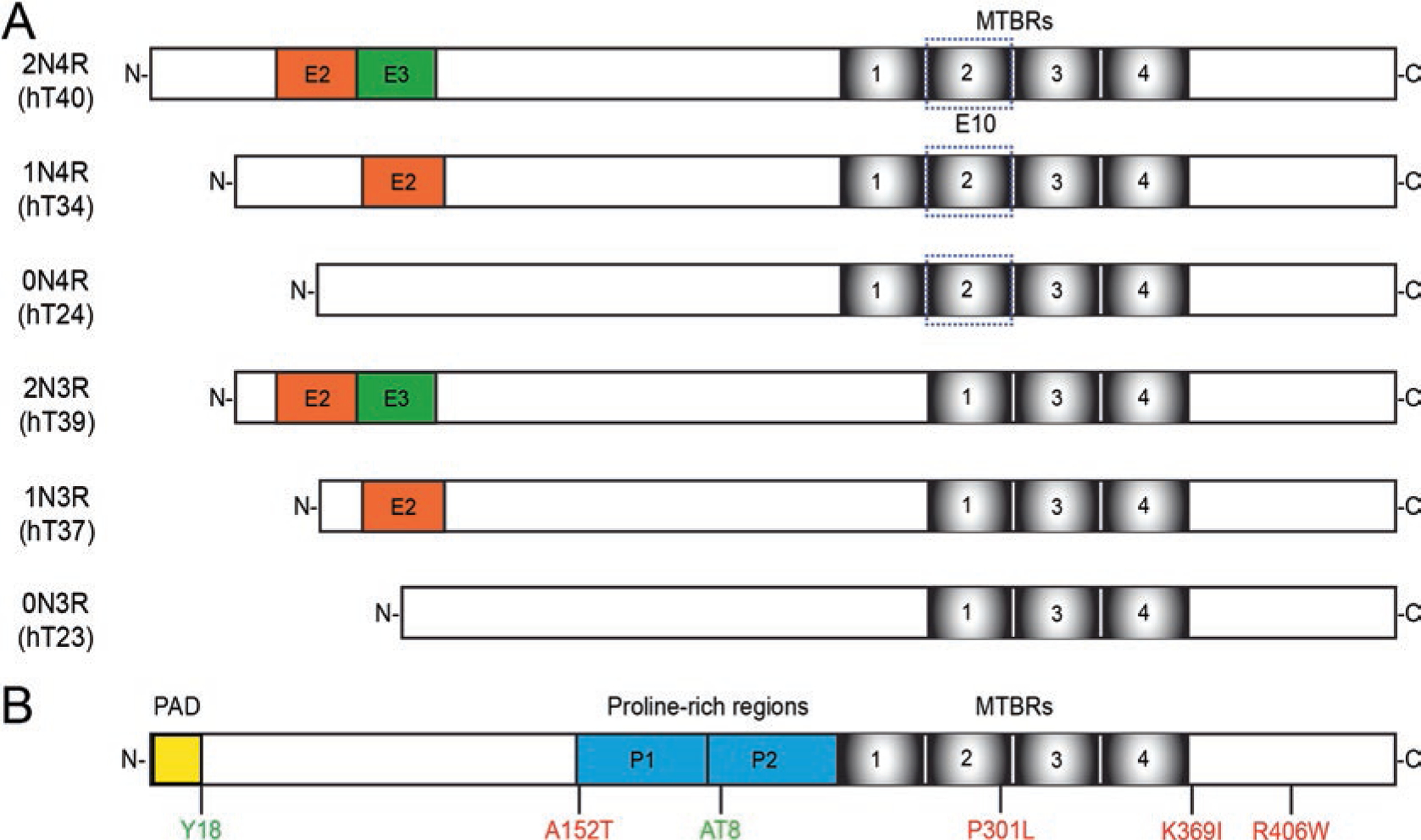
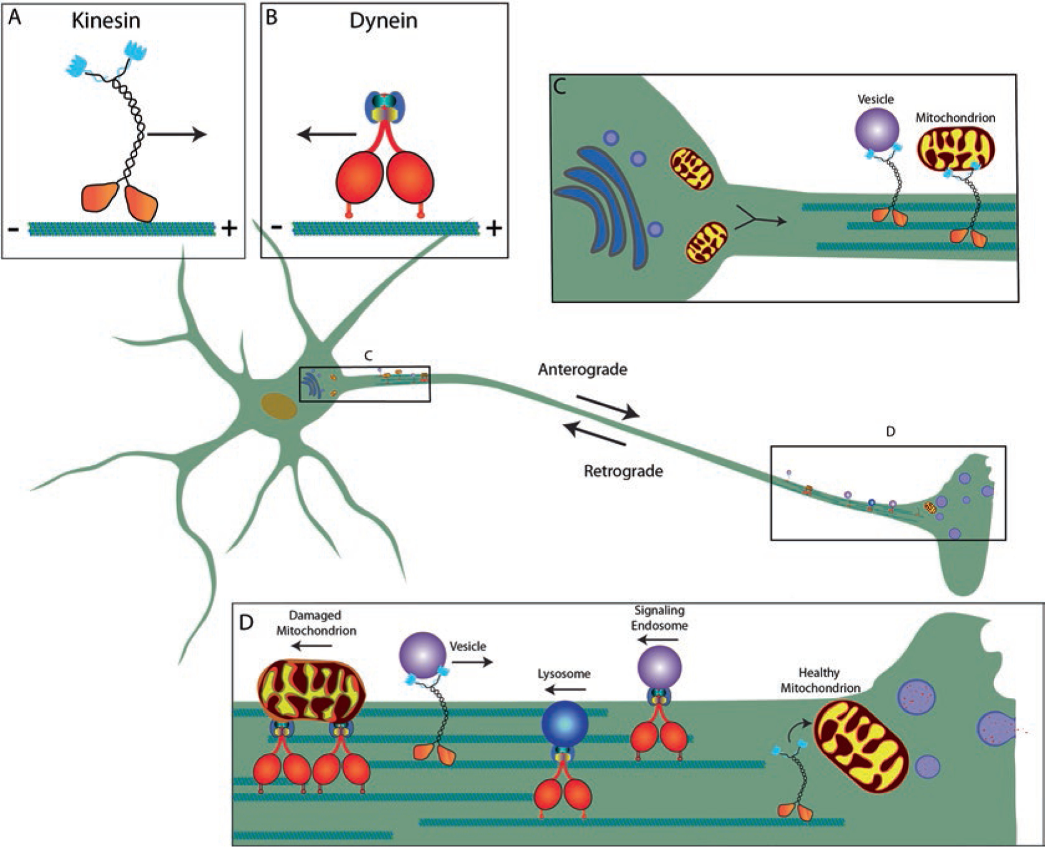
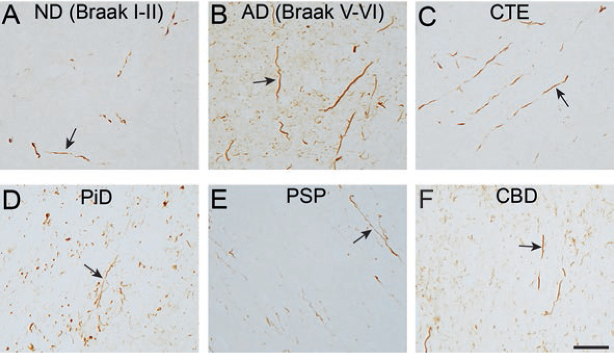
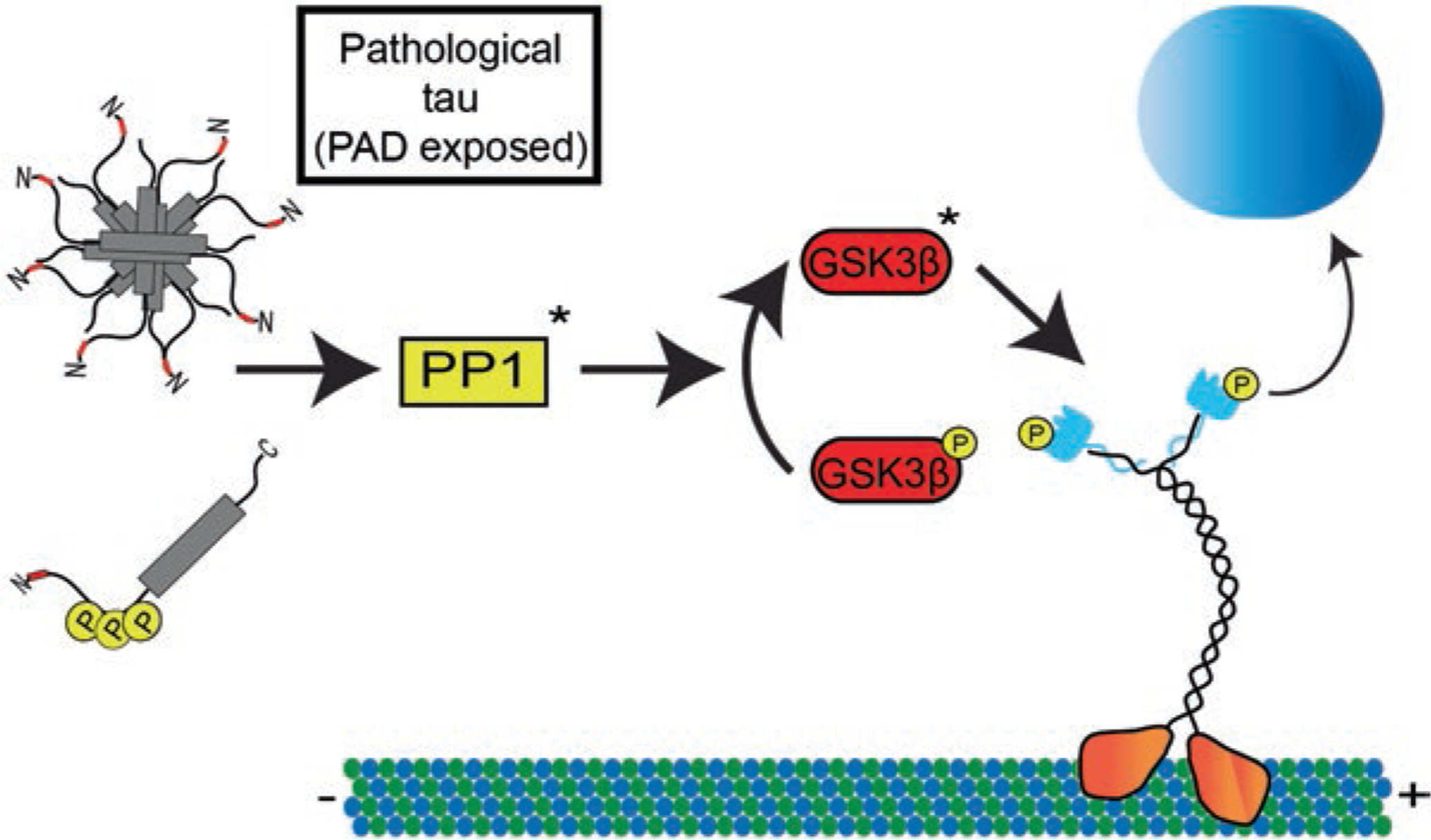
Tau is a microtubule-associated protein that is involved in both normal and pathological processes in neurons. Since the discovery and characterization of tau over 40 years ago, our understanding of tau's normal functions and toxic roles in neurodegenerative tauopathies has continued to expand. Fast axonal transport is a critical process for maintaining axons and functioning synapses, critical subcellular compartments underlying neuronal connectivity. Signs of fast axonal transport disruption are pervasive in Alzheimer's disease and other tauopathies and various mechanisms have been proposed for regulation of fast axonal transport by tau. Post-translational modifications of tau including phosphorylation at specific sites, FTDP-17 point mutations, and oligomerization, confer upon tau a toxic effect on fast axonal transport. Consistent with the well-established dependence of axons on fast axonal transport, these disease-related modifications are closely associated temporally and spatially with axonal degeneration in the early disease stages. These factors position tau as a potentially critical factor mediating the disruption of fast axonal transport that precedes synaptic dysfunction and axonal degeneration at later disease stages. In this chapter, we review the evidence that tau affects fast axonal transport and examine several potential mechanisms proposed to underlie this toxicity.

Keywords: Alzheimer’s disease; Axonal transport; Axons; Kinases; Neurodegeneration; Phosphatases; Tau protein; Tauopathies.


Figures

Fig. 7.1
Fig. 7.1
Tau isoforms and selected modifications. (a) In the adult human brain tau primarily exists as six isoforms generated through alternative mRNA splicing. The isoforms differ based on the inclusion of exons 2 and 3 in the N-terminus of the protein (2 N, 1 N, or 0 N) and exon 10. Exon 10 contains the second of four potential microtubule-binding repeat regions. Isoforms are referred to as 3R or 4R based on the number of repeat regions they contain. (b) Modifications to the protein can affect its function and induce dysfunction in disease. Some selected modifications discussed here include phosphorylation sites (green) at tyrosine 18 and the AT8 sites (serine 202 and threonine 205). FTDP-17 mutations can lead to inherited early-onset frontotemporal dementias (reviewed in [53]). Several of these mutations have been linked to FAT dysfunction including A152T, P301L, K369I, and R406W (red). Functional domains associated with transport include the phosphatase activating domain (yellow), a motif that is conformationally displayed in disease-related forms of tau and linked to changes in signaling pathways that regulate transport. The tau molecule contains many phosphorylation sites throughout the sequence, some of which are found in healthy tissues and others are associated with tau pathology. (Reviewed in [84])
Fig. 7.2
Fig. 7.2
Neurons depend on robust microtubule-based transport in axons. A healthy, functional neuron is dependent on the molecular motor complexes kinesin (a) and dynein (b), whose roles are to transport material along microtubules in the plus- (anterograde) or minus-end (retrograde) direction, respectively. Materials synthesized in the soma (e.g., cytoskeletal components, mitochondria and membrane-bound organelles) rely on kinesin for their delivery to the correct axonal compartment (c). Kinesin-driven anterograde transport is necessary for the delivery of synaptic components, including mitochondria and vesicles, to the axon terminal where they will aid in cell signaling as well as delivery of channels to axon to support propagations of the action potential. Dynein-driven retrograde transport is necessary for the transport of signaling endosomes and material undergoing breakdown and recycling, like damaged mitochondria, multivesicular bodies and lysosomes (d) back to the neuronal soma
Fig. 7.3
Fig. 7.3
Dystrophic axons containing tau pathology is a prominent feature in multiple tauopathies. The TNT1 antibody detects exposure of the phosphatase activating domain (PAD). (a, b) TNT1 pathology-containing axons are observed in the subcortical white matter in non-demented aged patients with early stages of tau deposition (a; ND (Braak stage I-II)) and robust TNT1 axonal pathology is seen in severe Alzheimer’s disease brains (b; AD (Braak stage V-VI). C-F) Axonal tau pathology in the subcortical white matter displays PAD exposure (i.e. TNT1 reactivity) in chronic traumatic encephalopathy (c; CTE), Pick’s disease (d; PiD), progressive supranuclear palsy (e; PSP) and corticobasal degeneration (f; CBD) as well. Scale bar is 50 μm
Fig. 7.4
Fig. 7.4
Tau alters kinesin-cargo interactions through modulation of a PP1-GSK3β signaling pathway. Tau contains a phosphatase-activating domain (PAD) within amino acids 2–18 at the extreme N-terminus of tau (shown in red). Under normal conditions this epitope is obscured allowing for normal kinesin-based transport along the microtubules. In disease conditions tau can undergo a variety of modifications including aggregation or specific phosphorylation events that can aberrantly expose the PAD epitope. In the proposed model, these forms of tau can disrupt normal kinesin-based transport by activating protein phosphatase 1 (PP1). This in turn dephosphorylates an inhibitory phosphate on GSK3β in order to activate it. GSK3β then phosphorylates kinesin light chain inducing a release of cargo and disruption of FAT

References

    1. Adalbert R, Milde S, Durrant C, Ando K, Stygelbout V, Yilmaz Z, Gould S, Brion JP, Coleman MP. Interaction between a MAPT variant causing frontotemporal dementia and mutant APP affects axonal transport. Neurobiol Aging. 2018;68:68–75. - PMC - PubMed
    1. Ahmed Z, Josephs KA, Gonzalez J, DelleDonne A, Dickson DW. Clinical and neuropathologic features of progressive supranuclear palsy with severe pallido-nigro-luysial degeneration and axonal dystrophy. Brain. 2008;131:460–72. - PubMed
    1. Amaratunga A, Morin PJ, Kosik KS, Fine RE. Inhibition of kinesin synthesis and rapid anterograde axonal transport in vivo by an antisense oligonucleotide. J Biol Chem. 1993;268:17427–30. - PubMed
    1. Beevers JE, Lai MC, Collins E, Booth HDE, Zambon F, Parkkinen L, Vowles J, Cowley SA, Wade-Martins R, Caffrey TM. MAPT genetic variation and neuronal maturity Alter isoform expression affecting axonal transport in iPSC-derived dopamine neurons. Stem Cell Rep. 2017;9:587–99. - PMC - PubMed
    1. Binder LI, Frankfurter A, Rebhun LI. The distribution of tau in the mammalian central nervous system. J Cell Biol. 1985;101:1371–8. - PMC - PubMed

LinkOut - more resources