. 2020 Jun;29(6):1459-1472.
doi: 10.1002/pro.3845. Epub 2020 Mar 3.

Nucleocytoplasmic transport of intrinsically disordered proteins studied by high-speed super-resolution microscopy

Affiliations

Nucleocytoplasmic transport of intrinsically disordered proteins studied by high-speed super-resolution microscopy

Samuel L Junod et al. Protein Sci. 2020 Jun.

Abstract

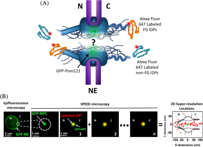
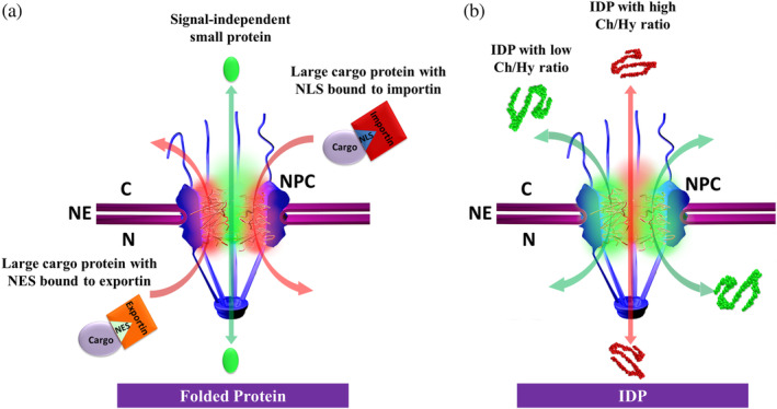
Both natively folded and intrinsically disordered proteins (IDPs) destined for the nucleus need to transport through the nuclear pore complexes (NPCs) in eukaryotic cells. NPCs allow for passive diffusion of small folded proteins while barricading large ones, unless they are facilitated by nuclear transport receptors. However, whether nucleocytoplasmic transport of IDPs would follow these rules remains unknown. By using a high-speed super-resolution fluorescence microscopy, we have measured transport kinetics and 3D spatial locations of transport routes through native NPCs for various IDPs. Our data revealed that the rules executed for folded proteins are not well followed by the IDPs. Instead, both large and small IDPs can passively diffuse through the NPCs. Furthermore, their diffusion efficiencies and routes are differentiated by their content ratio of charged (Ch) and hydrophobic (Hy) amino acids. A Ch/Hy-ratio mechanism was finally suggested for nucleocytoplasmic transport of IDPs.

Keywords: intrinsically disordered proteins (IDPs); nuclear pore complex (NPC); nucleocytoplasmic transport; super-resolution.


Conflict of interest statement

The authors declare no conflict of interest.

Figures

Figure 1
Figure 1
Nuclear transport of IDPs through the NPC illuminated by SPEED microscopy. (a) The fundamental mechanism for nuclear transport of IDPs through the NPCs remains unknown. Native NPCs embedded into the NE were labeled through genetically tagging green fluorescent protein (GFP) to a scaffold Nup POM121, in which the fluorescence of GFP‐POM121was utilized to localize the NPC. Alexa Fluor 647 dye molecules were used to label FG‐ and non‐FG‐IDPs. N, nucleus; C, cytoplasm. (b) Wide‐field epifluorescence image shown a green fluorescent ring of NE in a permeabilized HeLa cell expressing GFP‐POM121. SPEED microscopy illuminated only a single NPC on the NE. Labeled IDPs were tracked as they diffused through the NPC and their dwelling positions (red dots) were localized and then superimposed to obtain a 2D super‐resolution distribution around the NPC centroid (green dot). N, nucleus; C, cytoplasm. IDP, intrinsically disordered protein; NE, nuclear envelope; NPC, nuclear pore complex; SPEED, single‐point edge‐excitation sub‐diffraction
Figure 2
Figure 2
Two‐dimensional super‐resolution spatial distributions and 3D transport routes of various IDPs with different molecular weights. The molecular weight (i), disordered level (ii), 2D super‐resolution spatial distribution superposed with the 2D physical schematic of NPC in the (X, Y) coordinate plane (iii), and 3D spatial probability density map superposed with the 3D physical schematic of NPC (a cut‐away view) in the (X, R, Ɵ) cylindrical coordinate system for each IDP candidate were provided. In addition, a histogram of spatial probability density for the radial dimension was shown in iv to highlight either a single peak representing a central axial transport route or two peaks representing a peripheral transport route. (a) Nsp1 (1–603) refers an FG segment (AA 1–603) of yeast Nsp1 with a molecular weight of 60 kDa (i). With a high disordered level (ii), Nsp 1 (1–603) was found to mainly diffuse through the NPC's central axial channel, based on its 2D super‐resolution spatial distribution (black dots in iii) and 3D spatial probability density map (red clouds in NPC's axial and radial views shown in iv). N, nucleus; C, cytoplasm. (b–g) Defined similarly as in A, the major information and results for Nup159 (441–881), Nup42 (1–372), Nup116 (348–458), SLD2 (1–453), Coilin (1–576), and CREST (1–402) were presented. IDP, intrinsically disordered protein; NE, nuclear envelope; NPC, nuclear pore complex
Figure 3
Figure 3
IDP's Ch/Hy ratio determines its spatial transport route through the NPC. The 3D spatial probability density map overlaid with the 3D physical schematic of NPC (a cut‐away view) in the (X, R, Ɵ) cylindrical coordinate system (i) and the AA sequence (ii) for each IDP candidate were provided. Additionally, a histogram of spatial probability density for the radial dimension was shown in (ii) to highlight either a single peak representing a central axial transport route or two peaks representing a peripheral transport route. (a, b) WT Nup116 (348–458) and its charge mutant separate their 3D transport routes through the NPC after mutating 31 more neutral AAs adjacent to FG motifs into highly charged residues (k in red and d, e in blue). N, nucleus; C, cytoplasm. (c, d) Two segments of WT Nsp1 (1–603), Nsp1 (1–172), and Nsp1 (173–603), have different charged AAs (k in red and d, e in light blue) and Ch/Hy ratios, which differentiates them into two distinct 3D diffusion routes through the NPCs. AA, amino acid; Ch/Hy, charged/hydrophobic; IDP, intrinsically disordered protein; NPC, nuclear pore complex
Figure 4
Figure 4
A Ch/Hy ratio model for nucleocytoplasmic transport of IDPs. (a) A size‐exclusion model for folded proteins. Small signal‐independent proteins (<40 kDa) can passively diffuse through the NPC in both directions (green double‐headed arrow); however, larger protein cargos can be repelled by the NPC unless carrying an NLS for import or an NES for export. These signals will be recognized by transport receptors (importins for NLS or exportins for NES) to form complexes and then the complexes transport through the NPC in the mode of facilitated translocation (red single‐headed arrows). Emerging evidence suggests that passive and facilitated transports mainly adopt their spatial routes through the NPC's central axial channel (green regions) and the peripheral areas (red areas), respectively. (b) A Ch/Hy ratio model for IDPs. Both large (>40 kDa) and small (<40 kDa) IDPs can passively diffuse through the NPC (double‐headed arrows). Moreover, the IDPs with high Ch/Hy ratio (> ~0.37) prefer diffusing through the central axial channel (red arrow and area), while the IDPs with smaller Ch/Hy ratio (< ~0.37) mainly diffuse through the peripheral area around the central channel (green arrows and areas). Ch/Hy, charged/hydrophobic; IDP, intrinsically disordered protein; NES, nuclear export signal; NLS, nuclear localization sequence; NPC, nuclear pore complex

References

    1. Oldfield CJ, Cheng Y, Cortese MS, Brown CJ, Uversky VN, Dunker AK. Comparing and combining predictors of mostly disordered proteins. Biochemistry. 2005;44:1989–2000. - PubMed
    1. Dunker AK, Obradovic Z, Romero P, Garner EC, Brown CJ. Intrinsic protein disorder in complete genomes. Intrinsic protein disorder in complete genomes. Genome Inform Ser Workshop Genome Inform. 2000;11:161–171. - PubMed
    1. Iakoucheva LM, Brown CJ, Lawson JD, Obradović Z, Dunker AK. Intrinsic disorder in cell‐signaling and cancer‐associated proteins. J Mol Biol. 2002;323:573–584. - PubMed
    1. Frege T, Uversky VN. Intrinsically disordered proteins in the nucleus of human cells. Biochem Biophys Rep. 2015;1:33–51. - PMC - PubMed
    1. Love JJ, Li X, Chung J, Dyson HJ, Wright PE. The LEF‐1 high‐mobility group domain undergoes a disorder‐to‐order transition upon formation of a complex with cognate DNA. Biochemistry. 2004;43:8725–8734. - PubMed

Publication types

Substances