

Comparison of Abbreviated Breast MRI vs Digital Breast Tomosynthesis for Breast Cancer Detection Among Women With Dense Breasts Undergoing Screening
- PMID: 32096852
- PMCID: PMC7276668
- DOI: 10.1001/jama.2020.0572
Comparison of Abbreviated Breast MRI vs Digital Breast Tomosynthesis for Breast Cancer Detection Among Women With Dense Breasts Undergoing Screening
Erratum in
-
Errors in Author Affiliations.JAMA. 2020 Mar 24;323(12):1194. doi: 10.1001/jama.2020.2991. JAMA. 2020. PMID: 32207778 Free PMC article. No abstract available.
Abstract
Importance: Improved screening methods for women with dense breasts are needed because of their increased risk of breast cancer and of failed early diagnosis by screening mammography.
Objective: To compare the screening performance of abbreviated breast magnetic resonance imaging (MRI) and digital breast tomosynthesis (DBT) in women with dense breasts.
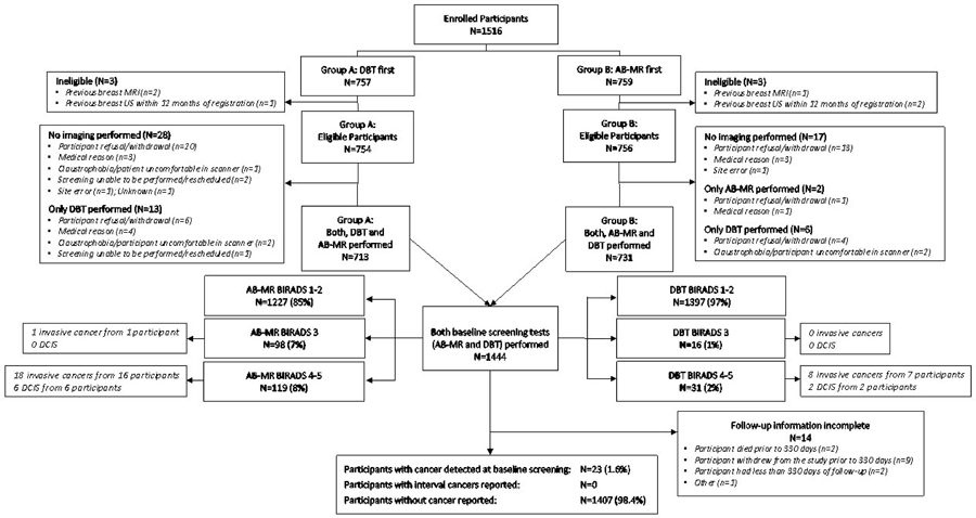
Design, setting, and participants: Cross-sectional study with longitudinal follow-up at 48 academic, community hospital, and private practice sites in the United States and Germany, conducted between December 2016 and November 2017 among average-risk women aged 40 to 75 years with heterogeneously dense or extremely dense breasts undergoing routine screening. Follow-up ascertainment of cancer diagnoses was complete through September 12, 2019.
Exposures: All women underwent screening by both DBT and abbreviated breast MRI, performed in randomized order and read independently to avoid interpretation bias.
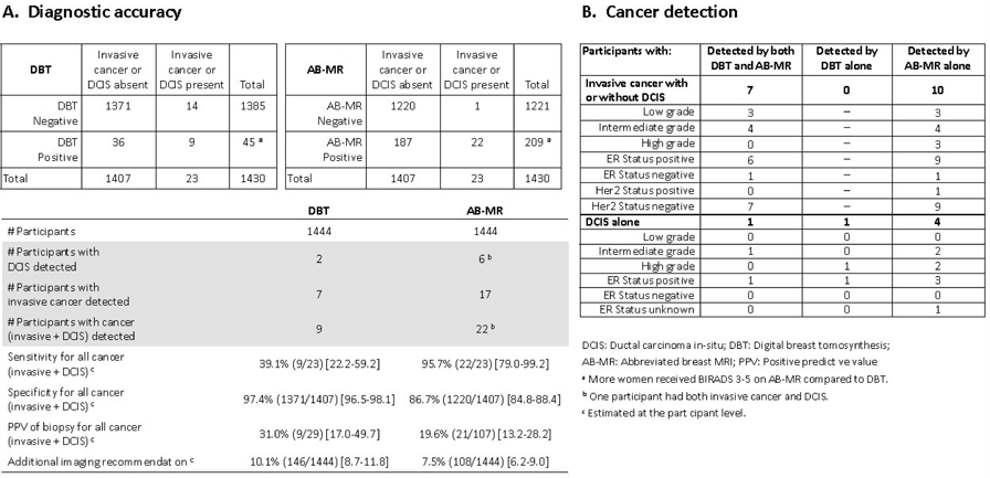
Main outcomes and measures: The primary end point was the invasive cancer detection rate. Secondary outcomes included sensitivity, specificity, additional imaging recommendation rate, and positive predictive value (PPV) of biopsy, using invasive cancer and ductal carcinoma in situ (DCIS) to define a positive reference standard. All outcomes are reported at the participant level. Pathology of core or surgical biopsy was the reference standard for cancer detection rate and PPV; interval cancers reported until the next annual screen were included in the reference standard for sensitivity and specificity.
Results: Among 1516 enrolled women, 1444 (median age, 54 [range, 40-75] years) completed both examinations and were included in the analysis. The reference standard was positive for invasive cancer with or without DCIS in 17 women and for DCIS alone in another 6. No interval cancers were observed during follow-up. Abbreviated breast MRI detected all 17 women with invasive cancer and 5 of 6 women with DCIS. Digital breast tomosynthesis detected 7 of 17 women with invasive cancer and 2 of 6 women with DCIS. The invasive cancer detection rate was 11.8 (95% CI, 7.4-18.8) per 1000 women for abbreviated breast MRI vs 4.8 (95% CI, 2.4-10.0) per 1000 women for DBT, a difference of 7 (95% CI, 2.2-11.6) per 1000 women (exact McNemar P = .002). For detection of invasive cancer and DCIS, sensitivity was 95.7% (95% CI, 79.0%-99.2%) with abbreviated breast MRI vs 39.1% (95% CI, 22.2%-59.2%) with DBT (P = .001) and specificity was 86.7% (95% CI, 84.8%-88.4%) vs 97.4% (95% CI, 96.5%-98.1%), respectively (P < .001). The additional imaging recommendation rate was 7.5% (95% CI, 6.2%-9.0%) with abbreviated breast MRI vs 10.1% (95% CI, 8.7%-11.8%) with DBT (P = .02) and the PPV was 19.6% (95% CI, 13.2%-28.2%) vs 31.0% (95% CI, 17.0%-49.7%), respectively (P = .15).
Conclusions and relevance: Among women with dense breasts undergoing screening, abbreviated breast MRI, compared with DBT, was associated with a significantly higher rate of invasive breast cancer detection. Further research is needed to better understand the relationship between screening methods and clinical outcome.
Trial registration: ClinicalTrials.gov Identifier: NCT02933489.
Conflict of interest statement
Conflict of interest disclosures:
Dr. Kuhl, Dr. Bergin, Dr. Gareen, Dr. Holt, Dr. Miller, Dr. Newstead, Dr. Nicholson, Dr. Prather, Mr. Snyder, Dr. Sung and Dr. Ward have nothing to report. Dr Comstock reports grants from ECOG-ACRIN during the conduct of the study; personal fees from Bracco Diagnostics and personal fees from Bayer Inc outside the submitted work. Dr Gareen reports grants from National Cancer Institute and grants from Bracco Imaging during the conduct of the study. Dr Gatsonis reports grants from EA Cancer Research Group during the conduct of the study. Dr Harvey reports other from Hologic, Inc and non-financial support and other from Volpara Solutions, LLC outside the submitted work. Dr Rahbar reports grants from GE Healthcare outside the submitted work. Dr Schnall reports grants from National Cancer Institute and grants from Bracco Diagnostics during the conduct of the study; grants from Siemens Healthineers outside the submitted work.
Figures
Comment in
-
Options for Addressing the Dilemma of Managing Dense Breasts.JAMA Surg. 2020 Apr 1;155(4):279-280. doi: 10.1001/jamasurg.2020.0280. JAMA Surg. 2020. PMID: 32096827 No abstract available.
-
An Abbreviated MRI Protocol for Breast Cancer Screening in Women With Dense Breasts: Promising Results, but Further Evaluation Required Prior to Widespread Implementation.JAMA. 2020 Feb 25;323(8):719-721. doi: 10.1001/jama.2020.0357. JAMA. 2020. PMID: 32096832 No abstract available.
-
Commentary on "Comparison of Abbreviated Breast MRI vs Digital Breast Tomosynthesis for Breast Cancer Detection Among Women With Dense Breasts Undergoing Screening".AJR Am J Roentgenol. 2021 Jan;216(1):37. doi: 10.2214/AJR.20.23674. Epub 2020 Nov 10. AJR Am J Roentgenol. 2021. PMID: 32812772 No abstract available.
-
Preclinical Assessment of the Safety of an 18F-labeled MCT1/MCT4 Inhibitor in a Swine Model for PET/CT Imaging of Cancer Metabolism.Radiol Imaging Cancer. 2020 Jul 31;2(4):e204022. doi: 10.1148/rycan.2020204022. eCollection 2020 Jul. Radiol Imaging Cancer. 2020. PMID: 33778728 Free PMC article. No abstract available.
-
Breast MRI Finds More Invasive Cancers than Digital Breast Tomosynthesis in Women with Dense Breasts Undergoing Screening.Radiol Imaging Cancer. 2020 Jul 31;2(4):e204023. doi: 10.1148/rycan.2020204023. eCollection 2020 Jul. Radiol Imaging Cancer. 2020. PMID: 33778729 Free PMC article. No abstract available.
References
-
- Boyd NF, Guo H, Martin LJ, et al. Mammographic density and the risk and detection of breast cancer. N Engl J Med. 2007;356:227–36 - PubMed
-
- Holm J, Humphreys K, Li J et al. Risk factors and tumor characteristics of interval cancers by mammographic density. J Clin Oncol. 2015;33:1030–7 - PubMed
-
- Ohuchi N, Suzuki A, Sobue T et al. ; J-START investigator groups. Sensitivity and specificity of mammography and adjunctive ultrasonography to screen for breast cancer in the Japan Strategic Anti-cancer Randomized Trial (J-START): a randomised controlled trial. Lancet. 2016;387:341–348. - PubMed
Publication types
MeSH terms
Associated data
Grants and funding
- U10 CA180868/CA/NCI NIH HHS/United States
- UG1 CA189956/CA/NCI NIH HHS/United States
- U10 CA180795/CA/NCI NIH HHS/United States
- UG1 CA232760/CA/NCI NIH HHS/United States
- UG1 CA189828/CA/NCI NIH HHS/United States
- U10 CA180790/CA/NCI NIH HHS/United States
- U10 CA180828/CA/NCI NIH HHS/United States
- UG1 CA189819/CA/NCI NIH HHS/United States
- UG1 CA233328/CA/NCI NIH HHS/United States
- UG1 CA233290/CA/NCI NIH HHS/United States
- UG1 CA233270/CA/NCI NIH HHS/United States
- U10 CA180836/CA/NCI NIH HHS/United States
- UG1 CA189860/CA/NCI NIH HHS/United States
- P30 CA008748/CA/NCI NIH HHS/United States
- U10 CA180791/CA/NCI NIH HHS/United States
- U10 CA180847/CA/NCI NIH HHS/United States
