. 2020 Apr 8;28(4):1200-1213.
doi: 10.1016/j.ymthe.2020.02.003. Epub 2020 Feb 7.

HucMSC-Derived Exosomes Mitigate the Age-Related Retardation of Fertility in Female Mice

Affiliations

HucMSC-Derived Exosomes Mitigate the Age-Related Retardation of Fertility in Female Mice

Weijie Yang et al. Mol Ther. .

Abstract

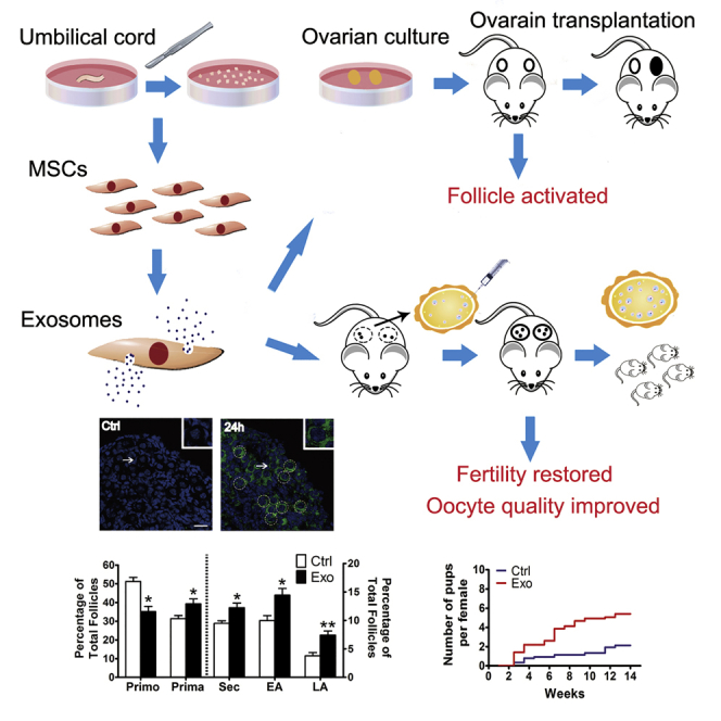
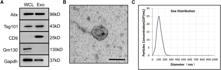
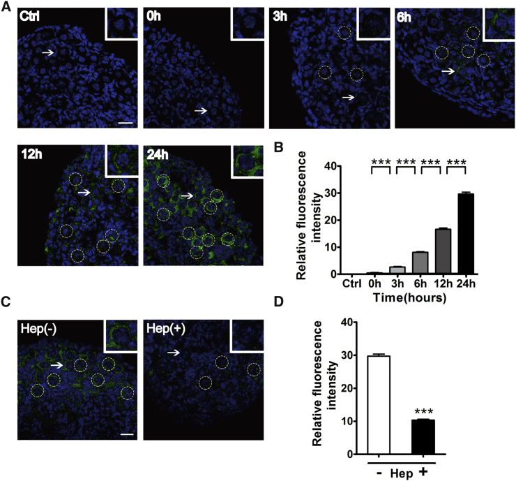
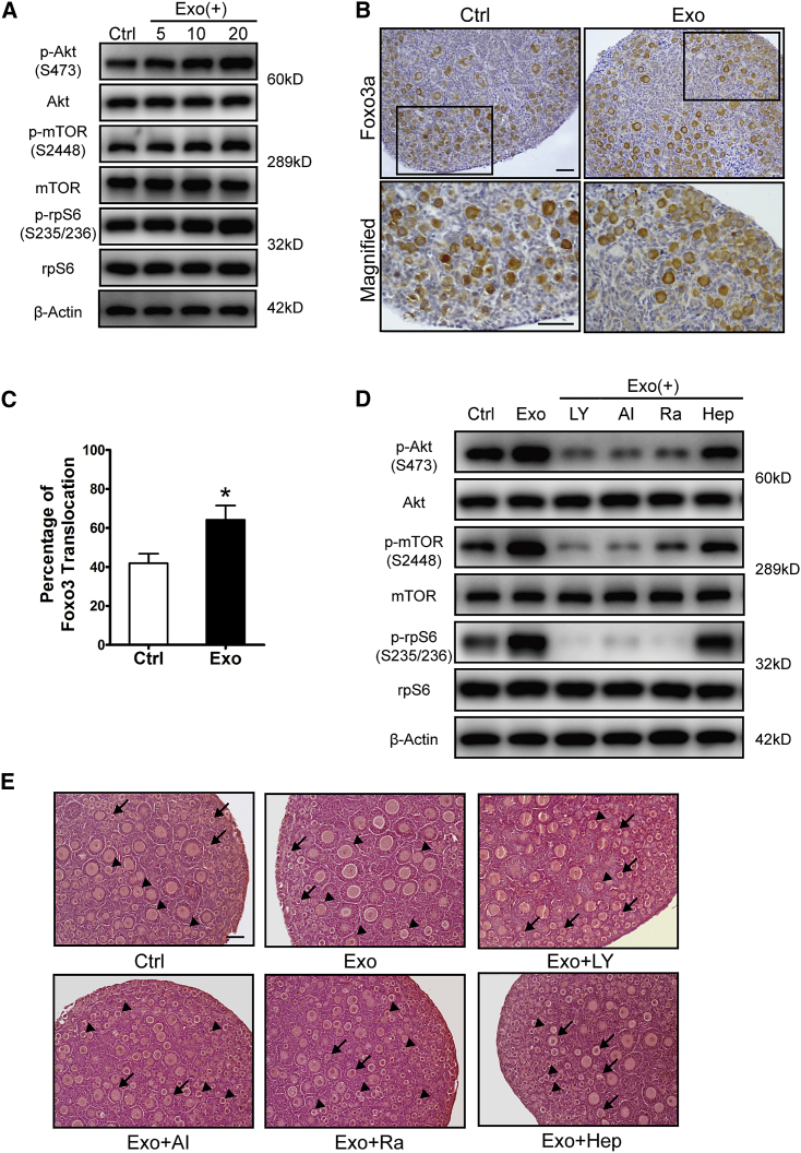
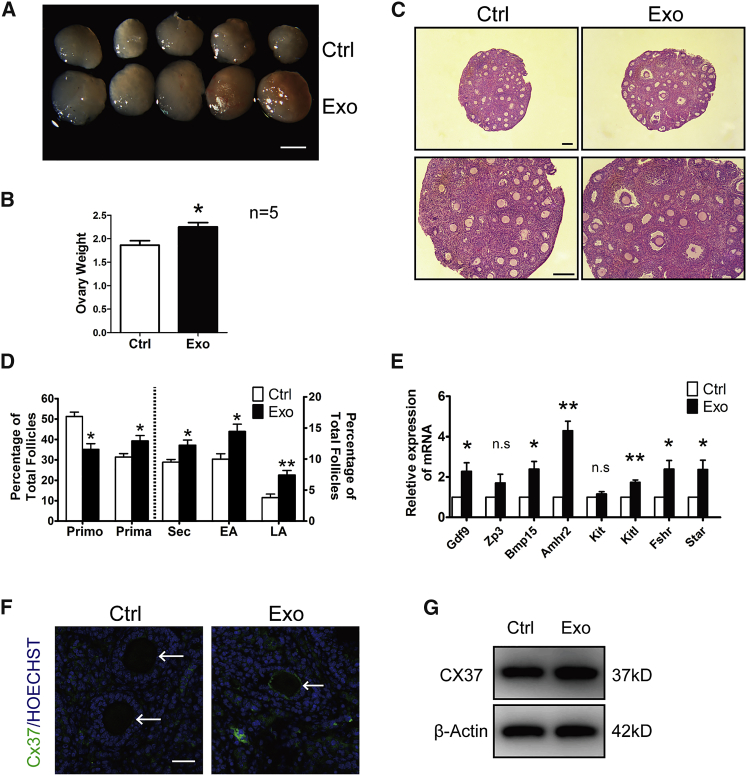
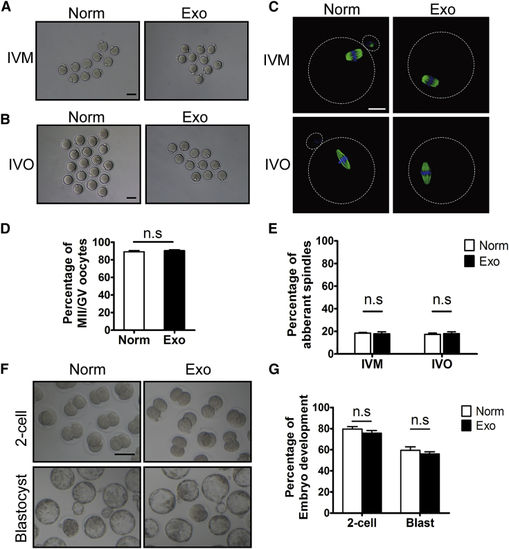
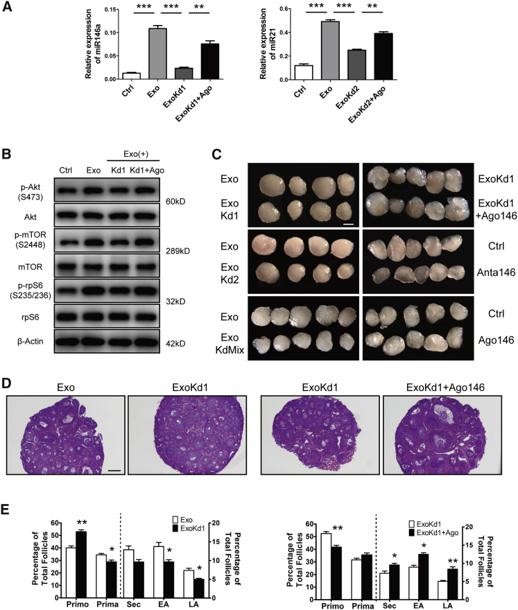
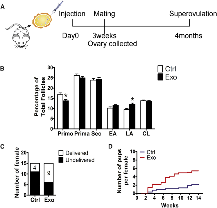
In mammals, resting primordial follicles serve as the ovarian reserve. The decline in ovarian function with aging is characterized by a gradual decrease in both the quantity and quality of the oocytes residing within the primordial follicles. Many reports show that mesenchymal stem cells have the ability to recover ovarian function in premature ovarian insufficiency (POI) or natural aging animal models; however, the underlying mechanism remains unclear. In this study, using exosomes derived from human umbilical cord mesenchymal stem cells (HucMSC-exos), we found the specific accumulation of exosomes in primordial oocytes. The stimulating effects of exosomes on primordial follicles were manifested as the activation of the oocyte phosphatidylinositol 3-kinase (PI3K)/mTOR signaling pathway and the acceleration of follicular development after kidney capsule transplantation. Further analysis revealed the stimulatory effects of HucMSC-exos on primordial follicles were through carrying functional microRNAs, such as miR-146a-5p or miR-21-5p. In aged female mice, the intrabursal injection of HucMSC-exos demonstrated the recovery of decreased fertility with increased oocyte production and improved oocyte quality. Although assisted reproductive technologies have been widely used to treat infertility, their overall success rates remain low, especially for women in advanced maternal age. We propose HucMSC-exos as a new approach to mitigate the age-related retardation of fertility in women.

Keywords: exosomes; mesenchymal stem cells; ovarian aging; premature ovarian insufficiency; primordial follicle.


Figures

None
Graphical abstract
Figure 1
Figure 1
Characterization of HucMSC-exos (A) Western blot analysis of exosome-related markers Alix, Tsg101, and CD9. Plasma protein Gm130 was used as a negative control. Both lanes were loaded with 20 μg of proteins as measured by Qubit 3.0. WCL, whole-cell lysate; Exo, exosome. (B) Representative image of HucMSC-exos observed by transmission electron microscope (TEM). Scale bar, 100 μm. (C) Nanoparticle tracking analysis of size distribution and concentration of HucMSC-exos. The mean protein concentration and mean particle concentration of the HucMSC-exos were 0.79 mg/mL and 2.7 × 1010 particles/mL, respectively.
Figure 2
Figure 2
Uptake of HucMSC-exos by Cultured Newborn Ovaries Newborn ovaries (P2.5) were incubated with PKH67-labeled exos for 0, 3, 6, 12, and 24 h. (A) Gradual accumulation of HucMSC-exos in primordial follicles. Ovaries were collected for frozen sections and nuclei were stained with Hoechst 33342. Green indicates exos; blue shows Hoechst 33342. For the control (Ctrl), ovaries were incubated with PBS for 24 h. (B) Fluorescence intensity analysis of labeled HucMSC-exos. n = 4/group. (C) Internalization of HucMSC-exos after ovaries were cultured for 24 h in the presence (left panel) or absence (right panel) of 5,000 IU of heparin (Hep). (D) Fluorescence intensity analysis of internalized HucMSC-exos. n = 4/group. Insets represent magnification of representative primordial follicles marked by white arrows. Dotted circles indicate primordial follicles. Data are shown as mean ± SEM. ***p < 0.001. Scale bars, 20 μm.
Figure 3
Figure 3
Activation of Primordial Follicles after HucMSC-exo Treatment Newborn ovaries were treated for 24 h with HucMSC-exos at 5, 10, or 20 μg/mL (3 × 108, 6 × 108, 1.2 × 109 particles/mL, respectively). (A) Dose-dependent activation of the PI3K-Akt-mTOR signaling pathway in ovaries with increased expression of p-AKT (Ser473), p-mTOR (Ser2448), and p-rpS6 (Ser235/236). The expression of rpS6, mTOR, and β-tubulin were used as internal controls. (B) Immunostaining of Foxo3a in primordial follicles after newborn ovaries were incubated with 20 μg/mL HucMSC-exos for 24 h. The lower panel represents the magnified images of black frames in the upper panel. (C) Percentage of activated primordial follicles with nucleus exclusion of Foxo3a in control and exo-treated ovaries. n = 3/group. (D) Blockage of the PI3K-Akt-mTOR signaling pathway by its specific inhibitors LY294002, Akt VIII, and rapamycin and the exosome internalization inhibitor heparin. LY, LY294002; AI, Akt VIII; Ra, rapamycin; Hep, heparin. (E) Histology of ovaries collected at 96 h in each group. After 24 h of treatment, ovaries in each group were further cultured in control media for 72 h. Arrowhead indicates primordial follicles, and arrow indicates activated follicles. Data are shown as mean ± SEM. *p < 0.05. Scale bars, 50 μm.
Figure 4
Figure 4
Acceleration of Follicular Development after HucMSC-exo Treatment Paired ovaries were treated with or without HucMSC-exos for 24 h and transplanted into kidney capsules of the same recipient by ovariectomy. (A) Ovaries at 14 days after transplantation. Isolated ovaries from control (Ctrl) versus HucMSC-exos (Exo) treatment. Scale bar, 1 mm. (B) Comparison of ovarian weight between control and exo-treated ovaries (n = 5). (C) Ovarian histology showing more antral follicles in HucMSC-exo-treated ovaries. Scale bar, 50 μm. (D) Distribution of different stages of follicles with or without exposure to HucMSC-exos. Primo, primordial follicle; Prima. primary follicle; Sec, secondary follicle; EA, early antral follicle; LA, late antral follicle. n = 5/group. (E) Relative expression of follicular growth and development-related genes in ovaries of each group. The levels of all tested mRNAs in control group were set to 1. n = 3/group. (F) Immunofluorescence of Cx37 in ovaries treated with or without HucMSC-exos. Arrows point to oocytes. Green represents Cx37; blue represents Hoechst 33342. Scale bar, 20 μm. (G) Western blot of Cx37 expressions in ovaries treated with or without HucMSC-exos. The expression of β-actin was used as an internal control. Data are shown as mean ± SEM. *p < 0.05, **p < 0.01.
Figure 5
Figure 5
Evaluation of Oocyte Quality by In Vitro Maturation (IVM), In Vitro Fertilization (IVF), and Early Embryonic Development Newborn ovaries (P2.5) treated with HucMSC-exos were transplanted into the kidney capsules of recipient mice for 18 days. GV oocytes for IVM were collected by directly puncturing fully grown follicles under the microscope, and mature MII oocytes were retrieved after a single injection of hCG into recipient mice (IVO). (A and B) Morphology of MII oocytes after IVM (A) and IVO (B). Oocytes from superovulated ovaries of 3-week-old mice were used as normal controls (Norm). (C) Immunofluorescence of β-tubulin on spindle of MII oocytes. Green indicates β-tubulin; blue indicates nuclear staining with Hoechst 33342. (D and E) Percentages of MII oocytes with aberrant spindles after IVM (D) and IVO (E) treatment. (F) Representative two-cell embryos and blastocysts after IVO oocytes being fertilized in vitro (IVF). (G) Efficiency of early embryonic development. Percentage of IVO mature oocytes in control and exo-treated group capable of developing into two-cell embryos and blastocysts. All experiments were performed with at least three replicates. Data are shown as mean ± SEM. n.s., not significant; *p < 0.05. White scale bar, 20 μm; black scale bar, 50 μm.
Figure 6
Figure 6
Effects of Exo-Carrying MicroRNAs on Follicular Activation and Development (A) Expression of miR-146a-5p and miR-21-5p in ovaries after 24 h of different treatments. Crtl, control (PBS); Exo, HucMSC-exos (20 μg/mL); ExoKd1, exos carrying Antagomir-146a-5p; ExoKd2, exos carrying Antagomir-21-5p; ExoKd1+Ago, Agomir-146a-5p; ExoKd2+Ago, Agomir-21-5p. (B) Ovarian expression of p-Akt (Ser473), p-mTOR (Ser2448), and p-rpS6 (Ser235/236) after 24 h of treatments. The expressions of Akt, mTOR, rpS6, and β-actin were used as internal controls. (C) Comparisons of ovarian development after various treatment combinations. Paired ovaries (P2.5) were separated for different treatments with 24-h incubation and then were transplanted under kidney capsules of recipient mice for 14 days. Exo KdMix, exos containing both Antagomirs; Anta 146, Antagomir-146a-5p; Ago 146, Agomir-146a-5p. Scale bar, 1 mm. (D) Ovarian histology between paired ovaries. Left, Exo versus ExoKd1; Right, ExoKd1 versus ExoKd1+Ago 146. Scale bar, 50 μm. (E) Distribution of different stages of follicles in paired ovaries with treatments corresponding to (D). n = 5/group. *p < 0.05, **p < 0.01, ***p < 0.001.
Figure 7
Figure 7
Stimulation of Follicular Development in Old Mice by HucMSC-exos (A) Timeline of HucMSC-exos treatment. Mice at 10 months of age were intrabursally injected with 20 μL of HucMSC-exos or the same volume of PBS. For some mice, one lateral ovary was given HucMSC-exos and the other one received PBS. Ovaries were collected for morphology 3 weeks later. Some mice were injected with HucMSC-exos or PBS bilaterally for a 4-month fertility test. (B) Distribution of follicles at different developmental stages in ovaries treated with PBS (Ctrl) or HucMSC-exos (Exo). Primo, primordial follicle; Prima, primary follicle; Sec, secondary follicle; EA, early antral follicle; LA, late antral follicle; CL, corpus luteum. n = 5/group. (C) Number of mice that gave birth to pups during the test. (D) Comparison of the cumulative numbers of pups per female in the control group (blue) and HucMSC-exo-treated (red) group. Data are shown as mean ± SEM. *p < 0.05.
Figure 8
Figure 8
Evaluation of Oocyte Quality after HucMSC-exos Treatment In Vivo Oocytes were collected from old mice by superovulation after a fertility test. (A) Representative morphology of MII oocytes with normal (Norm) or aberrant (Abn) spindles in PBS (Ctrl)- or HucMSC-Exo (Exo)-treated group. Green indicates β-tubulin; blue indicates nuclear staining with Hoechst 33342. (B) Percentage of aberrant oocytes in the two groups. n = 4 mice/group. (C and D) ROS fluorescence staining (green) (C) and relative fluorescence intensity ratio (D) in MII oocytes from control and HucMSC-exo-treated groups. Each group included 10 oocytes for analysis. (E) Oocyte mitochondrial membrane potential (ΔΨm) shown by JC-1 staining between the two groups. Red indicates higher ΔΨm; green indicates lower ΔΨm. (F) The ratio of red/green fluorescence intensity reflects the increase in oocyte mitochondrial activity in HucMSC-exo-treated mice. *p < 0.05, **p < 0.01 compared with controls. Scale bars, 20 μm.

References

    1. Skinner M.K. Regulation of primordial follicle assembly and development. Hum. Reprod. Update. 2005;11:461–471. - PubMed
    1. McLaughlin E.A., McIver S.C. Awakening the oocyte: controlling primordial follicle development. Reproduction. 2009;137:1–11. - PubMed
    1. Reddy P., Zheng W., Liu K. Mechanisms maintaining the dormancy and survival of mammalian primordial follicles. Trends Endocrinol. Metab. 2010;21:96–103. - PubMed
    1. Macklon N.S., Fauser B.C. Aspects of ovarian follicle development throughout life. Horm. Res. 1999;52:161–170. - PubMed
    1. Broekmans F.J., Soules M.R., Fauser B.C. Ovarian aging: mechanisms and clinical consequences. Endocr. Rev. 2009;30:465–493. - PubMed

Publication types