. 2020 Apr 22;106(2):246-255.e6.
doi: 10.1016/j.neuron.2020.01.030. Epub 2020 Feb 24.

Posterior Neocortex-Specific Regulation of Neuronal Migration by CEP85L Identifies Maternal Centriole-Dependent Activation of CDK5

Affiliations

Posterior Neocortex-Specific Regulation of Neuronal Migration by CEP85L Identifies Maternal Centriole-Dependent Activation of CDK5

Andrew Kodani et al. Neuron. .

Abstract

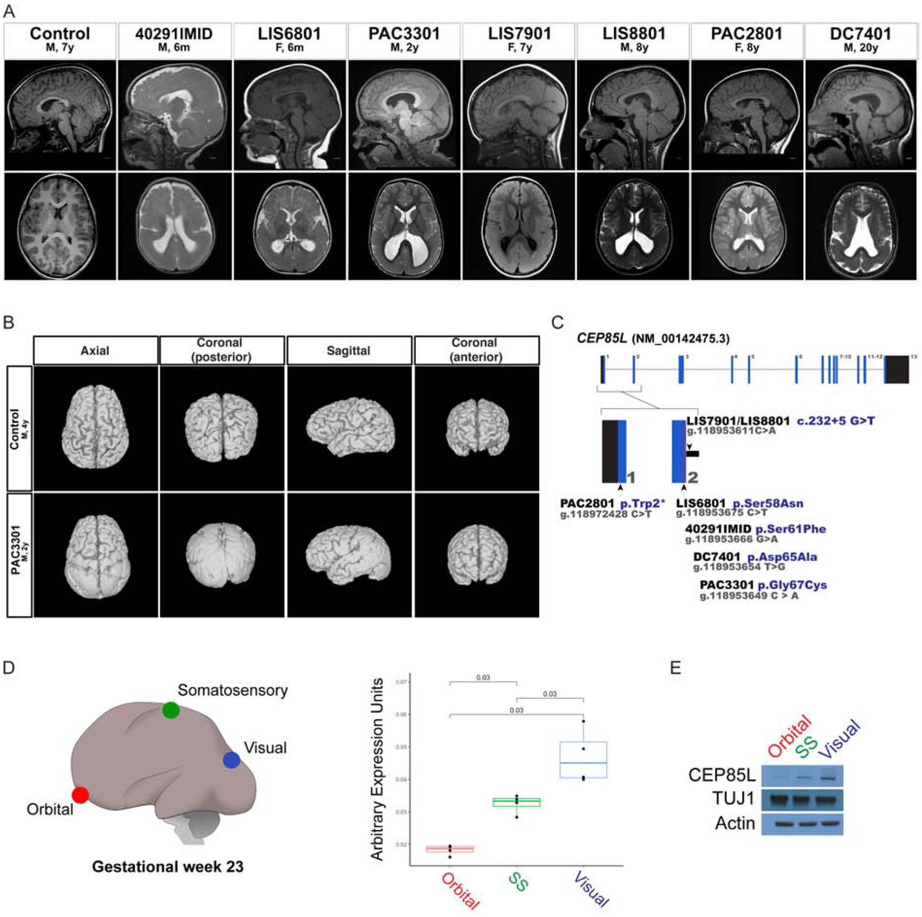
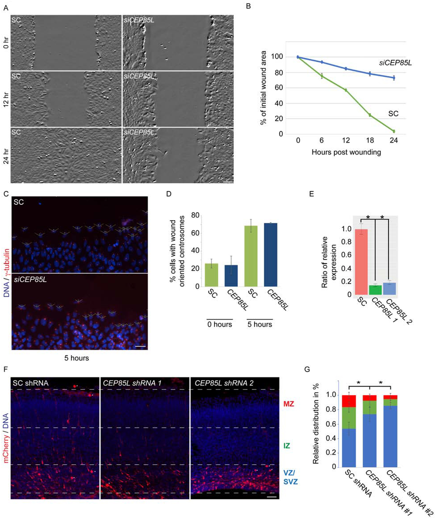
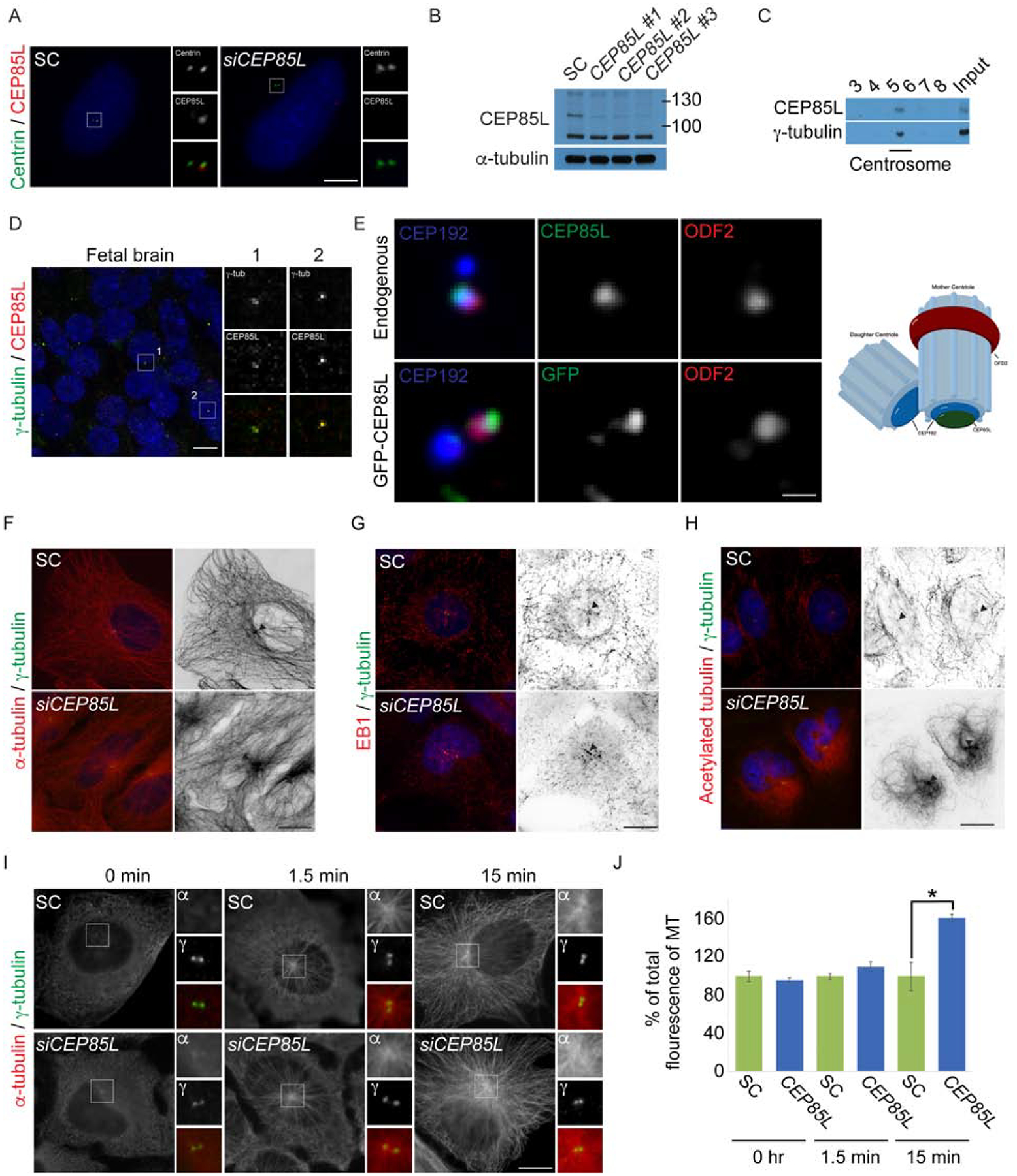
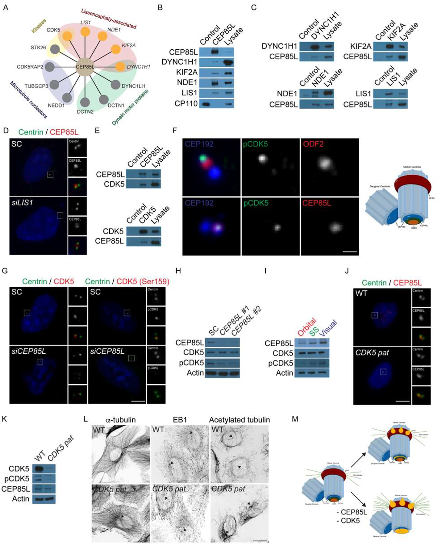
Genes mutated in human neuronal migration disorders encode tubulin proteins and a variety of tubulin-binding and -regulating proteins, but it is very poorly understood how these proteins function together to coordinate migration. Additionally, the way in which regional differences in neocortical migration are controlled is completely unknown. Here we describe a new syndrome with remarkably region-specific effects on neuronal migration in the posterior cortex, reflecting de novo variants in CEP85L. We show that CEP85L is required cell autonomously in vivo and in vitro for migration, that it localizes to the maternal centriole, and that it forms a complex with many other proteins required for migration, including CDK5, LIS1, NDE1, KIF2A, and DYNC1H1. Loss of CEP85L disrupts CDK5 localization and activation, leading to centrosome disorganization and disrupted microtubule cytoskeleton organization. Together, our findings suggest that CEP85L highlights a complex that controls CDK5 activity to promote neuronal migration.

Keywords: CDK5; CEP85L; Centrosome; De novo; Lissencephaly; Pachygyria; genetics; neurodevelopment.


Conflict of interest statement

Declaration of Interests C.A.W. serves on advisory boards for the Allen Brain Institute, Third Rock Ventures, and Maze Therapeutics and on editorial boards for Annals of Neurology, Trends in Neurosciences, and neuroDEVELOPMENTS. M.S. received research funding from Roche, Novartis, Pfizer, LAM Therapeutics, and Quadrant Biosciences; has served on the scientific advisory boards of Sage Therapeutics, Roche, Takeda, Celgene, and the PTEN Research Foundation; and serves on the Board of the Tuberous Sclerosis Alliance. All of these activities are outside of the submitted manuscript. J.S.C. is a consultant for Invitae. D.V. serves as a consultant to SK Life Science and Otsuka Pharmaceuticals, is on the speaker’s bureaus for UCB and Greenwich Pharmaceuticals, and conducts industry-supported clinical drug trials for SK Life Science, Biogen, and UCB Pharmaceuticals. K.M. is an employee of GeneDX, Inc.

Figures

Figure 1:
Figure 1:. Variants in CEP85L cause posterior-specific pachygyria.
A. Sagittal and axial plane MRI images of a control and affected individuals with posterior reduced gyral folding. B. Three-dimensional MRI presentation of a control and PAC3301 patient with a de novo CEP85L variant. C. Schematic representation of exons of CEP85L shown as blue bars. The variants in CEP85L are found in exons 1 and 2. D. Brain region-specific qPCR of gestational week 23 cortex (GW), demonstrating the increasing rostral-to-caudal expression pattern of CEP85L normalized to β-actin. Orbital (red), somatosensory (green), and visual (blue) cortex. For quantifications, one brain region was analyzed in triplicate or quadruplicate (n=1). p < 0.03 (Student T-test). E. Whole cell lysate from the posterior frontal, parietal and occipital lobes of a GW 23 fetus blotted for CEP85L and the lissencephaly-associated protein, LIS1. Actin and TUJ1 served as a loading control and neuron-specific sampling control, respectively.
Figure 2:
Figure 2:. CEP85L is required for neuronal migration.
A. Time-lapse stills from scratch wound assays of scrambled control (SC) and CEP85L siRNA-transfected U2-OS cells. Confluent monolayers were wounded using a P200 tip and imaged over 24 hours using the Zeiss Celldiscoverer 7. Scale bar represents 200μm for all images. B. Quantifications of the areas of migration at the indicated time points of SC and CEP85L-depleted cells. For all quantifications, three distinct experiments were performed. C. Immunostaining of γ-tubulin (red) and DNA (blue) in SC and CEP85L siRNA transfected cells along the wound edge. Open-faced triangles are facing the wound. Scale bar represents 200mm for all images. D. Percentage of cells along the wound edge with centrosomes oriented toward the wound at 0 and 5 hrs. For all quantifications, 100 cells were analyzed per experiment (n=3). P<0.005 (Student’s t test). E. qRT-PCR of scrambled control (SC), CEP85L #1, CEP85L #2 shRNA transfected cells normalized to β-actin and represented as a ratio of the control. F. Embryonic day 14.5 mice were electroporated with mCherry and a SC, or with Cep85L #1 or #2 shRNAs and collected at E17.5. Scale bar 100mm. G. Percentage of electroporated SC or Cep85L shRNA transfected mCherry-positive cells in the ventricular and subventricular zone (VZ/SVZ), intermediate zone (IZ), or cortical plate (CP). At least three electroporated brains from each condition were quantified (n>3). *p < 0.005 (Student’s t-test).
Figure 3:
Figure 3:. CEP85L localizes to the mother centriole and regulates microtubule cytoskeletal organization.
A. U2-OS cells treated with scrambled control (SC) or CEP85L siRNA co-stained with antibodies to Centrin (green) and CEP85L (red). Scale bar represents 5mm for all images. B. Whole-cell lysate from SC, CEP85L #1, #2 or #3 siRNA-treated U2-OS cells immunoblotted for CEP85L. Actin served as a loading control. C. Fractions from sucrose gradient-separated U2-OS cell lysates immunoblotted for CEP85L and γ-tubulin to identify the centrosomal fraction. D. Fresh frozen gestational week 23 fetal brains were co-stained for γ-tubulin (green) and CEP85L (red) Scale bar is 5mm for all images. E. Airyscan microscopy of U2-OS cells co-stained for CEP192 (blue) to mark the proximal centrioles, ODF2 (red) to mark subdistal appendages, and CEP85L (green) or GFP-CEP85L (green). Scale bar represents 1μm for Airyscan images. F-H. Immunofluorescence analysis of SC and CEP85L siRNA-treated U2-OS cells co-stained with γ-tubulin (green) and α-tubulin, EB1, or acetylated tubulin (red). Figures right of the merged image are inverted images of α-tubulin, EB1 or acetylated tubulin. Triangles denote the centrosome. Scale bars represent 10mm for all images. I. SC and CEP85L siRNA-treated U2-OS cells were subjected to a microtubule regrowth assay, fixed at the indicated time points and co-stained with α-tubulin (red) and γ-tubulin (green). Scale bar indicates 5mm for all images. J. Quantification of the mean fluorescence intensities ± s.d. of centrosomal α-tubulin in SC and CEP85L siRNA treated cells as expressed as the mean percentage ± s.d. of the fluorescence intensities of SC cells. For all quantifications, 10 cells were analyzed per experiment (n=3). * − p < 0.005 (Student’s t-test).
Figure 4:
Figure 4:. CEP85L is required to localize and activate the lissencephaly protein, CDK5.
A. Schematic of centrosomal CEP85L interacting proteins identified by endogenous immunoprecipitation of CEP85L followed by LC-MS/MS analysis. Interactors were sorted and prioritized based on centrosomal localization and disease-association. B. Immunoprecipitated endogenous CEP85L and CP110 from HeLa cell lysates was immunoblotted for co-precipitating proteins for CEP85L, DYNC1H1, KIF2A, NDE1 and LIS1. CP110 served as a negative control throughout. Lysate represents 5% of the total cell lysate used in the immunoprecipitation assays. C. HeLa cell lysate was subjected to immunoprecipitation of DYNC1H1, KIF2A, NDE1 and LIS1. Precipitating proteins were immunoblotted for CEP85L, DYNC1H1, KIF2A, NDE1 and LIS1. D. U2-OS cells transfected with siRNA to SC or LIS1 co-stained with Centrin (green) and CEP85L (red). E. HeLa cell lysate was subjected to immunoprecipitation of CEP85L, CDK5, and CP110, which served as a negative control. Precipitating proteins were immunoblotted for CEP85L and CDK5. F. Airyscan maximum projections of U2-OS cells co-stained with antibodies to pCDK5 (green), ODF2 (red, to mark the subdistal appendages of mother centrioles), CEP85L (red), and CEP192 (blue) to mark the proximal domains of the centrioles. Scale bar represents 1μm for Airyscan images. G. Immunofluorescence of SC and CEP85L siRNA-transfected U2-OS cells co-stained for Centrin (green) and CDK5 (red) or pCDK5 (red). Scale bars represent 5mm for all images. H. Total cell lysates from U2-OS cells transfected with SC, CEP85L #1 or #2 probed with antibodies to CEP85L, CDK5, and pCDK5. Actin served as a loading control. I. Whole cell lysate from the posterior frontal, parietal and occipital lobes of a GW 23 human fetus blotted for CEP85L, CDK5 and pCDK5. Actin served as a loading control. J. WT and CDK5 patient fibroblasts (p.V162fsX19, CDK5 pat) co-stained with antibodies to Centrin (green) and pCDK5 (red). K. Whole cell lysate from WT or CDK5 patient fibroblasts probed with antibodies to CDK5, pCDK5, and CEP85L. Actin served as a loading control. L. Inverted images of WT and CDK5 patient cells stained with α-tubulin, EB1 or acetylated tubulin. Triangles denote the centrosome. Scale bars represent 10μm for all images. M. CEP85L (green) localizes CDK5 (beige) to the proximal end of mother centrioles (CEP192, blue) to be activated. At the centrosome CDK5 activity restricts the accumulation of LIS-associated proteins (orange) that localize to the proximal mother centriole and its subdistal appendages. Consequently, loss of CEP85L or CDK5 causes the excessive localization of LIS-proteins resulting in excessive anchoring of microtubules at the mother centriole leading to cells incapable of migrating.

References

    1. Alkuraya FS, Cai X, Emery C, Mochida GH, Al-Dosari MS, Felie JM, Hill RS, Barry BJ, Partlow JN, Gascon GG, et al. (2011). Human mutations in NDE1 cause extreme microcephaly with lissencephaly [corrected]. Am J Hum Genet 88, 536–547. - PMC - PubMed
    1. Askham JM, Vaughan KT, Goodson HV, and Morrison EE (2002). Evidence that an interaction between EB1 and p150(Glued) is required for the formation and maintenance of a radial microtubule array anchored at the centrosome. Mol Biol Cell 13, 3627–3645. - PMC - PubMed
    1. Bakircioglu M, Carvalho OP, Khurshid M, Cox JJ, Tuysuz B, Barak T, Yilmaz S, Caglayan O, Dincer A, Nicholas AK, et al. (2011). The essential role of centrosomal NDE1 in human cerebral cortex neurogenesis. Am J Hum Genet 88, 523–535. - PMC - PubMed
    1. Choi YK, Liu P, Sze SK, Dai C, and Qi RZ (2010). CDK5RAP2 stimulates microtubule nucleation by the gamma-tubulin ring complex. J Cell Biol 191, 1089–1095. - PMC - PubMed
    1. Dammermann A, and Merdes A (2002). Assembly of centrosomal proteins and microtubule organization depends on PCM-1. J Cell Biol 159, 255–266. - PMC - PubMed

Publication types

LinkOut - more resources