. 2020 Feb 21;21(4):1469.
doi: 10.3390/ijms21041469.

Interactome and F-Actin Interaction Analysis of Dictyostelium discoideum Coronin A

Affiliations

Interactome and F-Actin Interaction Analysis of Dictyostelium discoideum Coronin A

Tohnyui Ndinyanka Fabrice et al. Int J Mol Sci. .

Abstract

Coronin proteins are evolutionary conserved WD repeat containing proteins that have been proposed to carry out different functions. In Dictyostelium, the short coronin isoform, coronin A, has been implicated in cytoskeletal reorganization, chemotaxis, phagocytosis and the initiation of multicellular development. Generally thought of as modulators of F-actin, coronin A and its mammalian homologs have also been shown to mediate cellular processes in an F-actin-independent manner. Therefore, it remains unclear whether or not coronin A carries out its functions through its capacity to interact with F-actin. Moreover, the interacting partners of coronin A are not known. Here, we analyzed the interactome of coronin A as well as its interaction with F-actin within cells and in vitro. Interactome analysis showed the association with a diverse set of interaction partners, including fimbrin, talin and myosin subunits, with only a transient interaction with the minor actin10 isoform, but not the major form of actin, actin8, which was consistent with the absence of a coronin A-actin interaction as analyzed by co-sedimentation from cells and lysates. In vitro, however, purified coronin A co-precipitated with rabbit muscle F-actin in a coiled-coil-dependent manner. Our results suggest that an in vitro interaction of coronin A and rabbit muscle actin may not reflect the cellular interaction state of coronin A with actin, and that coronin A interacts with diverse proteins in a time-dependent manner.

Keywords: Actin; Dictyostelium; coronin A; interactome analysis.


Conflict of interest statement

The authors declare no conflict of interest.

Figures

Figure 1
Figure 1
Coronin A interactome analysis. Growing Dictyostelium cells were collected, lysed and subjected to AP-MS as described in Materials and Methods. Shown are all interacting proteins with log2ratio > 1.5 and q-value < 0.05. (A). Volcano plot; (B). Log2ratiorank. See also Tables S1 and S2.
Figure 2
Figure 2
Coronin A-F-actin interaction within cells. Cells were either left untreated (left) or treated with Jasplakinolide (middle) or Latrunculin A (right). The cells were lysed in 200 μL F-actin stabilization buffer and F-actin and G-actin were separated by ultracentrifugation of the lysate as described in Materials and Methods. The pellet was resuspended in the exact same volume as the original lysis volume. Proteins in the supernatant (S) or pellet (P) fractions were separated by SDS-PAGE and immunoblotted for coronin A and actin. Shown are representative results from at least four independent experiments.
Figure 3
Figure 3
Co-purification of Dictyostelium coronin A and actin.Dictyostelium cells expressing the constructs indicated were lysed in lysis buffer and homogenized using a glass Tenbroek homogenizer followed by low speed centrifugation. (A,B). FLAG-CorA was purified using an anti-FLAG column. Fractions were collected, separated by SDS-PAGE, and tested for the presence of coronin A (A) and actin (B) by Western blotting. (C,D). Coronin A fused to a Histidine-tag was purified using Nickel beads. Cells were lysed in the absence of NaCl, fractions were collected, separated by SDS-PAGE, and tested for the presence of coronin A (C) and actin (D) by Western blotting. (E,F). Coronin A fused to a Histidine-tagged myosin heavy chain fragment was purified using Nickel beads. Fractions were collected, separated by SDS-PAGE, and tested for the presence of coronin A (E) and actin (F) by Western blotting. Shown are representative results from at least three independent experiments.
Figure 4
Figure 4
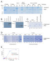
Coronin A-F-actin interaction with muscle F-actin in vitro. (A). Purified FLAG-CorA and/or equimolar amounts of rabbit muscle G-actin or F-actin was subjected to ultracentrifugation at 100,000× g, for 1h at 4 °C in the presence of rabbit muscle G-actin and F-actin and different NaCl concentrations. The supernatant was removed and the pellet resuspended in 2× SDS-PAGE sample buffer. Samples were separated by SDS-PAGE and the gel stained using Coomassie blue. Lanes 1–4: controls; lanes 5–8: coronin A and G-actin; lanes 9–14: Sedimentation analysis was performed in the presence of the NaCl concentrations indicated. (B). Rabbit muscle F-actin or G-actin were incubated in the absence or presence of S. cerevisiae Crn1, incubated for 20 min at room temperature and the samples were then processed as described in the Materials and Methods. Pellets (P) and supernatants (S) were separated by SDS-PAGE and immunoblotted for Crn1 (left panel) or actin (right panel) as described above. The lower band most likely represents a degradation product of Crn1. (C). Purified actin, FLAG-CorA or FLAG-CorAΔCC were analyzed as in A, separated by SDS-PAGE and the gel stained using Coomassie blue. (D). Interaction of the indicated amounts of purified FLAG-CorA (top) or FLAG-CorAΔCC (bottom) with rabbit muscle G- and F-actin was carried out as described in the Materials and Methods. Samples were separated by SDS-PAGE and the gel stained using Coomassie blue. (E). Plot of the ratio of rabbit muscle F-actin-bound (co-pelleting, P) to non-bound (S) for FLAG-CorA or FLAG-CorAΔCC determined from the mean grey values of the bands from the Coomassie blue stained gels. Curve fitting shows an apparent Kd of 8.9 μg (CI 5–20 μg). Shown are representative results from at least three (two in the case of panels D, E) independent experiments.
Figure 5
Figure 5
Time-dependent analysis of the coronin A interactome. Cells expressing either FLAG-CorA or FLAG-CorAΔCC were sampled at 24, 48 or 110 h, lysed, and subjected to AP-MS as described in Materials and Methods. The 25 most significant interactors (log2ratio > 1; q-value < 0.05) are shown. For clarity, proteins with unknown function are not represented. Note that the expression of FLAG-CorAΔCC is ~40-fold enriched relative to FLAG-CorA. See also Table S1.
Figure 6
Figure 6
Phagocytosis in the presence and absence of coronin A. (A): Histogram profiles of the distribution and percentage of wild type and corA cells that have phagocytosed (S1) live E. coli after different time points of incubation. Incubation at 4 °C (control) showed significant reduction of phagocytosis. Shown are representative results from at least 3 independent experiments. (BE): Plot of the percentage of wild type, corA, corA + CorA, corA + FLAG-CorA and corA + FLAG-CorAΔCC DH1-10 cells that have taken up beads of the indicated sizes (B) live E. coli (C), heat-killed bacteria (D), or heat-killed yeast (E), respectively (error bar = standard error; ** p < 0.002).

References

    1. Pieters J., Muller P., Jayachandran R. On guard: Coronin proteins in innate and adaptive immunity. Nat. Rev. Immunol. 2013;13:510–518. doi: 10.1038/nri3465. - DOI - PubMed
    1. de Hostos E.L. A brief history of the coronin family. Subcell. Biochem. 2008;48:31–40. - PubMed
    1. Eckert C., Hammesfahr B., Kollmar M. A holistic phylogeny of the coronin gene family reveals an ancient origin of the tandem-coronin, defines a new subfamily, and predicts protein function. BMC Evolut. Biol. 2011;11:268. doi: 10.1186/1471-2148-11-268. - DOI - PMC - PubMed
    1. Goode B.L., Wong J.J., Butty A.C., Peter M., McCormack A.L., Yates J.R., Drubin D.G., Barnes G. Coronin promotes the rapid assembly and cross-linking of actin filaments and may link the actin and microtubule cytoskeletons in yeast. J. Cell Biol. 1999;144:83–98. doi: 10.1083/jcb.144.1.83. - DOI - PMC - PubMed
    1. Tardieux I., Liu X., Poupel O., Parzy D., Dehoux P., Langsley G. A Plasmodium falciparum novel gene encoding a coronin-like protein which associates with actin filaments. FEBS Lett. 1998;441:251–256. doi: 10.1016/S0014-5793(98)01557-9. - DOI - PubMed

LinkOut - more resources