

Choroidal Vascularity Index: An In-Depth Analysis of This Novel Optical Coherence Tomography Parameter
- PMID: 32098215
- PMCID: PMC7074450
- DOI: 10.3390/jcm9020595
Choroidal Vascularity Index: An In-Depth Analysis of This Novel Optical Coherence Tomography Parameter
Abstract
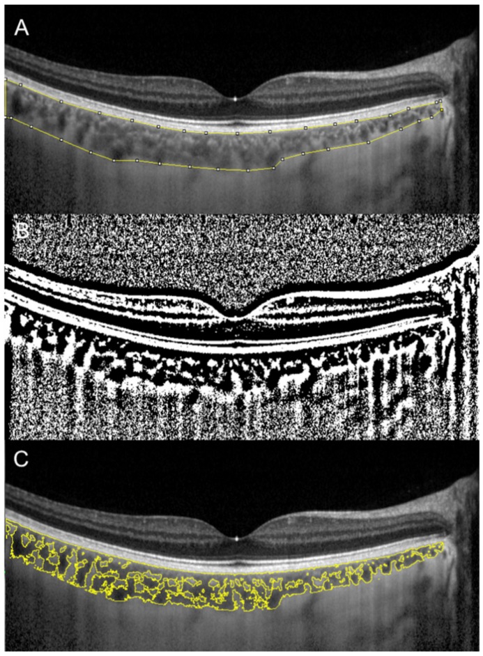
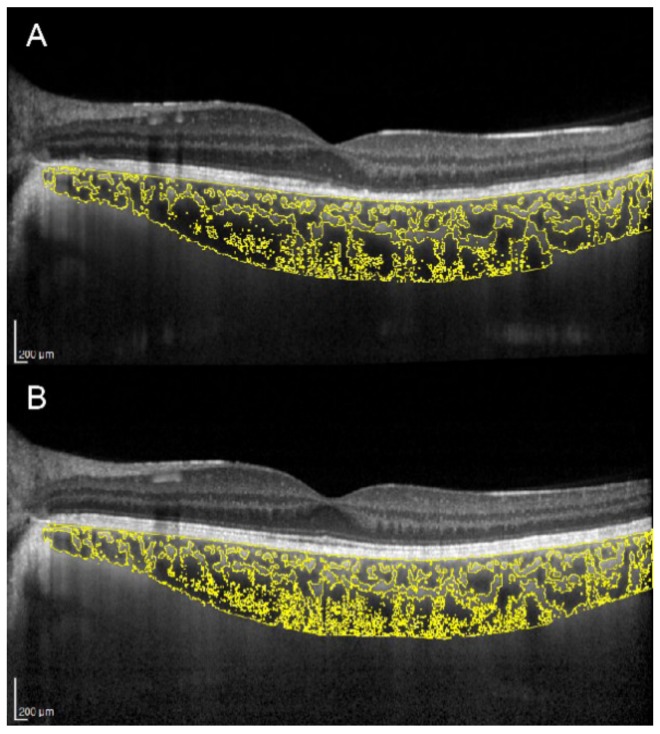
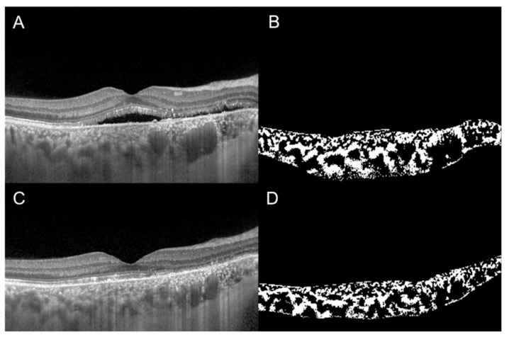
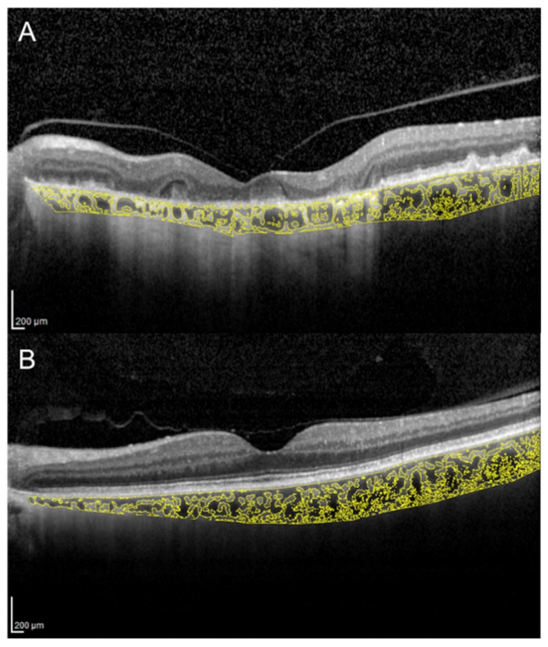
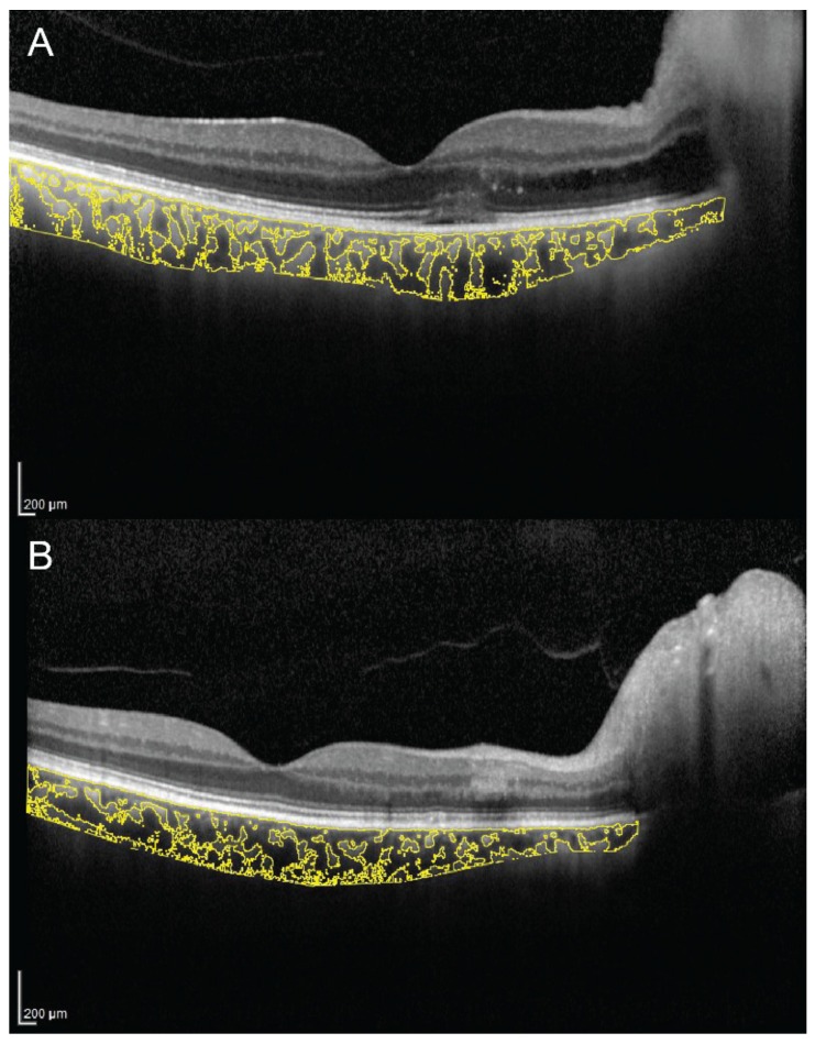
Remarkable improvements in optical coherence tomography (OCT) technology have resulted in highly sophisticated, noninvasive machines allowing detailed and advanced morphological evaluation of all retinal and choroidal layers. Postproduction semiautomated imaging analysis with dedicated public-domain software allows precise quantitative analysis of binarized OCT images. In this regard, the choroidal vascularity index (CVI) is emerging as a new imaging tool for the measurement and analysis of the choroidal vascular system by quantifying both luminal and stromal choroidal components. Numerous reports have been published so far regarding CVI and its potential applications in healthy eyes as well as in the evaluation and management of several chorioretinal diseases. Current literature suggests that CVI has a lesser variability and is influenced by fewer physiologic factors as compared to choroidal thickness. It can be considered a relatively stable parameter for evaluating the changes in the choroidal vasculature. In this review, the principles and the applications of this advanced imaging modality for studying and understanding the contributing role of choroid in retinal and optic nerve diseases are discussed. Potential advances that may allow the widespread adoption of this tool in the routine clinical practice are also presented.
Keywords: choroidal imaging biomarkers; choroidal vascularity index; optical coherence tomography; retinal imaging.
Conflict of interest statement
The authors declare no conflict of interest.
Figures
References
-
- Linsenmeier R.A., Padnick-Silver L. Metabolic dependence of photoreceptors on the choroid in the normal and detached retina. Investig. Ophthalmol. Vis. Sci. 2000;41:3117–3123. - PubMed
-
- Alm A., Bill A. Ocular and optic nerve blood flow at normal and increased intraocular pressures in monkeys (Macaca irus): A study with radioactively labelled microspheres including flow determinations in brain and some other tissues. Exp. Eye Res. 1973;15:15–29. doi: 10.1016/0014-4835(73)90185-1. - DOI - PubMed
Publication types
LinkOut - more resources
Full Text Sources
Other Literature Sources
