Review
. 2020 Feb 21;21(4):1483.
doi: 10.3390/ijms21041483.

Lumbar Degenerative Disease Part 1: Anatomy and Pathophysiology of Intervertebral Discogenic Pain and Radiofrequency Ablation of Basivertebral and Sinuvertebral Nerve Treatment for Chronic Discogenic Back Pain: A Prospective Case Series and Review of Literature

Affiliations
Review

Lumbar Degenerative Disease Part 1: Anatomy and Pathophysiology of Intervertebral Discogenic Pain and Radiofrequency Ablation of Basivertebral and Sinuvertebral Nerve Treatment for Chronic Discogenic Back Pain: A Prospective Case Series and Review of Literature

Hyeun Sung Kim et al. Int J Mol Sci. .

Abstract

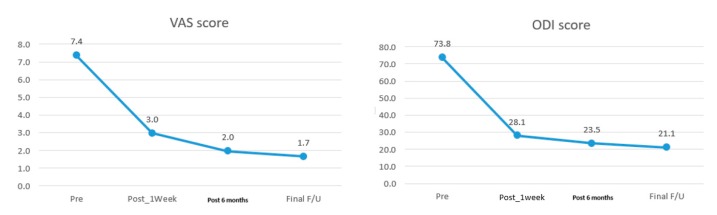
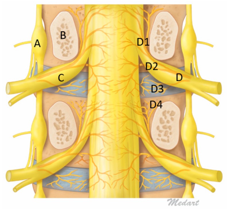
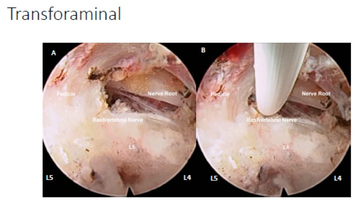
Degenerative disc disease is a leading cause of chronic back pain in the aging population in the world. Sinuvertebral nerve and basivertebral nerve are postulated to be associated with the pain pathway as a result of neurotization. Our goal is to perform a prospective study using radiofrequency ablation on sinuvertebral nerve and basivertebral nerve; evaluating its short and long term effect on pain score, disability score and patients' outcome. A review in literature is done on the pathoanatomy, pathophysiology and pain generation pathway in degenerative disc disease and chronic back pain. 30 patients with 38 levels of intervertebral disc presented with discogenic back pain with bulging degenerative intervertebral disc or spinal stenosis underwent Uniportal Full Endoscopic Radiofrequency Ablation application through either Transforaminal or Interlaminar Endoscopic Approaches. Their preoperative characteristics are recorded and prospective data was collected for Visualized Analogue Scale, Oswestry Disability Index and MacNab Criteria for pain were evaluated. There was statistically significant Visual Analogue Scale improvement from preoperative state at post-operative 1wk, 6 months and final follow up were 4.4 ± 1.0, 5.5 ± 1.2 and 5.7 ± 1.3, respectively, p < 0.0001. Oswestery Disability Index improvement from preoperative state at 1week, 6 months and final follow up were 45.8 ± 8.7, 50.4 ± 8.2 and 52.7 ± 10.3, p < 0.0001. MacNab criteria showed excellent outcomes in 17 cases, good outcomes in 11 cases and fair outcomes in 2 cases Sinuvertebral Nerve and Basivertebral Nerve Radiofrequency Ablation is effective in improving the patients' pain, disability status and patient outcome in our study.

Keywords: basivertebral nerve; degenerative disc disease; discogenic back pain; endoscopic spine surgery; lumbar spondylosis; pain management; pathophysiology of back pain; prolapsed intervertebral disc; radiofrequency ablation; sinuvertebral nerve.


Conflict of interest statement

The authors declare no conflict of interest. The funders had no role in the design of the study; in the collection, analyses, or interpretation of data; in the writing of the manuscript, or in the decision to publish the results.

Figures

Figure 1
Figure 1
Mean Visual Analogue Scale Pre-operative, 1 week, 6 months post-operatively and Oswestry Disability Index at 1 week, 6 months post-operatively and final follow up showing clinically significant decrease in disability.
Figure 2
Figure 2
Figure of Coronal Mid Pedicle Cut of Lumbar Spine. A: Sympathetic ganglion, B: Pedicle, C: Dorsal Root Ganglion, D: Sinuvertebral Nerve giving rise to branches D1: Ascending branch which goes intraosseous and give rise to Basivertebral Nerve near the pedicle D4, D2: Descending Branch supplying adjacent to Posterior longitudinal ligament and disc, D3: Direct branches to intervertebral disc.
Figure 3
Figure 3
Radiofrequency ablation of the sinuvertebral nerve and basivertebral nerve. In (A), normal disc, there is a presence of sinuvertebral nerve branches but no pathological neurotization and no neovascularization. (B) Pathological neovascularization and neurotization around the disc with hyperalgesia from signals transmitted by the sinuvertebral nerves and basivertebral nerves. (C) When radiofrequency is applied to the pathological hyperalgesic nerve fibers, there is twitching of the patient’s buttock muscles. (D) As radiofrequency ablation continues, the pathological neurotized nerve fibers are coagulated and twitching of the buttock muscles stopped in our case series. The patients performed better in clinical outcomes if they had initial buttock twitching and subsequent stoppage of twitching after radiofrequency ablation.
Figure 4
Figure 4
Provocative discogram at L1/2. The left picture shows the landmark of the Antero Posterior view of the L1/2 disc entry point. The right picture shows the lateral view with provocative discogram, the patient had immediate axial back pain and left buttock pain which is similar in nature to her chronic back pain.
Figure 5
Figure 5
(A) Degenerative disc disease with Pfirrmann grade III disc, showing inhomogeneous structure, and an unclear distinction of nucleus and annulus and intermediate signal intensity in T2 image with slightly decreased disc height and disc bulge. (B) Radiofrequency ablation of the disc, sinuvertebral nerve and basivertebral nerve showing shrinkage of degenerative disc and there is an increase in the signal of Modic changes in the adjacent vertebra body.
Figure 6
Figure 6
(A) Uniportal interlaminar endoscopic approach to disc showing the relationship of the lumbar five (L5) traversing nerve root which is retracted away by the working channel, exposing the disc of L4/5, the basivertebral nerve is located above pedicle of left L5, there is grade 3 neovascularization and inflammatory granulation tissue with adhesion around the basivertebral nerve region. In most circumstances, the basivertebral nerve is too fine to be seen by endoscopic vision. (B) Same region in the same patient after radiofrequency was applied to shrink the degenerative disc, and to ablate the pathological neovascularization with underlying inflammatory tissues and the basivertebral nerve, the typical response is twitching of the buttock when the correct location of the basivertebral nerve is ablated. (C) Another patient with similar steps in retraction of traversing nerve root and exposing neovascularized tissue and the location of the basivertebral nerve. (D) Radiofrequency ablation applied to neovascularized tissue, disc and basivertebral nerve.
Figure 7
Figure 7
(A) Right transforaminal endoscopic approach of L4/5 disc space, under endoscopic view we can assess the traversing nerve root of L5 to be decompressed, L5 pedicle and location of basivertebral nerve above the pedicle are observed. (B) Application of radiofrequency ablation on the basivertebral nerve.
Figure 8
Figure 8
Drawings of the neovascularization grading system. Left picture showing a grade 1 normal appearance with sparse epidural vessels around the disc. The middle picture showed a grade 2 increased neovascularization of epidural vessels with vascularization. The right picture showed grade 3 with increased neovascularization of epidural vessels with vascularization and adhesion on neural tissues.
Figure 9
Figure 9
Sagittal cuts across L5/S1 in a patient with spinal canal stenosis, degenerative disc disease of L5/S1 with disc protrusion into the spinal canal and vertebral bodies of L4 and L5, there are Type 1 Modic changes signifying bone marrow edema and inflammation. The corresponding axial and sagittal cuts are labelled (AC) accordingly. On axial cuts, we can see preoperative spinal canal stenosis with disc bulge and ligamentum hypertrophy with Schiaz grade A3 with rootlets lying dorsally occupying more than half of the dura sac area. This patient suffered buttock pain and both leg pain especially right leg pain for 5 months. The patient visited the clinic with visual analogue scale (VAS) score of 8 that was aggravated from 3 months ago, especially buttock pain and right leg pain, in spite of the preoperative MRI and spinal stenosis was not severe (Schiaz Grade A3). We can check the severe Modic change (Type 1) in the adjacent vertebrae. The patient had decompressed and radiofrequency ablation to sinuvertebral and basivertebral nerves in MRI postoperative day 1 which had been maintained in a 1-year follow up. There is shrinkage of the disc and less marrow changes in bone edema. The patient’s final follow-up VAS was 1 and returned to work as per normal.

References

    1. Vos T.P., Flaxman A.D.P., Naghavi M.P., Lozano R.P., Michaud C.M.D., Ezzati M.P., Shibuya K.P., Salomon J.A.P., Abdalla S.M., Aboyans V.P., et al. Years lived with disability (YLDs) for 1160 sequelae of 289 diseases and injuries 1990–2010: A systematic analysis for the Global Burden of Disease Study 2010. Lancet. 2012;380:2163–2196. doi: 10.1016/S0140-6736(12)61729-2. - DOI - PMC - PubMed
    1. Zhu Q., Gao X., Levene H.B., Brown M.D., Gu W. Influences of Nutrition Supply and Pathways on the Degenerative Patterns in Human Intervertebral Disc. Spine (Phila Pa 1976) 2016;41:568–576. doi: 10.1097/BRS.0000000000001292. - DOI - PMC - PubMed
    1. Zawilla N.H., Darweesh H., Mansour N., Helal S., Taha F.M., Awadallah M., El Shazly R. Matrix metalloproteinase-3, vitamin D receptor gene polymorphisms, and occupational risk factors in lumbar disc degeneration. J. Occup. Rehabil. 2014;24:370–381. doi: 10.1007/s10926-013-9472-7. - DOI - PubMed
    1. Willems P. Decision making in surgical treatment of chronic low back pain: The performance of prognostic tests to select patients for lumbar spinal fusion. Acta Orthop. 2013;84:1–35. doi: 10.3109/17453674.2012.753565. - DOI - PubMed
    1. Wong J.J., Cote P., Sutton D.A., Randhawa K., Yu H., Varatharajan S., Goldgrub R., Nordin M., Gross D.P., Shearer H.M., et al. Clinical practice guidelines for the noninvasive management of low back pain: A systematic review by the Ontario Protocol for Traffic Injury Management (OPTIMa) Collaboration. Eur. J. Pain. 2017;21:201–216. doi: 10.1002/ejp.931. - DOI - PubMed

MeSH terms